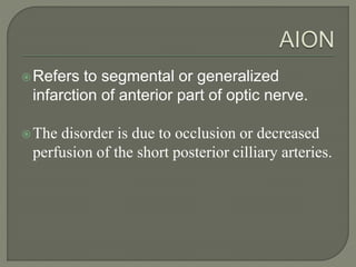
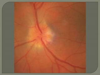
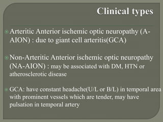
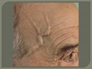
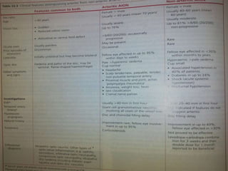
2. Refers to segmental or generalized
infarction of anterior part of optic nerve.
The disorder is due to occlusion or decreased
perfusion of the short posterior cilliary arteries.
3. Sudden moderate to severe painless loss of
vision
pupil:relative afferent pupillary defect(RAPD)
Fundus: pale disc swelling often involving only a
segment of the disc, flame-shaped hemorrhages,
optic atrophy after the edema resolves
Visual field: altitudinal hemianopia involving
superior or inferior half or central visual field
defect
4.
5. Arteritic Anterior ischemic optic neuropathy (A-
AION) : due to giant cell arteritis(GCA)
Non-Arteritic Anterior ischemic optic neuropathy
(NA-AION) : may be associated with DM, HTN or
atherosclerotic disease
GCA: have constant headache(U/L or B/L) in temporal area
with prominent vessels which are tender, may have
pulsation in temporal artery
6.
7.
8. ESR & C-reactive protein raised in GCA
Temporal artery biopsy confirmatory in
GCA
VFA: altitudinal hemianopia
FFA: hypo-perfusion, poor filling of a
portion of optic disc
9. Arteritic AION :
* treat as medical emergency
* do not wait for biopsy results
* prompt steroid therapy can prevent bilateral
blindness ( treat
with systemic corticosteroids → start IV
methyl prednisolone 500mg followed by high dose
oral prednisolone(1mg/kg/day), taper when ESR
falls, maintenance dose 5-15mg 6-12 months
10. Non – arteritic AION : unlike A-AION, there is no
established treatment
Levodopa- carbidopa combination may be given
11. Similar features of AION except site of
involvement
Due to disorder affecting small pial vessels
which supply intraorbital portion of optic
nerve
Occurs in GCA, SLE, or conditions which
produce acute systemic hypotension