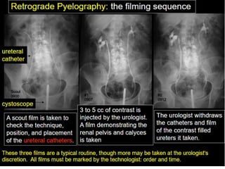
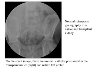
Retrograde Pyelography
Dr. Yash Kumar Achantani
OSR
DEFINITION
It is the roentgenographic demonstration of the renal
pelvis and ureter by the retrograde injection of radio-
opaque material through the ureters.
INDICATIONS
• Absent or unsatisfactory visualisation of the
collecting system on IVU.
• Unexplained hematuria, when the ureters have not
been completely visualised by IVU.
• Evaluating persistent intraureteral or intrapelvic
filling defects on IVU.
• Demonstrating the exact site of ureteral fistula.
• Brushing and/ or biopsy of suspected lesions.
• Evaluating the collecting system in patients who
cannot receive intravenous contrast medium
CONTRAINDICATIONS
• Acute urinary tract infection.
• Recent instrumentation.
• Pregnancy
CONTRAST MEDIUM
• Ionic contrast media can be used safely, however if
there is any specific contraindication like known
hypersensitivity etc., Non ionic contrast media may
be used. The Ionic contrast media is preferred due to
its low cost. The strength of contrast media should be
150-200 mg/ml.
• Contrast media should not be too dense as it will
obscure small lesions in the ureters and the pelvis
PROCEDURE
Patient Preparation
• Bowel preparation with cathartics is not routinely
performed.
Preliminary Film
• Full length supine AP abdomen before the
examination is started.
Anaesthesia
• May be performed under local anaesthesia although
general anesthesia is often required. Sterile
precautions are mandatory.
Technique
• The surgeon catheterizes the ureter via a cystoscope
and advances the ureteric catheter to the desired level.
Contrast medium is injected under fluoroscopic
control and spot films are exposed.
• Urine is aspirated and under fluoroscopic control
contrast medium is slowly injected. About 3-5 ml is
usually enough to fill the pelvis but the injection
should be terminated before this if the patient
complains of pain or fullness in the loin.
Films
(a) Supine PA film of the kidney
(b) Both 35° anterior obliques of the kidneys. Low kVp
(65-75 kVp) technique is used to visualise calculi and
contrast medium.
(c) If there is pelvi-ureteric junction obstruction, the
contrast medium in the pelvis is aspirated. The films are
examined and if satisfactory, the catheter is withdrawn,
first to 10 cm below the renal pelvis and then to lie
above the ureteric orifice. About 2ml of contrast
medium is injected at each of these levels and films
taken.
AFTER CARE
1. Postanaesthetic observation.
2. Prophylatic antibiotics may be used.
COMPLICATIONS
• Complications of general anaesthesia.
• Contrast medium can be absorbed from the renal
pelvis, giving rise to adverse reactions. However, the
risks are much less than with excretory urography.
• Chemical pyelitis-if there is stasis of contrast
medium.
• Extravasation due to overdistension of the pelvis.
• Introduction of infection.
• Mucosal damage to the ureter.
• Perforation of the ureter or pelvis by the catheter.
On the scout image, there are ureteral catheter positioned in the
transplant ureter (right) and native left ureter.
Normal retrograde
pyelography of a
native and transplant
kidney
The transplant ureter, the ureteral anastomosis, and the distal right
ureter are normal in appearance.
Other than mild blunting of the left renal calyces, the left ureter and
upper collecting system are also normal.
Retrograde pyelogram demonstrating
the pelvicalyceal system and ureter down
to the vesicoureteric junction.
Retrograde pyelogram with pyelosinus extravasation due to
overdistension of the collecting system.
Retrograde pyelogram shows rounded filling defects in many
calyces representing papillary necrosis.
Goblet sign or
Champagne glass sign:
• irregular ureteric
narrowing with proximal
shouldering termed the
goblet sign
• Focally dilated
intraluminal mass
Causes of goblet cells
• TCC of ureter
• Metastatic disease in to
ureter
• Endometriosis involving
ureter
A case of TCC
Filling defect is seen in the
right ureter –
RAT BITE SIGN
URETERAL
STRICTURE
Retrograde pyelogram shows the area of narrowing at the site
of ureteral implantation into the bladder (arrow).
URETERAL DIVERTICULA
URETERAL STENOSIS
Two views of the retrograde pyelography showing stenosis of the
lower portion of the right ureter at the level of the iliac vessels. There
is severe dilatation of the medial and proximal portions of the right
ureter and hydronephrosis.
Retrograde Pyelography

Retrograde Pyelography

  • 1.
  • 2.
    DEFINITION It is theroentgenographic demonstration of the renal pelvis and ureter by the retrograde injection of radio- opaque material through the ureters.
  • 3.
    INDICATIONS • Absent orunsatisfactory visualisation of the collecting system on IVU. • Unexplained hematuria, when the ureters have not been completely visualised by IVU. • Evaluating persistent intraureteral or intrapelvic filling defects on IVU. • Demonstrating the exact site of ureteral fistula. • Brushing and/ or biopsy of suspected lesions. • Evaluating the collecting system in patients who cannot receive intravenous contrast medium
  • 4.
    CONTRAINDICATIONS • Acute urinarytract infection. • Recent instrumentation. • Pregnancy
  • 5.
    CONTRAST MEDIUM • Ioniccontrast media can be used safely, however if there is any specific contraindication like known hypersensitivity etc., Non ionic contrast media may be used. The Ionic contrast media is preferred due to its low cost. The strength of contrast media should be 150-200 mg/ml. • Contrast media should not be too dense as it will obscure small lesions in the ureters and the pelvis
  • 6.
    PROCEDURE Patient Preparation • Bowelpreparation with cathartics is not routinely performed. Preliminary Film • Full length supine AP abdomen before the examination is started. Anaesthesia • May be performed under local anaesthesia although general anesthesia is often required. Sterile precautions are mandatory.
  • 7.
    Technique • The surgeoncatheterizes the ureter via a cystoscope and advances the ureteric catheter to the desired level. Contrast medium is injected under fluoroscopic control and spot films are exposed. • Urine is aspirated and under fluoroscopic control contrast medium is slowly injected. About 3-5 ml is usually enough to fill the pelvis but the injection should be terminated before this if the patient complains of pain or fullness in the loin.
  • 8.
    Films (a) Supine PAfilm of the kidney (b) Both 35° anterior obliques of the kidneys. Low kVp (65-75 kVp) technique is used to visualise calculi and contrast medium. (c) If there is pelvi-ureteric junction obstruction, the contrast medium in the pelvis is aspirated. The films are examined and if satisfactory, the catheter is withdrawn, first to 10 cm below the renal pelvis and then to lie above the ureteric orifice. About 2ml of contrast medium is injected at each of these levels and films taken.
  • 9.
    AFTER CARE 1. Postanaestheticobservation. 2. Prophylatic antibiotics may be used.
  • 10.
    COMPLICATIONS • Complications ofgeneral anaesthesia. • Contrast medium can be absorbed from the renal pelvis, giving rise to adverse reactions. However, the risks are much less than with excretory urography. • Chemical pyelitis-if there is stasis of contrast medium. • Extravasation due to overdistension of the pelvis. • Introduction of infection. • Mucosal damage to the ureter. • Perforation of the ureter or pelvis by the catheter.
  • 12.
    On the scoutimage, there are ureteral catheter positioned in the transplant ureter (right) and native left ureter. Normal retrograde pyelography of a native and transplant kidney
  • 13.
    The transplant ureter,the ureteral anastomosis, and the distal right ureter are normal in appearance. Other than mild blunting of the left renal calyces, the left ureter and upper collecting system are also normal.
  • 14.
    Retrograde pyelogram demonstrating thepelvicalyceal system and ureter down to the vesicoureteric junction.
  • 15.
    Retrograde pyelogram withpyelosinus extravasation due to overdistension of the collecting system.
  • 16.
    Retrograde pyelogram showsrounded filling defects in many calyces representing papillary necrosis.
  • 17.
    Goblet sign or Champagneglass sign: • irregular ureteric narrowing with proximal shouldering termed the goblet sign • Focally dilated intraluminal mass Causes of goblet cells • TCC of ureter • Metastatic disease in to ureter • Endometriosis involving ureter
  • 18.
    A case ofTCC Filling defect is seen in the right ureter – RAT BITE SIGN
  • 19.
  • 20.
    Retrograde pyelogram showsthe area of narrowing at the site of ureteral implantation into the bladder (arrow).
  • 21.
  • 22.
    URETERAL STENOSIS Two viewsof the retrograde pyelography showing stenosis of the lower portion of the right ureter at the level of the iliac vessels. There is severe dilatation of the medial and proximal portions of the right ureter and hydronephrosis.