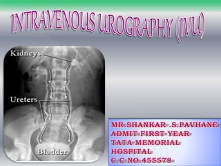
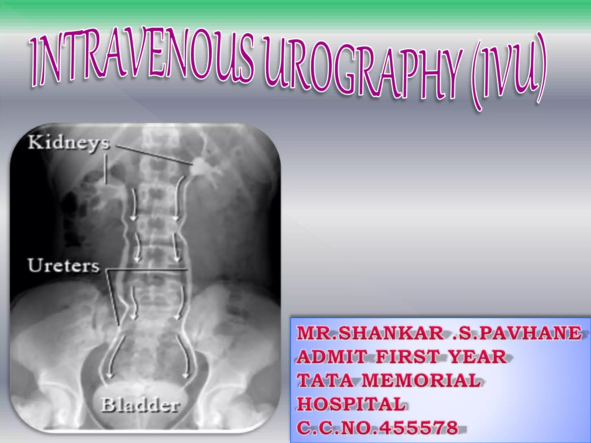
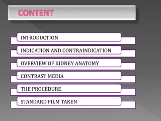
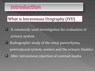
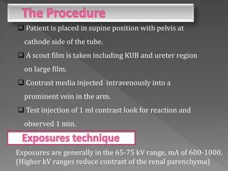
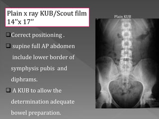
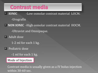
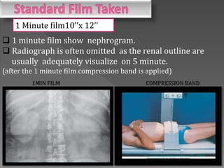
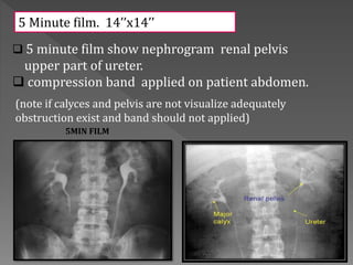
INTRODUCTION
INDICATION AND CONTRAINDICATION
OVERVIEW OF KIDNEY ANATOMY
CONTRAST MEDIA
THE PROCEDURE
STANDARD FILM TAKEN
What is Intravenous Urography (IVU)
 It commonly used investigation for evaluation of
urinary system.
 Radiographic study of the renal parenchyma,
pelvicalyceal system, ureters and the urinary bladder.
 After intravenous injection of contrast media.
 In suspected obstructive uropathies like stone etc.
 Congenital anomalies involving kidney.
 Investigation of hypertension.
 Suspected abdominal mass lesion arising from
kidney.
 In Blunt injuries of abdomen with Hematuria.
 Function of kidney.
 Bladder pathology –diverticulum fistula.
 Iodine sensitivity or previous reaction to
contrast media.
 Intractable cardial or renal failure.
 Multiple myeloma.
 Renal insufficiency.
 Thyrotoxicosis.
 The kidneys are a pair of fist-sized organs located at the
bottom of the rib cage.
 The organs of Urinary system.
-Kidneys
-Ureters
-Bladder and Urethra.
 Ask the patient history diabetics renal disease or allergy
to drugs
 Fasting for 4 hours.
 explain the exam to patient in layman language.
 Check the patient creatine level. (RFT. Normal range: 0.6-1.2mg)
Bowel Preparation
 Low residue diet.
 Bowel wash is given till bowel is clear of feacal
matter on previous night.
 Laxative Dulcolax is given 2-4 tablet at bedtime for
two day prior to exam .
 Patient is placed in supine position with pelvis at
cathode side of the tube.
 A scout film is taken including KUB and ureter region
on large film.
 Contrast media injected intravenously into a
prominent vein in the arm.
 Test injection of 1 ml contrast look for reaction and
observed 1 min.
Exposures are generally in the 65-75 kV range, mA of 600-1000.
(Higher kV ranges reduce contrast of the renal parenchyma)
Correct positioning .
 supine full AP abdomen
include lower border of
symphysis pubis and
diphrams.
 A KUB to allow the
determination adequate
bowel preparation.
Plain KUB
Plain x ray KUB/Scout film
14’’x 17’’
Contrast media is usually given as a IV bolus injection
within 30-60 sec.
 IONIC -Low osmolar contrast material LOCM.
-Urografin
 NON IONIC -High osmolar contrast material HOCM.
-Ultravist and Omnipaque.
 Adult dose
1-2 ml for each 1 kg.
 Pediatric dose
-1 ml for each 1 kg.
Mode of Injection
 1 minute film show nephrogram.
 Radiograph is often omitted as the renal outline are
usually adequately visualize on 5 minute.
(after the 1 minute film compression band is applied)
1 Minute film10’’x 12’’
 5 minute film show nephrogram renal pelvis
upper part of ureter.
 compression band applied on patient abdomen.
(note if calyces and pelvis are not visualize adequately
obstruction exist and band should not applied)
5 Minute film. 14’’x14’’
5MIN FILM
If compression is applied
10 minute film
center on kidney to
demonstrate distended
collecting system and
proximal ureter.
 supine position.
 All film taken in
expiratory phase.
10 Minute film. 14’’x 14’’
10MIN FILM
 Visualize the ureter in
prone position as they
fill better.
 Supine full length AP
 Compression is
released when
satisfactory
Demonstration of
pelvicalyceal system
has been achived.
15 Minute film. 14’’x 17’’
15MIN FILM
 Its give complete overview
of urinary track.
 Bladder distention can be
evaluated.
 Taken immediately after
voiding.
 its use to assess for
-Residual urine
-Bladder mucosal lesions
- Diverticula
- Bladder tumor
- Outlet obstruction.
Post Void film. 8’’x 10’’
35 Minute film. 14’’x 17’’
35MIN FILM
Oblique film:
 better visualize the calyaceal system.
 filling defect that may overlap in routine AP View
Prone film:
 better imaging the ureter.
Upright film:
 Layering of contrast media is in severely
hydronephrotic system.
Post void film:
 Filling defect in bladder post wall.
 Diverticula.
INTRAVENOUS UROGRAM

INTRAVENOUS UROGRAM

  • 2.
    INTRODUCTION INDICATION AND CONTRAINDICATION OVERVIEWOF KIDNEY ANATOMY CONTRAST MEDIA THE PROCEDURE STANDARD FILM TAKEN
  • 3.
    What is IntravenousUrography (IVU)  It commonly used investigation for evaluation of urinary system.  Radiographic study of the renal parenchyma, pelvicalyceal system, ureters and the urinary bladder.  After intravenous injection of contrast media.
  • 4.
     In suspectedobstructive uropathies like stone etc.  Congenital anomalies involving kidney.  Investigation of hypertension.  Suspected abdominal mass lesion arising from kidney.  In Blunt injuries of abdomen with Hematuria.  Function of kidney.  Bladder pathology –diverticulum fistula.
  • 5.
     Iodine sensitivityor previous reaction to contrast media.  Intractable cardial or renal failure.  Multiple myeloma.  Renal insufficiency.  Thyrotoxicosis.
  • 6.
     The kidneysare a pair of fist-sized organs located at the bottom of the rib cage.  The organs of Urinary system. -Kidneys -Ureters -Bladder and Urethra.
  • 8.
     Ask thepatient history diabetics renal disease or allergy to drugs  Fasting for 4 hours.  explain the exam to patient in layman language.  Check the patient creatine level. (RFT. Normal range: 0.6-1.2mg) Bowel Preparation  Low residue diet.  Bowel wash is given till bowel is clear of feacal matter on previous night.  Laxative Dulcolax is given 2-4 tablet at bedtime for two day prior to exam .
  • 9.
     Patient isplaced in supine position with pelvis at cathode side of the tube.  A scout film is taken including KUB and ureter region on large film.  Contrast media injected intravenously into a prominent vein in the arm.  Test injection of 1 ml contrast look for reaction and observed 1 min. Exposures are generally in the 65-75 kV range, mA of 600-1000. (Higher kV ranges reduce contrast of the renal parenchyma)
  • 10.
    Correct positioning . supine full AP abdomen include lower border of symphysis pubis and diphrams.  A KUB to allow the determination adequate bowel preparation. Plain KUB Plain x ray KUB/Scout film 14’’x 17’’
  • 11.
    Contrast media isusually given as a IV bolus injection within 30-60 sec.  IONIC -Low osmolar contrast material LOCM. -Urografin  NON IONIC -High osmolar contrast material HOCM. -Ultravist and Omnipaque.  Adult dose 1-2 ml for each 1 kg.  Pediatric dose -1 ml for each 1 kg. Mode of Injection
  • 12.
     1 minutefilm show nephrogram.  Radiograph is often omitted as the renal outline are usually adequately visualize on 5 minute. (after the 1 minute film compression band is applied) 1 Minute film10’’x 12’’
  • 13.
     5 minutefilm show nephrogram renal pelvis upper part of ureter.  compression band applied on patient abdomen. (note if calyces and pelvis are not visualize adequately obstruction exist and band should not applied) 5 Minute film. 14’’x14’’ 5MIN FILM
  • 14.
    If compression isapplied 10 minute film center on kidney to demonstrate distended collecting system and proximal ureter.  supine position.  All film taken in expiratory phase. 10 Minute film. 14’’x 14’’ 10MIN FILM
  • 15.
     Visualize theureter in prone position as they fill better.  Supine full length AP  Compression is released when satisfactory Demonstration of pelvicalyceal system has been achived. 15 Minute film. 14’’x 17’’ 15MIN FILM
  • 16.
     Its givecomplete overview of urinary track.  Bladder distention can be evaluated.  Taken immediately after voiding.  its use to assess for -Residual urine -Bladder mucosal lesions - Diverticula - Bladder tumor - Outlet obstruction. Post Void film. 8’’x 10’’ 35 Minute film. 14’’x 17’’ 35MIN FILM
  • 17.
    Oblique film:  bettervisualize the calyaceal system.  filling defect that may overlap in routine AP View Prone film:  better imaging the ureter. Upright film:  Layering of contrast media is in severely hydronephrotic system. Post void film:  Filling defect in bladder post wall.  Diverticula.