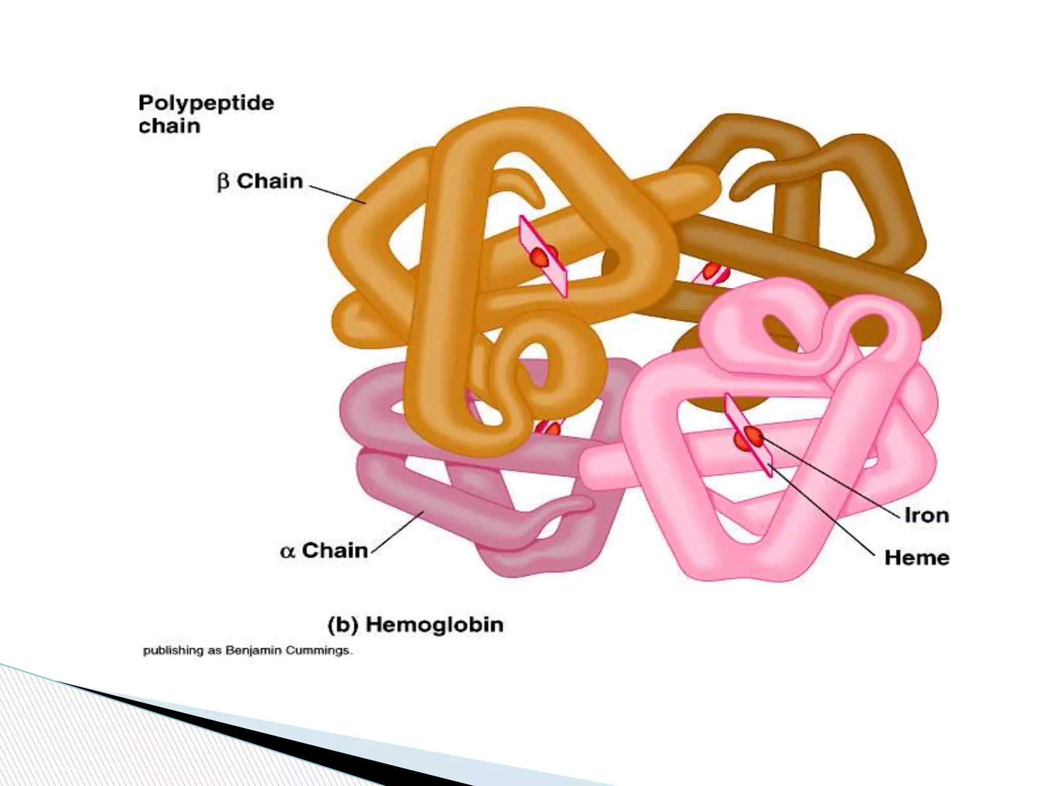
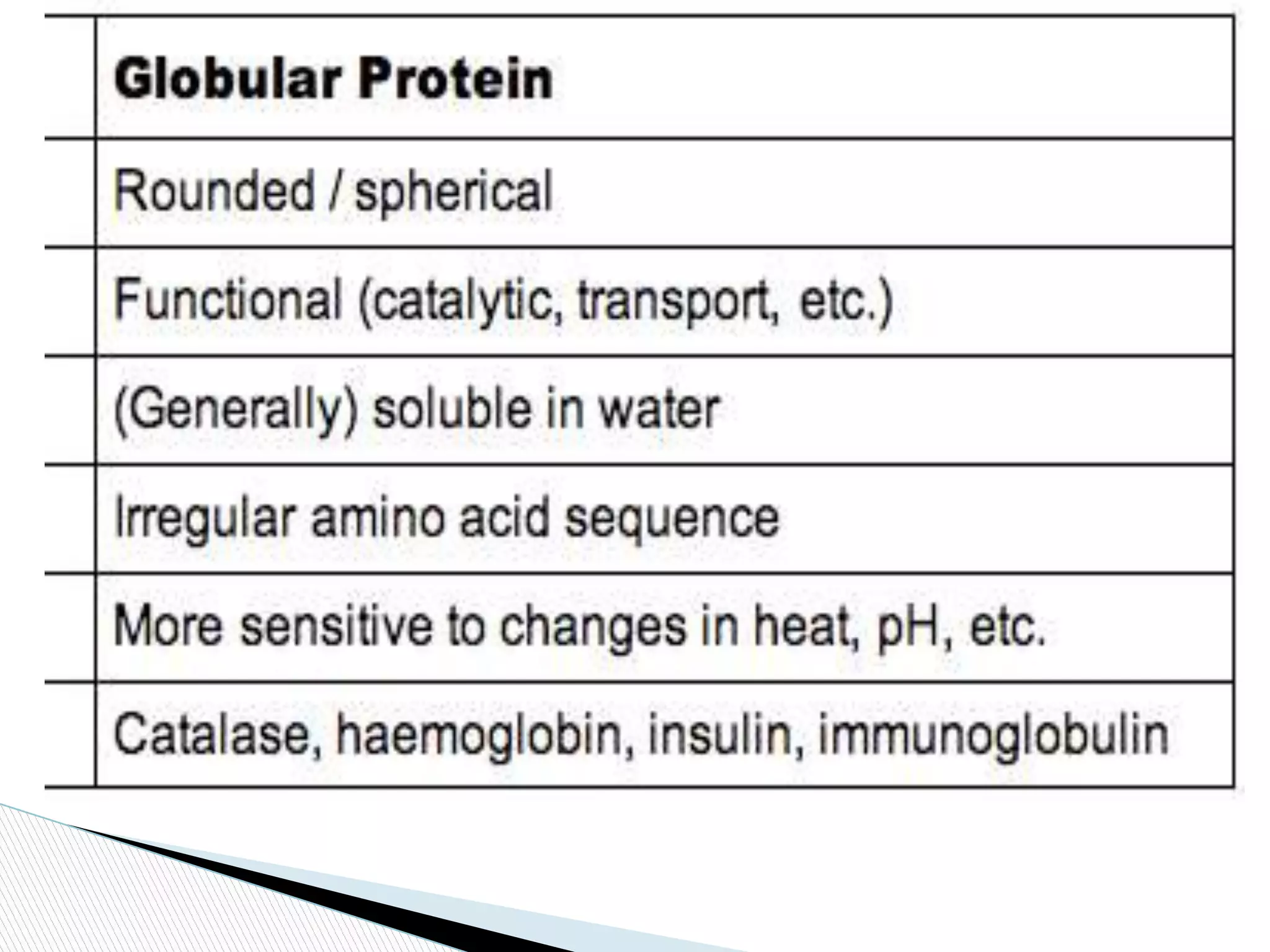
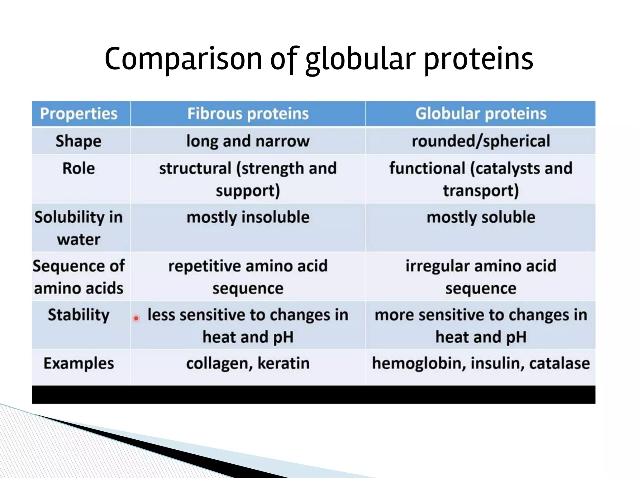
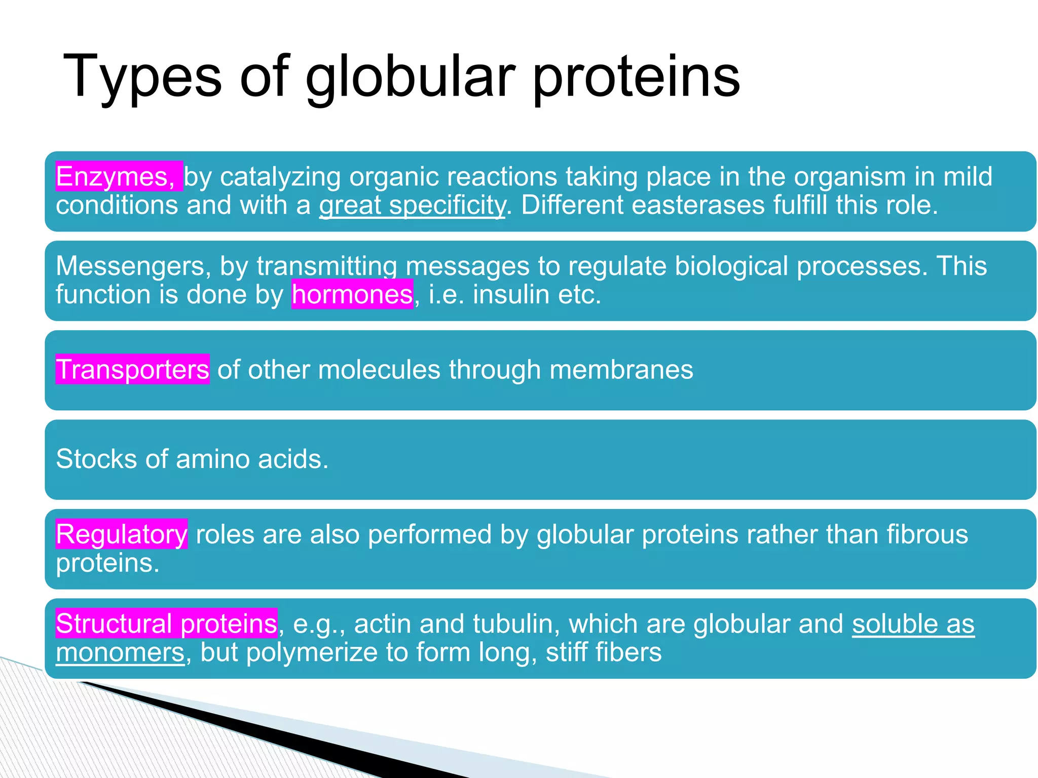
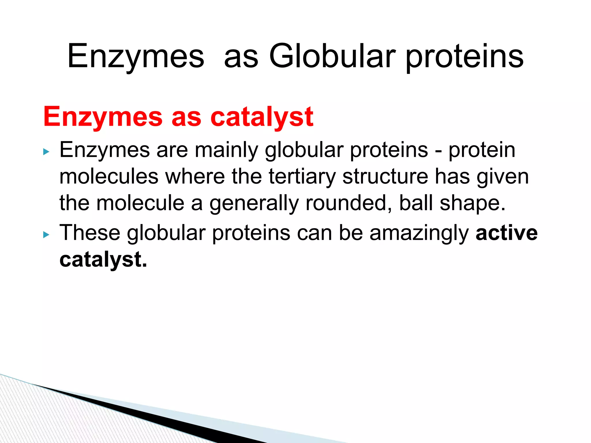
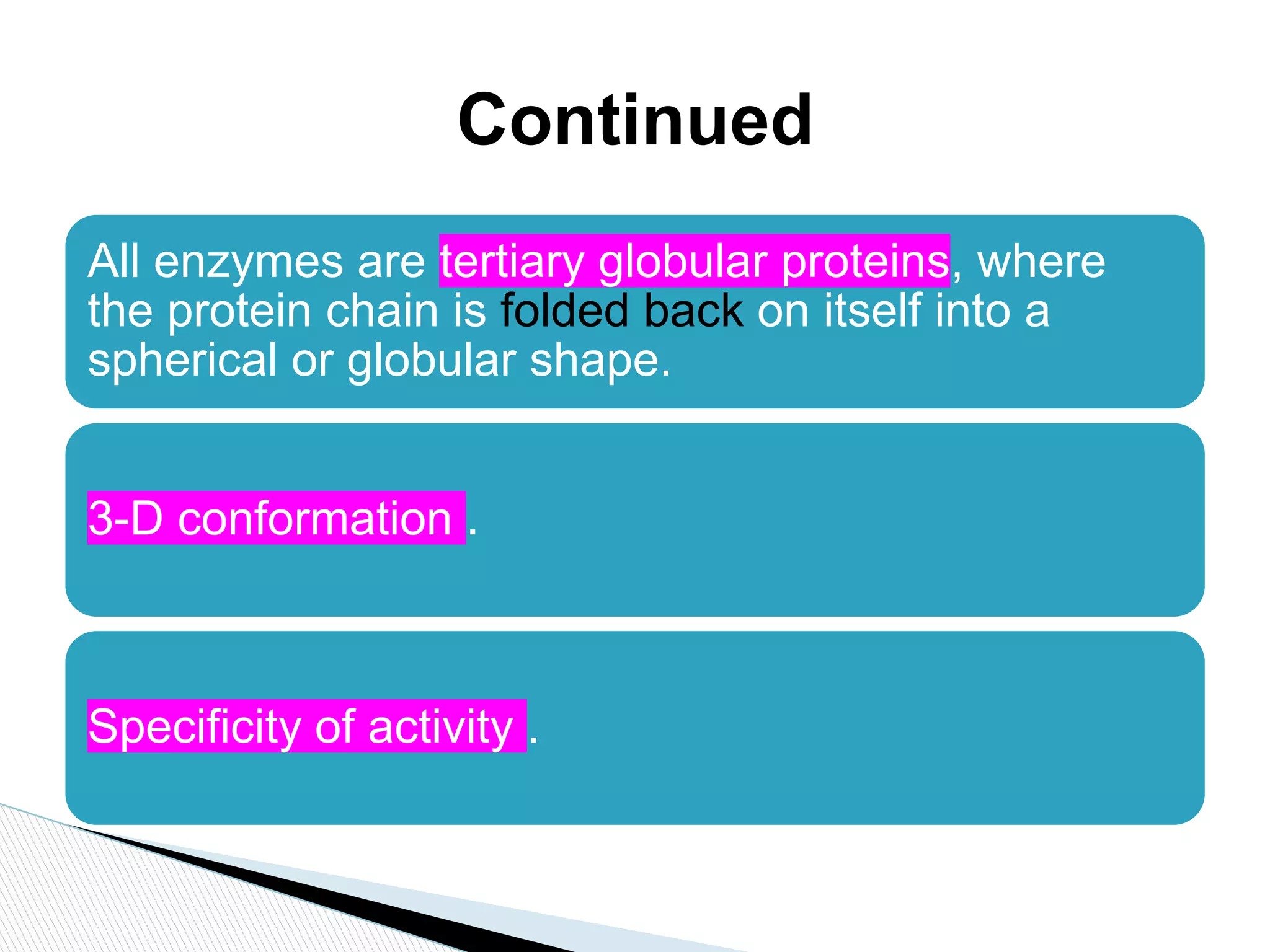
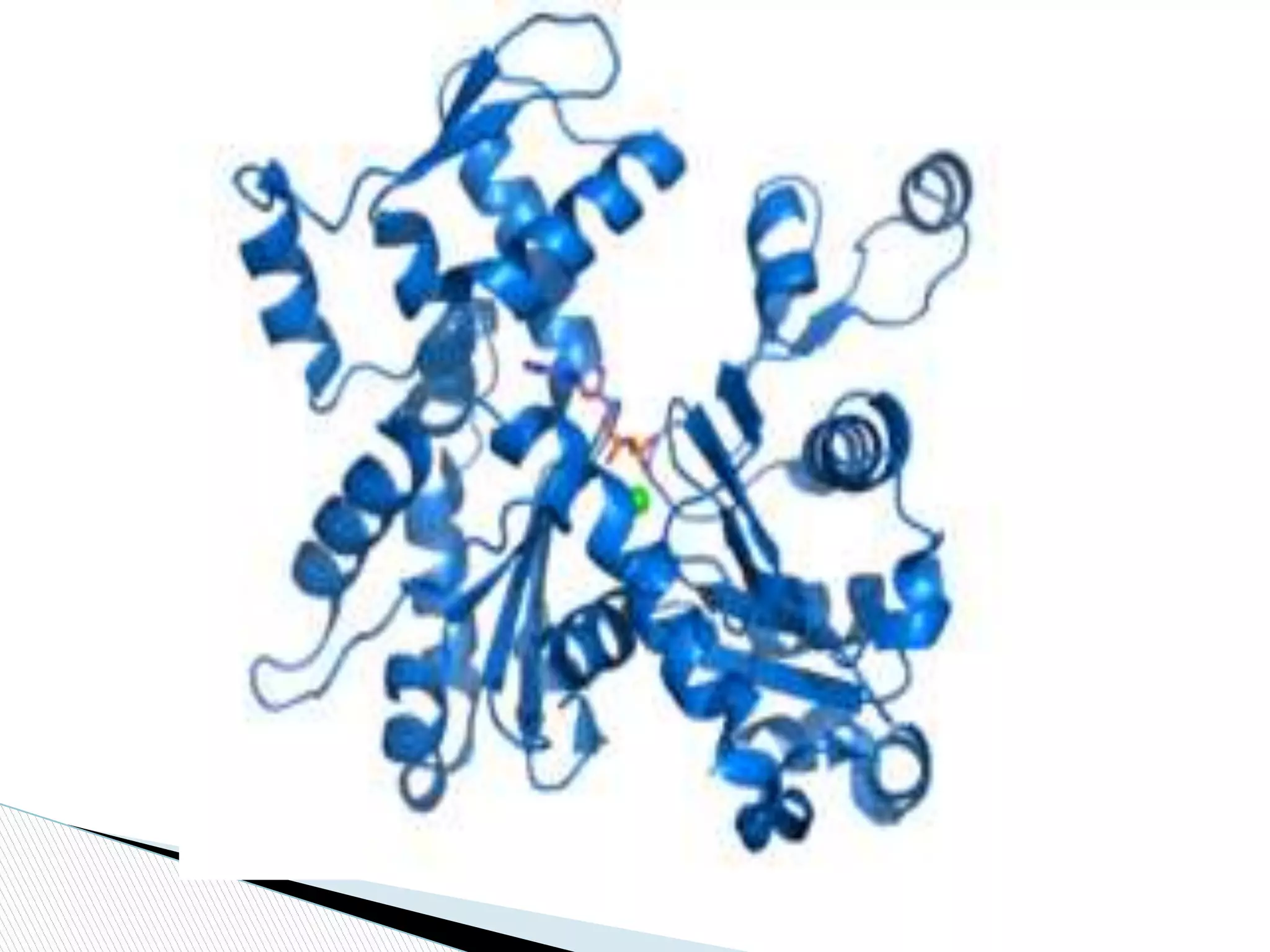
PRESENTED BY:
SABAHAT ALI(16-ARID-2569)
M.AHMED(16-ARID-2550)
GLOBULAR PROTEIN:
▶ Introduction to globular proteins
▶ Factor effecting the structure of Globular proteins
stability
▶ Denaturations
▶ Types of globular proteins
▶ Functions of different types of globular proteins
▶ Conclusion
▶ References
Contents
▶ A Globular protein is spherical in shape and has
the property of forming colloids with water.
▶ Water soluble
▶ Globular proteins are also called as
spheroproteins owing to their shape.
▶ globular proteins are soluble in water, acids and
bases.
▶ globular proteins they are held together by weak
intermolecular hydrogen bond.
Introduction to globular
protein
▶ Globular proteins are made up of not only
primary, secondary but also tertiary and
occasionally quaternary structures.
▶ Globular proteins consist of straight chains of
secondary structures which
abruptly join polypeptide chains and
change directions.
▶ more compact and rounded shape and have
functional roles.
▶ Carry out metabolism ,catalysis , synthesis and
storage reaction in cell.
Continued
➢ Loss of native conformations of tertiary structure.
➢ Denaturing proteins experience either the
destruction/disruption of internal tertiary or
secondary structure.
➢ Denaturation does not break the peptide bond
between adjacent amino acids, thus not affecting
the primary structure of the protein.
➢ Denaturation will interfere the normal alpha-helix
and beta sheets in a protein which ultimately
distort its 3D shape.
protein denaturation
▶ Hydrogen bonds
▶ Salt bridges
▶ Cofactors
▶ Disulphide bond
▶ Hydrophobic interactions
Factor effecting the structure of
Globular proteins stability
▶ Denaturation causes the disruption of hydrogen
bonding between close proximity amino acid.
▶ interfering a protein's secondary and tertiary
structure not primary.
▶ In tertiary structure there are four types of
bonding interactions between "side chains"
including: hydrogen bonding, ionic bridges,
disulfide bonds, and hydrophobic intermolecular
interactions.
Continued ………………..
▶ 1. Extreme pH (pH < 4 or pH > 9) : alters H-
bonding
▶ 2. Heat (temp >70oC): thermal effect, disrupts
weak forces of non-covalent bonds
▶ 3. Detergents or organic solvents : disrupts
hydrophobic interaction
▶ 4. Chaotropic agents (high concentrations) : e.g.,
urea and guanidinium chloride
Denaturation conditions
Comparison of globular proteins
Types of globular proteins
Enzymes, by catalyzing organic reactions taking place in the organism in mild
conditions and with a great specificity. Different easterases fulfill this role.
Messengers, by transmitting messages to regulate biological processes. This
function is done by hormones, i.e. insulin etc.
Transporters of other molecules through membranes
Stocks of amino acids.
Regulatory roles are also performed by globular proteins rather than fibrous
proteins.
Structural proteins, e.g., actin and tubulin, which are globular and soluble as
monomers, but polymerize to form long, stiff fibers
Function of protein
Enzymes as catalyst
▶ Enzymes are mainly globular proteins - protein
molecules where the tertiary structure has given
the molecule a generally rounded, ball shape.
▶ These globular proteins can be amazingly active
catalyst.
Enzymes as Globular proteins
All enzymes are tertiary globular proteins, where
the protein chain is folded back on itself into a
spherical or globular shape.
3-D conformation .
Specificity of activity .
Continued
Continued
❏ A hexokinase is an enzyme that phosphorylates
a six-carbon sugar, a hexose, to a hexose
phosphate
❏ In most tissues and organisms, glucose is the
most important substrate of hexokinases, and
glucose 6-phosphate the most important
product.
❏ Hexokinase is found in all mammalian tissues,
and is considered a "housekeeping enzyme.
Hexokinase:-
❏ Hexokinase II/B constitutes the principal regulated
isoform in many cell types and is increased in many
cancers.
❏ -Hexokinase III/C is substrate-inhibited by glucose
at physiologic concentrations. Little is known about the
regulatory characteristics of this isoform.
❏ -Hexokinase IV/D is also known as glucokinase
Continue…...
❏ The tertiary structure of hexokinase includes
an open alpha/beta sheet.
❏ The ATP-binding domain is composed of five beta
sheets and three alpha helices.
❏ four of the beta sheets are parallel and one is in the
anitparallel directions.
❏ .The alpha helices and beta loops connect the beta
sheets to produce this open alpha/beta sheet.
❏ The molecular weights of hexokinases are around 100
kD.
❏ Each consists of two similar 50kD halves, but only in
hexokinase II do both halves have functional active
sites
Hexokinase structure:-
❏ insulin is a protein composed of two chains
❏ A chain with 21 amino acids and a B chain
with 30 amino acids,
❏ these are linked together by sulfur atoms.
❏ Insulin is derived from a 74-amino-acid
prohormone molecule called proinsulin.
❏ Proinsulin is relatively inactive.
❏ Under normal conditions only a small
amount of it is secreted.
structure of insulin:-
❏ In the endoplasmic reticulum of beta cells the proinsulin
molecule is cleaved in two molecules
❏ The A and B chains of insulin and inactive C peptide is
produced.
❏ The A and B chains become linked together by two
sulfur-sulfur (disulfide) bonds.
Continue…….
❏ . Proinsulin, insulin, and C peptide are stored as
granules in the beta cells,
❏ then they are released into the capillaries of the islets
in response to appropriate stimuli.
❏ These capillaries empty into the portal vein,
Continue…….
❏ The pancreas of a normal adult contains
approximately 200 units of insulin.
❏ and the average daily secretion of insulin into the
circulation in healthy individuals ranges from 30 to 50
units.
Continue…...
Insulin, hormone that regulates the level of sugar (glucose) in
the blood
It is produced by the beta cells of the islets of Langerhans in the
pancreas.
Insulin is secreted when the level of blood glucose rises—as
after a meal.
When the level of blood glucose falls, secretion of insulin stops,
then the liver releases glucose into the blood.
function of insulin:-
Hemoglobin is the oxygen transporting protein of
red blood cells
hemoglubin is a globular protein with quaternary
structure.
Hemoglobin consists of four polypeptide subunits
2 alpha chains and two beta chains
structure of hemoglobin
Haemoglobin is found in red blood cells and
carries oxygen efficiently from the lungs to the
tissues in body.
It also aids in transporting hydrogen ions and
carbon dioxide back to the lungs.
The main component that binds with oxygen is the
iron.
Function……..
Similar to all the other proteins, haemoglobin is coded by DNA
. Mutations and changes in haemoglobin result in several blood
related diseases like sickle cell anemia,
A disease where the structure of the cell gets distorted
and do not have the capacity to carry much oxygen in the
required manner like a normal blood cell would.
structure determines function.
continue…..
❏ Actin is a family of globular multi-functional
proteins.
❏ It form microfilaments.
❏ It is found in essentially all eukaryotic cells
ACTIN:-
An actin protein is the monomeric
subunit of two types of filaments in cells
microfilaments, one of the three major
components of the cytoskeleton,
thin filaments, part of the contractile
apparatus in muscle cells.
Structure of Actin
❏ It can be present as either a free monomer called G-
actin
(globular) or as part of a linear polymer microfilament
called F-actin (filamentous),
both of which are essential for such important cellular
functions as the mobility and contraction of cells during cell
division.
Continue…...
Actin participates in many important cellular processes.
muscle contraction.
cell motility.
cell division
cytokinesis,
vesicle and organelle movement,
cell signaling,
the establishment and maintenance of cell junctions and cell shape
function of actin

Globular proteins

  • 1.
  • 2.
    ▶ Introduction toglobular proteins ▶ Factor effecting the structure of Globular proteins stability ▶ Denaturations ▶ Types of globular proteins ▶ Functions of different types of globular proteins ▶ Conclusion ▶ References Contents
  • 3.
    ▶ A Globularprotein is spherical in shape and has the property of forming colloids with water. ▶ Water soluble ▶ Globular proteins are also called as spheroproteins owing to their shape. ▶ globular proteins are soluble in water, acids and bases. ▶ globular proteins they are held together by weak intermolecular hydrogen bond. Introduction to globular protein
  • 4.
    ▶ Globular proteinsare made up of not only primary, secondary but also tertiary and occasionally quaternary structures. ▶ Globular proteins consist of straight chains of secondary structures which abruptly join polypeptide chains and change directions. ▶ more compact and rounded shape and have functional roles. ▶ Carry out metabolism ,catalysis , synthesis and storage reaction in cell. Continued
  • 5.
    ➢ Loss ofnative conformations of tertiary structure. ➢ Denaturing proteins experience either the destruction/disruption of internal tertiary or secondary structure. ➢ Denaturation does not break the peptide bond between adjacent amino acids, thus not affecting the primary structure of the protein. ➢ Denaturation will interfere the normal alpha-helix and beta sheets in a protein which ultimately distort its 3D shape. protein denaturation
  • 8.
    ▶ Hydrogen bonds ▶Salt bridges ▶ Cofactors ▶ Disulphide bond ▶ Hydrophobic interactions Factor effecting the structure of Globular proteins stability
  • 9.
    ▶ Denaturation causesthe disruption of hydrogen bonding between close proximity amino acid. ▶ interfering a protein's secondary and tertiary structure not primary. ▶ In tertiary structure there are four types of bonding interactions between "side chains" including: hydrogen bonding, ionic bridges, disulfide bonds, and hydrophobic intermolecular interactions. Continued ………………..
  • 10.
    ▶ 1. ExtremepH (pH < 4 or pH > 9) : alters H- bonding ▶ 2. Heat (temp >70oC): thermal effect, disrupts weak forces of non-covalent bonds ▶ 3. Detergents or organic solvents : disrupts hydrophobic interaction ▶ 4. Chaotropic agents (high concentrations) : e.g., urea and guanidinium chloride Denaturation conditions
  • 12.
  • 13.
    Types of globularproteins Enzymes, by catalyzing organic reactions taking place in the organism in mild conditions and with a great specificity. Different easterases fulfill this role. Messengers, by transmitting messages to regulate biological processes. This function is done by hormones, i.e. insulin etc. Transporters of other molecules through membranes Stocks of amino acids. Regulatory roles are also performed by globular proteins rather than fibrous proteins. Structural proteins, e.g., actin and tubulin, which are globular and soluble as monomers, but polymerize to form long, stiff fibers
  • 14.
  • 15.
    Enzymes as catalyst ▶Enzymes are mainly globular proteins - protein molecules where the tertiary structure has given the molecule a generally rounded, ball shape. ▶ These globular proteins can be amazingly active catalyst. Enzymes as Globular proteins
  • 16.
    All enzymes aretertiary globular proteins, where the protein chain is folded back on itself into a spherical or globular shape. 3-D conformation . Specificity of activity . Continued
  • 17.
  • 18.
    ❏ A hexokinaseis an enzyme that phosphorylates a six-carbon sugar, a hexose, to a hexose phosphate ❏ In most tissues and organisms, glucose is the most important substrate of hexokinases, and glucose 6-phosphate the most important product. ❏ Hexokinase is found in all mammalian tissues, and is considered a "housekeeping enzyme. Hexokinase:-
  • 19.
    ❏ Hexokinase II/Bconstitutes the principal regulated isoform in many cell types and is increased in many cancers. ❏ -Hexokinase III/C is substrate-inhibited by glucose at physiologic concentrations. Little is known about the regulatory characteristics of this isoform. ❏ -Hexokinase IV/D is also known as glucokinase Continue…...
  • 20.
    ❏ The tertiarystructure of hexokinase includes an open alpha/beta sheet. ❏ The ATP-binding domain is composed of five beta sheets and three alpha helices. ❏ four of the beta sheets are parallel and one is in the anitparallel directions. ❏ .The alpha helices and beta loops connect the beta sheets to produce this open alpha/beta sheet. ❏ The molecular weights of hexokinases are around 100 kD. ❏ Each consists of two similar 50kD halves, but only in hexokinase II do both halves have functional active sites Hexokinase structure:-
  • 22.
    ❏ insulin isa protein composed of two chains ❏ A chain with 21 amino acids and a B chain with 30 amino acids, ❏ these are linked together by sulfur atoms. ❏ Insulin is derived from a 74-amino-acid prohormone molecule called proinsulin. ❏ Proinsulin is relatively inactive. ❏ Under normal conditions only a small amount of it is secreted. structure of insulin:-
  • 23.
    ❏ In theendoplasmic reticulum of beta cells the proinsulin molecule is cleaved in two molecules ❏ The A and B chains of insulin and inactive C peptide is produced. ❏ The A and B chains become linked together by two sulfur-sulfur (disulfide) bonds. Continue…….
  • 24.
    ❏ . Proinsulin,insulin, and C peptide are stored as granules in the beta cells, ❏ then they are released into the capillaries of the islets in response to appropriate stimuli. ❏ These capillaries empty into the portal vein, Continue…….
  • 25.
    ❏ The pancreasof a normal adult contains approximately 200 units of insulin. ❏ and the average daily secretion of insulin into the circulation in healthy individuals ranges from 30 to 50 units. Continue…...
  • 27.
    Insulin, hormone thatregulates the level of sugar (glucose) in the blood It is produced by the beta cells of the islets of Langerhans in the pancreas. Insulin is secreted when the level of blood glucose rises—as after a meal. When the level of blood glucose falls, secretion of insulin stops, then the liver releases glucose into the blood. function of insulin:-
  • 28.
    Hemoglobin is theoxygen transporting protein of red blood cells hemoglubin is a globular protein with quaternary structure. Hemoglobin consists of four polypeptide subunits 2 alpha chains and two beta chains structure of hemoglobin
  • 30.
    Haemoglobin is foundin red blood cells and carries oxygen efficiently from the lungs to the tissues in body. It also aids in transporting hydrogen ions and carbon dioxide back to the lungs. The main component that binds with oxygen is the iron. Function……..
  • 31.
    Similar to allthe other proteins, haemoglobin is coded by DNA . Mutations and changes in haemoglobin result in several blood related diseases like sickle cell anemia, A disease where the structure of the cell gets distorted and do not have the capacity to carry much oxygen in the required manner like a normal blood cell would. structure determines function. continue…..
  • 32.
    ❏ Actin isa family of globular multi-functional proteins. ❏ It form microfilaments. ❏ It is found in essentially all eukaryotic cells ACTIN:-
  • 33.
    An actin proteinis the monomeric subunit of two types of filaments in cells microfilaments, one of the three major components of the cytoskeleton, thin filaments, part of the contractile apparatus in muscle cells. Structure of Actin
  • 34.
    ❏ It canbe present as either a free monomer called G- actin (globular) or as part of a linear polymer microfilament called F-actin (filamentous), both of which are essential for such important cellular functions as the mobility and contraction of cells during cell division. Continue…...
  • 36.
    Actin participates inmany important cellular processes. muscle contraction. cell motility. cell division cytokinesis, vesicle and organelle movement, cell signaling, the establishment and maintenance of cell junctions and cell shape function of actin