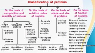
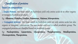
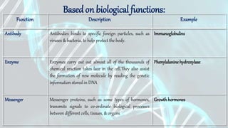
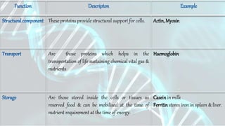
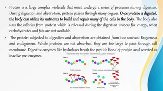
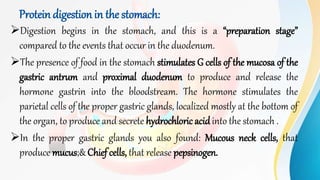
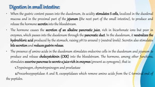
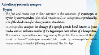
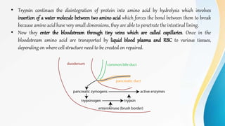
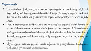
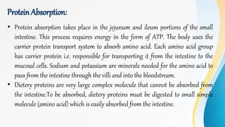
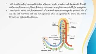
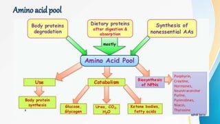
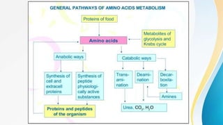
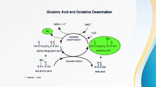
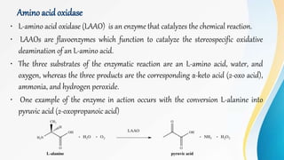
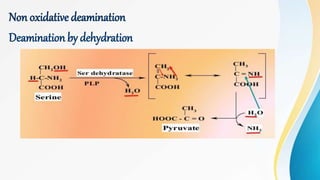
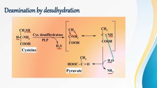
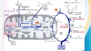
1. PROTEINS
1
College: G. Pulla reddy college of Pharmacy
Subject: Foodanalysis
Roll no: 170118885002
Name: K.Vaishali
Class : M. pharmacy, 1st year
Department:Ph.Analysis
2. Introduction
• Proteins are large biomolecules, or macromolecules, consisting of one or more long chains of amino acid
residues.
• Chemically, these are organic compounds made of carbon, hydrogen, nitrogen, oxygen or sulfur. Amino
acids are the building blocks of proteins, and proteins are the building blocks of muscle mass, according
to the NationalInstitutesof Health(NIH).
• These are most abundant organic molecules in living systems and are way
more diverse in structure and functions within organisms like catalysing
metabolic reactions, DNA replication, responding to stimuli, providing
structure to cells and organisms, and transporting molecules from one
location to another.
• A single cell can contain thousands of proteins, each with a unique
function. They do most of the work in cells
3. • A protein molecule consists of many amino acids joined together to form long chains,
much as beads are arranged on a string. There are about 20 different amino acids
that occur naturally in proteins.
• Protein is one of the essential nutrients, or macronutrients, in the human diet, but not
all the protein we eat converts into proteins in our body.
• Dietary sources of protein include both animals and plants: meats, dairy products,
fish and eggs, as well as grains, legumes and nuts.
4. • Proteins were recognized as a distinct class of biological molecules in the eighteenth century by Antoine
Fourcroy and others, distinguished by the molecules' ability to coagulate or flocculate under treatments
with heat or acid. Noted examples at the time included albumin from egg whites, blood serum albumin,
fibrin, and wheat gluten.
• Proteins were first described by the Dutch chemist Gerardus Johannes Mulder and named by the Swedish
chemist Jons Jacob Berzelius in 1838. The term "protein" is derived from the Greek word (proteios),
meaning "primary", "in the lead", or "standing in front".
• The first protein to be sequenced was insulin, by Frederick Sanger, in 1949. Sanger correctly determined
the amino acid sequence of insulin, thus conclusively demonstrating that proteins consisted of linear
polymers of amino acids rather than branched chains, colloids, or cyclols.
• The first protein structures to be solved were hemoglobin and myoglobin, by Max Perutz and Sir John
Cowdery Kendrew, respectively, in 1958.
5.
6. Basedon composition:
Classification of proteins
1. Simple Proteins: Are those which on hydrolysis yield only amino acids & no other organic
or inorganic hydrolysis products.
Eg: Albumins, Globulins, Gutelins, Protamines , Histones, Scleroproteins.
2. Conjugated proteins: Are those which on hydrolysis yield not only amino acids but also
organic or inorganic components. The non amino acid part is called prosthetic group. The
conjugated proteins are classified based on prosthetic group.
Eg: Nucleoproteins, Lipoproteins, Glycoproteins, Phosphoproteins, Metalloproteins,
Chromoproteins, Flavoproteins.
7. Basedon biological functions:
Function Description Example
Antibody Antibodies binds to specific foreign particles, such as
viruses & bacteria, to help protect the body.
Immunoglobulins
Enzyme Enzymes carry out out almost all of the thousands of
chemical reaction takes lace in the cell.They also assist
the formation of new molecule by reading the genetic
information stored in DNA
Phenylalanine hydroxylase
Messenger Messenger proteins, such as some types of hormones,
transmits signals to co-ordinate biological processes
between different cells, tissues, & organs
Growth hormones
8. Function Descripton Example
Structuralcomponent These proteins provide structural support for cells. Actin,Myosin
Transport Are those proteins which helps in the
transportation of life sustaining chemical vital gas &
nutrients
Haemoglobin
Storage Are those stored inside the cells or tissues as
reserved food & can be mobilised at the time of
nutrient requirement at the time of energy.
Caseinin milk
Ferritinstores iron in spleen & liver.
9. Basedon structure
A) FIBROUS PROTEIN: A Fibrous protein is a protein with an elongated shape. Fibrous proteins
provide structural support for cells and tissues. There are special types of helices present in two
fibrous proteins α-keratin and collagen. These proteins form long fibers that serve a structural role
in the human body.
Function: Perform the structural functions in cells.
Eg: Collagen,Myosin,Keratin & silk.
B) GLOBULAR PROTEIN: These are spherical or globular in shape. The polypeptide chain is
tightely folded into sherical shape. Physically they are soft than fibrous proteins.
Function: Forms enzymes, Antibodies & some hormones
Eg: Insulin, Haemoglobin,DNA & RNA polymerase
C) INTERMEDIATE PROTEINS: There structure is intermediate to linear & globular in
shape.They are short & less linear proteins.
Function: Blood clotting ptotein
Eg.Fibrinogen
10. Primary structure
• Primary structure, is simply
the sequence of amino acids in
a polypeptide chain. For
example, the hormone insulin
has two polypeptide chains, A
and B. Each chain has its own
set of amino acids, assembled
in a particular order.
11. Secondary structure
The next level of protein structure, secondary structure, refers to local folded structures
that form within a polypeptide due to interactions between atoms of the backbone. The
most common types of secondary structures are the α helix and the β pleated sheet. Both
structures are held in shape by hydrogen bonds, which form between the carbonyl O of
oneaminoacid and the aminoH of another.
12. Tertiary structure
The overall three-dimensional structure of a polypeptide is called its tertiary
structure. The tertiary structure is primarily due to interactions between the R
groups of the amino acidsthat make up the protein.
R group interactions that contribute to tertiary structure include hydrogen
bonding,ionic bonding,dipole-dipoleinteractions, and Londondispersionforces.
Eg: Growthhormone
13. Quaternary structure
Some proteins are made up
of multiple polypeptide
chains, also known as
subunits. When these
subunits come together,
they give the protein its
quaternary structure.
Eg: Haemoglobin
15. • Protein is a large complex molecule that must undergo a series of processes during digestion.
During digestion and absorption, protein passes through many organs. Once protein is digested,
the body can utilize its nutrients to build and repair many of the cells in the body. The body also
uses the calories from protein which is released during the digestion process for energy, when
carbohydrates and fats are not available.
• The protein subjected to digestion and absorption are obtained from two sources: Exogenous
and endogenous. Whole proteins are not absorbed; they are too large to pass through cell
membrane. Digestive enzymes like hydrolases break the peptide bond of protein and secreted as
inactive pre-enzymes.
16. Protein digestion in the stomach:
Digestion begins in the stomach, and this is a “preparation stage”
compared to the events that occur in the duodenum.
The presence of food in the stomach stimulates G cells of the mucosa of the
gastric antrum and proximal duodenum to produce and release the
hormone gastrin into the bloodstream. The hormone stimulates the
parietal cells of the proper gastric glands, localized mostly at the bottom of
the organ, to produce and secrete hydrochloric acidinto the stomach .
In the proper gastric glands you also found: Mucous neck cells, that
produce mucus;& Chief cells, that release pepsinogen.
17. All of these substances, together with others such as potassium ions and the
gastric lipase, are present in the gastric juice, which has a pH that ranges
between 1 and 2.5.
Some proteins rich in disulfide bonds, such as keratins, are resistant to
denaturation by low pH, and hence difficult to digest. On the contrary, most
of the globular proteins are almost completely hydrolyzed into constituent
amino acids.
Finally, the low pH of the gastric juice activates pepsinogen, a zymogen, to
pepsin, the first enzyme involved in protein digestion.
18. Role of gastricHCl:
It causes denaturation of protein, convert proteins into metaproteins, which are
easily digested, activates pepsinogen to pepsin and makes the pH in the stomach
suitable for the action of pepsin.
19. Pepsin:
• It is an endopeptidase acting on central peptide bond in which amino group is belongs to
aromatic amino acid. Eg: Phenylalanine, tyrosine and tryptophan. It is secreted in an inactive
formcalledpepsinogen. Its optimum pH- 1.5-2.2. It is activated by HCl.
• There are different isoenzymes of pepsinogen,
such as type I, synthesized by the cells of the
fundus of the stomach, and type II that is
produced in all the regions of the organ. All the
isoenzymes are converted to the active enzyme.
• Pepsin , at the duodenal level, stimulate the
secretion of cholecystokinin, to release pancratic
juices
20. Renin:
It is a milk clotting enzyme. It is present in stomach of infants and young
animals. Its optimum pH is 4. It acts on casein converting it to soluble
paracasein which in turns bind calcium ions forming insoluble Ca-
paracaseinate which is then digested by pepsin.
Gelatinase:
Its an enzyme that liquefies gelatine. The end products of protein digestion in
the stomach are proteoses, peptones and large polypeptides.
21. Digestionin small intestine:
• When the gastric content passes into the duodenum, its acidity stimulates S cells, localized in the duodenal
mucosa and in the proximal part of the jejunum (the next part of the small intestine), to produce and
release the hormone secretininto the bloodstream.
• The hormone causes the secretion of an alkaline pancreatic juice, rich in bicarbonate ions but poor in
enzymes, which passes into the duodenum through the pancreatic duct. In the duodenum, it neutralizes the
hydrochloric acid produced by the stomach, raising pH to around 7 (neutral levels). Secretin also stimulates
bilesecretionand reducesgastrinrelease.
• The presence of amino acids in the duodenum stimulates endocrine cells in the duodenum and jejunum to
produce and release cholecystokinin (CKK) into the bloodstream. The hormone, among other functions,
stimulates exocrinepancreasto secretea juicerichin enzymes(present as zymogens), that is:
1.Trypsinogen, chymotrypsinogen and proelastase
2.Procarboxypeptidase A and B, exopeptidases which remove amino acids from the C-terminal end of
the peptides.
22. Trypsin
• The first and master step in their activation is the conversion of trypsinogen to
trypsin by enteropeptidase (also called enterokinase), an endopeptidase produced by
cells of the duodenumafter cholecystokinin stimulation.
• Enteropeptidase catalyses the cleavage of a specific peptide bond between a lysine
residue and an isoleucine residue of the trypsinogen, with release of a hexapeptide.
This causes a conformational rearrangement of the protein that activates it, that is,
trypsin is formed.Trypsinogen converted into trypsin by enteropeptidase which
cleaves carboxy terminal of following amino acid; Phe, Tyr, Trp.
Activationof pancreaticzymogens
23. • Trypsin continues the disintegration of protein into amino acid by hydrolysis which involves
insertion of a water molecule between two amino acid which forces the bond between them to break
because amino acid have very small dimensions, they are able to penetrate the intestinal lining.
• Now they enter the bloodstream through tiny veins which are called capillaries. Once in the
bloodstream amino acid are transported by liquid blood plasma and RBC to various tissues,
depending on where cell structure need to be created on repaired.
24. Chymotrypsin
• The activation of chymotrypsinogen to chymotrypsin occurs through different
steps .In the first step, trypsin catalyzes the cleavage of a specific peptide bond, and
this causes the activation of chymotrypsinogen to π-chymotrypsin, which is fully
active.
• Then, π-chymotrypsin itself catalyzes the release of two dipeptides with formation
of the δ-chymotrypsin, a more stable form of the enzyme. δ-Chymotrypsin
undergoes two conformational changes, the first of which leads to the formation of
the κ-chymotrypsin, and the second of α-chymotrypsin, the final active form of the
enzyme.
• Chymotrypsin acts on peptide bonds adjacent to phenylalanine, tryptophan,
methionine, tyrosine and leucine residues.
25. Elastase:
• It is an endoeptidase that acts on peptide bonds formedby glycine,alanine& serine.
• It is secreted in inactive form proelastase. It is activated by trypsin.
• Optimum PH is 8.
• It digest elastin & collagen.
Procarboxypeptidase:
• Procarboxypeptidase A is activated to carboxypeptidase A;
the protease cleaves peptide bonds adjacent to amino acids
with branched or aromatic side chains, such as
phenylalanineandvaline.
• Procarboxypeptidase B is activated to carboxypeptidase B,
specific for amino acids with basic side chains, such as lysine
andarginine.
26. Intestinal juices
Aminopeptidase:
• It is an exopeptidase that acts on terminal peptide bond at the animo terminus of polypeptide
chain.
• It releases a single amino acid.
Tripeptidase:
• It acts on tripeptides.
• It releases a single amino acid & a dipeptide.
Dipetidase:
• It acts on dipeptides.
• It releases two amino acids.
27. Protein Absorption:
• Protein absorption takes place in the jejunum and ileum portions of the small
intestine. This process requires energy in the form of ATP. The body uses the
carrier protein transport system to absorb amino acid. Each amino acid group
has carrier protein i.e. responsible for transporting it from the intestine to the
mucosal cells. Sodium and potassium are minerals needed for the amino acid to
pass from the intestine through the villi and into the bloodstream.
• Dietery proteins are very large complex molecule that cannot be absorbed from
the intestine.To be absorbed, dietery proteins must be digested to small simple
molecule (amino acid) which is easily absorbed from the intestine.
28. • Villi, line the walls of our small intestine while even smaller structure called microvilli. The villi
and microvilli are series of folds that serve to increase the surface area available for absorption.
• The digested amino acid from the inside of our small intestine through the epithelial cells of
our villi and microvillli and into our capillaries. Once in capillaries the amino acid moves
through our body via bloodstream.
29. Mechanismof absorption
There are two mechanisms for amino acid absorption
1. Carrier protein transport system
2. Gluthathione transport system
Carrier protein transport system
• It is the main system of amino acid absorption.
• It is an active process needs energy which is derived from ATP molecules.
• Absorption of one amino acid needs one ATP molecule.
• Each carrier protein has one site for amino acid & one for Na+.
• It co transports amino acid & Na+ from intestinal lumen to cytosol of intestinal mucosal cell.
• The absorbed amino acid passes through the portal circulation , while Na+. extrudes out of the cell in
exchange with K+ by sodium pump
30.
31. Gluthathione transport system
• Gluthathione is used to transport amino acids from intestinal lumen to cytosol of
intestinal mucosal cells.
• It is an active transport that needs energy. Which is derived from ATP.
• Absorption of one amino acid molecule needs 3 ATP molecules.
• Gluthathione reacts with amino acid in the presence of glutamyl transpeptidase to form
glutamyl amino acid.
• Glutamyl amino acid releases amino acid in cytosol of intestinal mucosal cells with the
formation of 5-oxoproline that is used for regeneration of gluthathione to begin another
turn of cycle.
34. Metabolismof proteins:
A. Removal of ammoniaby
I. Oxidative deamination
i. Glutamate dehydrogenase
ii. Amino acid oxidase
2. Direct deamination( non oxidative)
i. Deamination by dehydration
ii. Deamination by desulhydration
3 Transamination
B. Fateof carbon skeleton of amino acid
C. Metabolismof ammonia
35.
36. Oxidative deamination
• Deamination is also an oxidative reaction that occurs under aerobic conditions
in all tissues but especially the liver.
• During oxidative deamination, an amino acid is converted into the
corresponding keto acid by the removal of the amine functional group as
ammonia and the amine functional group is replaced by the ketone group. The
ammonia eventually goes into the urea cycle.
• Oxidative deamination occurs primarily on glutamic acid because glutamic acid
was the end product of many transamination reactions.
• The glutamate dehydrogenase is allosterically controlled by ATP and ADP. ATP
acts as an inhibitor whereas ADP is an activator.
37.
38. Amino acid oxidase
• L-amino acid oxidase (LAAO) is an enzyme that catalyzes the chemical reaction.
• LAAOs are flavoenzymes which function to catalyze the stereospecific oxidative
deamination of an L-amino acid.
• The three substrates of the enzymatic reaction are an L-amino acid, water, and
oxygen, whereas the three products are the corresponding α-keto acid (2-oxo acid),
ammonia, and hydrogen peroxide.
• One example of the enzyme in action occurs with the conversion L-alanine into
pyruvic acid (2-oxopropanoic acid)
41. Transamination
• Transamination, a chemical reaction that transfers an amino group to a ketoacid
to form new amino acids. This pathway is responsible for the deamination of
most amino acids.
• Transamination in biochemistry is accomplished by enzymes called
transaminases or aminotransferases. α-ketoglutarate acts as the predominant
amino-group acceptor and produces glutamate as the new amino acid.
Aminoacid+ α-ketoglutarate↔ α-ketoacid + Glutamate
• Glutamate's amino group, in turn, is transferred to oxaloacetate in a second
transamination reaction yielding aspartate.
Glutamate+ oxaloacetate ↔ α-ketoglutarate+ aspartate
42. The most usual and major keto acid involved with transamination reactions is
alpha-ketoglutaric acid, an intermediate in the citric acid cycle. A specific
example is the transamination of alanine to make pyruvic acid and glutamic
acid.
43. Transdeamination
• Liver contains only glutamate dehydrogenase which deaminates Glutamate.
Thus all aminoacids are first Transaminated to Glutamate which is finally
deaminated.
• This coupling of Transamination and de-amination is called
Transdeamination.
44. The Fate of the Carbon Skeleton
• Any amino acid can be converted into an intermediate of the citric acid cycle. Once
the amino group is removed, usually by transamination, the α-keto acid that remains
is catabolized by a pathway
• Those amino acids that can form any of the intermediates of carbohydrate
metabolism can subsequently be converted to glucose via a metabolic pathway known
as gluconeogenesis. These amino acids are called glucogenicamino acids.
• Amino acids that are converted to acetoacetyl-CoA or acetyl-CoA, which can be used
for the synthesis of ketone bodies but not glucose, are called ketogenicaminoacids.
• Some amino acids fall into both categories. Leucine and lysine are the only amino
acids that are exclusively ketogenic.
45.
46. Metabolismof ammonia
• Ammonia is constantly liberated in the metabolism of amino acids & other nitrogen compounds.
• At physiological pH , it exists as NH4
+ ons
Formation of ammonia
• Amino acids - by transdeamination.
• Amino group of purines & pyrimide catabolism.
• By action of intestinal bacteria (urease) on urea.
Transportation of ammoia
• There is a regular & constant production of NH3 from various tissues & its
concentration in the circulation is low ( 10-20 mg /dl ).
• Body has an efficient mechanism for NH3 transportation & its utilization for urea
cycle.
47. • The transport of ammonia between various tissues & the liver mostly occurs in
the form of glutamate or alanine & not as free NH3.
• Alanine is important for NH3 transport from muscle to liver by glucose -
alanine cycle.
• Glutamine is a storehouse of NH3. It is present in highest concentration (8
mg/dl in adults) in blood among the amino acids.
• Synthesis mostly occurs in liver, brain & muscle.
• Glutamine is freely diffusible in tissues, hence easily transported.
• Glutamine synthase (mitochondrial enzyme) is responsible for synthesis of
glutamine from glutamate & ammonia, requires ATP & Mg2+.
• Glutamine can be deaminated by hydrolysis to release ammonia by
glutaminase.Glutaminase found in kidney & intestinal cells.
Role of glutamate
49. Metabolic disposal of ammonia
Ammoniotelic
The aquatic animals off
NH3 into the
surrounding water.
Uricotelic
Ammonia is converted
mostly to uric acid.
E.g.Reptiles & Birds
Ureotelic: The
mammals including
man convert NH3 to
urea.
50. Urea formation
• Urea is the principal end-product of protein metabolism in humans. It
is important route for detoxication of NH3 .
• It is operated in liver, released into blood and cleared by kidney.
• Urea is highly soluble, nontoxic and has a high nitrogen content
(46%), so, it represents about 80- 90% of the nitrogen excreted in
urine per day in man..
• Biosynthesis of urea in man is an energy- requiring process. It takes
place partially in mitochondria and partially in cytoplasm.
51. Urea cycle
• In ureotelic organisms, the ammonia in the mitochondria of hepatocytes is
converted to urea via the urea cycle. This pathway was discovered in 1932 by Hans
Krebs & a medical student associate, Kurt Henseleit.
• Krebs and Henseleit found that the rate of urea formation from ammonia was
greatly accelerated by adding any one of three α-amino acids: ornithine, citrulline,
or arginine. Each of these three compounds stimulated urea synthesis to a far
greater extent than any of the other common nitrogenous compounds
52. A molecule of ornithine combines with one molecule of ammonia and one of CO2 to form citrulline.
A second amino group is added to citrulline to form arginine, which is then hydrolyzed to yield urea, with regeneration of
ornithine.
Ureotelic animals have large amounts of the enzyme arginase in the liver. This enzyme catalyzes the
irreversible hydrolysis of arginine to urea and ornithine.
The ornithine is then ready for the next turn of the urea cycle. The urea is passed via the bloodstream to the kidneys
and is excreted into the urine.