Downloaded 115 times


![Measuring FMRI
• Haemoglobin has oxygen, and increase neuronal
activity increases demand for oxygen rendering in local
increase of blood flow near the area of activity.
• Haemoglobin is diamagnetic when oxygenated but
paramagnetic when deoxygenated.
• Difference in magnetic properties leads to differences
in MR signal of blood depending on the degree of
oxygenation.
• Since blood oxygenation varies according to the levels
of neural activity these differences can be used to
detect brain activity [blood oxygenation level
dependent (BOLD) imaging]](https://image.slidesharecdn.com/jmzo9v61qhepfesae2no-signature-b530d64c6ff48986dc7ef925b8cd6317122c3680f0a4a747901d8f2cddb16017-poli-140825195020-phpapp02/85/FMRI-7-320.jpg)


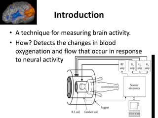
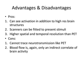
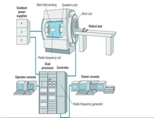
FMRI is a technique that measures brain activity by detecting changes in blood flow and oxygenation levels in the brain in response to neural activity. It has both advantages and disadvantages compared to other brain imaging methods like PET. FMRI uses strong magnetic fields to detect the magnetic signal from hydrogen atoms in water molecules in the brain. Changes in the magnetic properties of oxygenated versus deoxygenated hemoglobin allow FMRI to detect brain activity based on the blood oxygenation level dependent (BOLD) response to neural activation.