SlideShare a Scribd company logo
1 of 89
Download to read offline
1
LATIHAN SOAL DAN PEMBAHASAN
FARMAKOLOGIMOLEKULAR
1. Klasifikasi reseptor berdasarkan lokasi reseptornya pada sel ?
Jawab :
Reseptor Berdasarkan Lokasinya Reseptor
Membrane Bound Receptors 1) G-Protein-Linked Receptors
➔Serotonin, muskarinik, domaninergik,
noradrenergik
2) Enzyme Receptors
➔Tyrosine kinase
3) Ligand-Gated Ion Channel Receptors
➔Nikotinik, GABA,glutamat
Intracellularand NuclearReceptors 1) Hormone Receptors
➔Tiroid, estrogen, androgen glukokortikoid
2) Autocoid Receptors
3) Growth Factors Receptors
4) Insulin Receptors
2. Reseptor yang termasuk reseptor inti ?
Jawab :
- Reseptor glukokortikoid
- Reseptor mineralokortikoid
- Reseptor progesteron
- Reseptor androgen
- Reseptor estrogen
- RXR (Retinoid X Receptors)
➔ PPAR (Peroxisom Proliferators – Activated Receptor)
➔ T3R (Thyroid Hormone Receptor)
- Monomeric Tethered Orphan Receptors
➔ ROR (Retinoic Acid – Related Orphan Receptor)
➔ ERR (Estrogen – Related Receptor)
2
3. Mekanisme aksi glucocorticoid receptor (GC) ?
Jawab :
a) Aktivasi genomik
➔Membutuhkan aktivasi gen-gen tertentu. Mekanismenya yaitu kortisol masuk ke
dalam membran sel lalu berikatan dengan reseptor glukokortikoid yang terlebih
dahulu distabilkan oleh HSP (Heat Shock Protein). Setelah reseptor glukokortikoid
berikatan dengan kortisol, akan melepaskan HSp dan menduduki glukokortikoid.
Respon elemen dalam untaian DNA dalam inti kemudian mRNA ditranskripsi lalu
ditranslasi dan diterjemahkan membentuk protein antiinflamasi yang menghambat
inflamasi. Contoh : TGF- β, IL – 10, lipocortin.
b) Aktivasi non genomik
➔Kortisol masuk ke dalam membran sel lalu berikatan dengan reseptor
glukokortikoid. Sebelum kortisol diikat oleh reseptor glukokortikoid, distabilkan
oleh HSP. Setelah glukokortikoid berikatan dengan kortisol, HSP akan dilepaskan
dan akan aktivasi protein antiinflamasi yang menghambat inflamasi.
4. Apa saja yang termasuk ligan alamiah dan ligan sintetis glucocorticoid receptor (GC)?
Jawab :
- Ligan alamiah = kortisol, hidrokortison
- Ligan sintetis = deksametason, prednison, metil prednison, betametason
5. Bagaimanakah mekanisme molekular ikatan prednison dan reseptor GC menimbulkan
efek antiinflamasi ?
Jawab :
Prednison adalah senyawa sintetis berupa pro-drug dari prednisolon yang memiliki
aktivitas antiinflamasi dan imunomodulasi. Mekanisme kerjanya yaitu setelah
prednison melekat ke reseptor membran sel dan memasuki sel, prednison memasuki
nukleus lalu berikatan dan aktivasi reseptor nukleus (inti). Prednison di dalam darah
akan berubah menjadi bentuk aktif (prednisolon) lalu menghambat migrasi sel
polimorfonuklear (PMN) sehingga dapat inhibisi infiltrasi leukosit, supresi respon imun
humoral, dan mengurangi sekresi IL – 6 sehingga inflamasi tidak terjadi.
6. Bagaimanakah mekanisme aksi genomik pada kompleks ligan dan reseptor GC ?
3
Jawab :
Kompleks ligan dan reseptor GC, masuk ke inti dan mengikat GRE (Glucocorticoid
Response Elements) sehingga menyebabkan transrepresi gen pro inflamasi. GRE
sebagai suatu ‘landasan’ untuk reseptor glukokortikoid akan meregulas transkripsi gen
untuk menurunkan produksi sitokin pro inflamasi dan meningkatkan protein
antiinflamasi.
7. Bagaimanakah mekanisme molekular kortikosteroid memiliki efek samping
osteoporosis ?
Jawab :
*referensi dan jurnal terlampir
a) Efek Langsung
b) Efek tidak Langsung
➔Osteoporosis akibat terapi glukokortikoid juga dapat terjadi karena
hypogonadism, menurunkannya aktivitas fisik, menurunnya produksi growth
hormone, insulin-like growth factor 1 (IGF 1) dan IGF 1 binding protein (IGF-BP)
serta meningkatnya kehilangan kalsium di ginjal dan intestinal.
8. Apakah keterkaitan antara reseptor GC dengan HSP ?
4
Jawab :
HSP (Heat Shock Protein) menstabilkan bentuk molekul GC → selama tidak mengikat
ligan, reseptor GC dalam keadaan inaktif.
9. Bagaimanakah mekanisme kerja kompleks GC dan steroid dalam mengurangi
inflamasi?
Jawab :
Kompleks GC dansteroid dapat upregulates (meningkatkan eskpresi dari gen-gen yang
dikendalikan oleh lipocortin (annexin). Kortisol berikatan dengan reseptor GC
menghasilkan protein annexin 1 yang dapat menghambat aktivasi enzim c PLA 2α
sehingga fosfolipid tidak dapat diubah menjadi asam arachidonate → tidak dapat
produksi prostaglandin → inhibisi inflamasi.
Kortisol berikatan dengan reseptor GCinhibisi transkripsi NF-kB sehingga tidakterjadi
inflamasi → tidak terbentuk prostaglandin oleh enzim COX-2 → inhibisi inflamasi
Kortisol berikatan dengan glukokortikoid → aktivasi MAPK fosfatase 1 →inhibisi
terbentuknya MAPKs → inhibisi aktivasi enzim c PL 2α yang dapat mengubah
fosfolipid menjadi arachidonate → prostaglandin tidak terbentuk → inflamasi
berkurang.
10. Apakah yang dimaksud dengan NF-kB, AP-1, Jun Fos, dan MAPK ?
Jawab :
a) NF-kB
➔Nuclear factor kappa light chain enhancer of activated β cells
➔Faktor transkripsi yang bertanggung jawab padaproses inflamasi ; berupa protein
kompleks yang mengendalikan transkripsi DNA, produksi sitokin, dan pertahanan
sel.
b) AP-1
➔Activated Protein 1
➔Faktor transkripsi yang mengatur ekspresi gen sebagai respons terhadap stimulus
meliputi sitokin, growth factor, stres, dan infeksi mikroorganisme dengan
mengendalikan proses selular seperti diferensiasi, proliferasi, dan apoptosis.
5
c) Jun Fos
➔Onkogen sebagai informasi genetik yang bertanggung jawab dalam induksi
tumor oleh virus sarcoma murine FBJ dan virus 17 sarkoma avian yang diderivat
dari gen selular normal (c-fos).
d) MAPK
➔Mitogen Activated Protein Kinase
➔Enzim yang teraktivasi sebagai respon selular terhadap berbagai jenis hormon
faktor pertumbuhan, dan katalisis reaksi fosforilasi terhadap jenis protein tertentu.
Referensi :
1. Ayroldi, Emira et al. Mechanismes of the Antiinflammatory Effects of Glucocorticoids
: Genomic and Non-Genomic Interference with MAPK Signalling Pathways. The
FASEB Journal.
2. Curran, Tom. Fos and Jun : Oncogenic Transcription Factors. Tohoku J Exp Med.1992.
3. Compston, Juliet. Glucocorticoid – Induced Osteoporosis : An Update. Springer
Endocrine. 2018.
4. Lawrence, Toby. The Nuclear Factor NF-kB Pathway in Inflammation. Cold Spring
Harb Perspect Biol. 2009.
5. Schijvens et al. Pharmacology and Pharmacogenetics of Prednisone and Prednisolone
in Patients with Nephrotic Syndrome. Pediatric Nephrology. 2018.
6. Zenz, Rainer et al. Activator Protein 1 (Fos/Jun) Functions in Inflammatory Bone and
Skin Disease. Arthritis Research and Therapy. BMC Journal. 2008.
Endocrine
https://doi.org/10.1007/s12020-018-1588-2
REVIEW
Glucocorticoid-induced osteoporosis: an update
Juliet Compston 1
Received: 27 December 2017 / Accepted: 22 March 2018
© The Author(s) 2018
Abstract
Glucocorticoid-induced osteoporosis is the most common secondary cause of osteoporosis and the resulting fractures cause
significant morbidity. Following initiation of oral glucocorticoids, rapid bone loss occurs, and fracture risk increases within a
few months in a dose-dependent manner. These adverse effects are due to inhibition of bone formation accompanied by an
early but transient increase in bone resorption. Multiple mechanisms underlie these changes in bone remodeling; direct
effects include upregulation of PPARγR2, increased expression of sclerostin and increased RANKL/OPG ratio, whilst
hypogonadism, altered renal and intestinal calcium handling, and reduced production of insulin-like growth factor 1 also
contribute. Fracture risk assessment should be performed as soon as possible after glucocorticoids are initiated and bone
protective therapy started promptly in individuals at high-risk, with calcium and vitamin D supplements where appropriate.
Oral bisphosphonates are currently regarded as first line options on the grounds of their low cost. However, teriparatide has
been shown to be superior in its effects on BMD and vertebral fracture risk in glucocorticoid-treated individuals with
osteoporosis and should be considered as an alternative first line option in high-risk patients.
Key words Glucocorticoids ●
Bone density ●
Fracture ●
Bisphosphonates ●
Teriparatide
Introduction
Glucocorticoids are used in the treatment of a wide range of
diseases and it is estimated that 1–2% of the population is
receiving long-term glucocorticoid therapy [1–4]. The
adverse skeletal effects of glucocorticoid excess were first
described over 80 years ago, and today glucocorticoid-
induced osteoporosis is the most common secondary cause
of osteoporosis. Research into the epidemiology, patho-
physiology and clinical management of glucocorticoid-
induced osteoporosis has produced substantial advances, yet
the condition remains under-recognized and under-treated.
This review focuses on recent developments in the field and
their implications for clinical practice.
Epidemiology
Continuous oral glucocorticoid therapy is associated with
rapid bone loss and an increase in fracture risk that is seen
within 3–6 months of initiation and is dose-related. This
time course can be explained, at least in part, by the higher
doses of glucocorticoids and greater disease activity in the
early stages of treatment. Fracture risk remains elevated for
the duration of glucocorticoid therapy, but declines after its
withdrawal, although whether it reverts to baseline values is
unclear [5–8]. Vertebral fractures are particularly char-
acteristic of glucocorticoid-induced osteoporosis, although
the risk of non-vertebral fractures, including hip fractures, is
also increased.
Recent studies have confirmed the predilection for ver-
tebral fracture, the dose-related increase in fracture risk,
and decline in fracture risk with longer duration or dis-
continuation of glucocorticoid therapy. Amiche et al con-
ducted a Bayesian meta-analysis of fracture risk associated
with oral glucocorticoid use, drawing on data from the
control groups of clinical trials [9]. In individuals who had
initiated glucocorticoid therapy in the last 6 months, the
annual incidence of vertebral fracture was 5.1% [95%
credible intervals (CrI) 2.8–8.2] and of non-vertebral
fracture, 2.5% [95% CrI 1.2–4.2]. For those with duration
* Juliet Compston
jec1001@cam.ac.uk
1
Department of Medicine, Cambridge Biomedical Campus,
Cambridge CB2 0SL, UK
1234567890();,:
1234567890();,:
of glucocorticoid use ≥6 months, the corresponding figures
were 3.2% [95% CrI 1.8–5.0] and 3.0% [95% CrI 0.8–5.0].
Using a large administrative database, Balasubramanian
et al investigated the effects of initiating systemic (oral or
injected) glucocorticoid therapy on fracture risk in patients
with new-onset rheumatoid arthritis (mean age 49 years)
[10]. Fracture incidence rates were 5 to 9/1000 person
years at doses of <15 mg/day, 16 (11, 22.6) at doses
≥15 mg/day and 13.4 (10.7, 16.7) at cumulative doses
≥5400 mg. At 60-182 days after discontinuation of gluco-
corticoid therapy, fracture risk was 29% lower than in those
with ongoing use and by 12 months was similar to non-
glucocorticoid users.
The effects of inhaled or intravenous glucocorticoids on
fracture risk are less well documented. There is some evi-
dence that high doses of inhaled glucocorticoids may be
associated with increased fracture risk, although concurrent
use of oral glucocorticoids is often a confounding factor
[11–14]. In a large case control study from Denmark, no
increase in fracture risk was found in association with other
forms of topical steroids [15].
Variation in the severity of adverse effects of gluco-
corticoids, including bone loss, is well recognized but
poorly understood. Pre-receptor modulation of glucocorti-
coid activity by 11 beta-hydroxysteroid dehydrogenase
(11βHSD) enzymes, which interconvert inactive and active
cortisone/cortisol, may contribute to this variability through
the effects of pro-inflammatory cytokines [16, 17] and
genetic polymorphisms in the glucocorticoid receptor
gene [18].
Pathophysiology
Direct effects on bone
Glucocorticoid-induced osteoporosis is characterized by
decreased bone formation, with an additional early but
transient increase in bone resorption (Fig. 1). The initial
increase in remodeling rate is accompanied by reduced bone
formation at the level of the individual bone multicellular
unit (BMU), and this combination of increased bone turn-
over and a negative remodeling balance results in rapid
bone loss [19–23]. Subsequently, the decrease in bone
formation, both at tissue and BMU level, predominates
leading to a low turnover state. Direct effects of gluco-
corticoids on bone formation are mediated largely through
upregulation of peroxisome proliferator-activated receptor
gamma receptor 2 (PPARγ2) [24] and effects on the Wnt/β-
catenin signaling pathway [25, 26]. The former mechanism
favors the differentiation of pluripotent precursor cells to
adipocytes in preference to osteoblasts, resulting in
decreased numbers of osteoblasts. Increased expression of
sclerostin, which binds to the co-receptors for frizzled, Lrp4
and Lrp5, results in inhibition of Wnt sigalling leading to
reduced differentiation of osteoblast precursors to mature
osteoblasts and increased osteoblast and osteocyte apopto-
sis. The importance of sclerostin in mediating the effects of
glucocorticoids on bone formation is emphasized by the
demonstration, in mice with sclerostin deficiency, that bone
integrity is maintained in the presence of glucocorticoid
excess [27]. Furthermore, in a mouse model of
glucocorticoid-induced osteoporosis, treatment with an
antibody to sclerostin prevented the reduction in bone mass
and strength [28].
Glucocorticoids also have direct effects on bone resorp-
tion, increasing the production of macrophage colony sti-
mulating factor (M-CSF) and RANKL and decreasing
production of osteoprotegerin (OPG) by osteoblastic cells
and osteocytes, resulting in an increase both in the number
and activity of osteoclasts [29, 30]. This effect diminishes
with time, possibly as a result of the reduction in number of
osteoblasts and osteocytes. Finally, there is some evidence
from animal models that glucocorticoids affect osteocyte
morphology and mineralization [31].
Indirect effects on bone
Other mechanisms that may contribute to glucocorticoid-
induced bone loss through indirect effects on bone include
hypogonadism, reduced physical activity, increased renal
and intestinal losses of calcium, and reduced production of
growth hormone, insulin-like growth factor 1 (IGF1) and
IGF1 binding protein (IGF-BP) [32]. In addition, the
underlying diseases for which glucocorticoid therapy is
administered are often associated with increased inflam-
mation, which contributes to bone loss through increased
production of pro-inflammatory, pro-resorptive cytokines.
Whilst glucocorticoids suppress inflammation and hence
should mitigate the adverse effects of inflammation, disease
relapse despite therapy is associated with episodes of
increased bone resorption. Finally, glucocorticoid excess
has adverse effects on muscle mass and function, leading to
myopathy and increased risk of falls [33].
Changes in BMD and bone microarchitecture
Increased rates of bone loss in the hip, spine, and radius are
well documented in individuals treated with glucocorti-
coids. Data obtained from assessment of bone micro-
architecture using high-resolution peripheral computed
tomography (HRpQCT) are sparse. In a cross-sectional
study of 30 postmenopausal women who had received oral
glucocorticoids for longer than 3 months, despite similar
Endocrine
areal BMD values to 60 control subjects, significantly lower
total, cortical and trabecular volumetric BMD, thinner cor-
tices, increased trabecular separation and reduced trabecular
number were reported in the radius and tibia; whole bone
stiffness, assessed using finite element analysis, was also
significantly reduced in comparison to the controls [34].
Although the patients and controls were generally well
matched, however, bisphosphonate use was significantly
more common in the former (100% vs. 8.6%), so definite
attribution of the observed differences to glucocorticoid
therapy cannot be made.
Trabecular bone score (TBS) provides an indirect index
of trabecular bone architecture that can be obtained from
DXA images of the lumbar spine, and has predictive value
for fracture independent of BMD [35]. In 64 post-
menopausal women who had taken prednisolone in a dose
of ≥5 mg daily for >3 months, TBS was significantly lower
than in a group of non-glucocorticoid treated controls,
although lumbar spine BMD T-scores were not significantly
different [36]. Similar findings have been reported in 416
individuals on long-term glucocorticoids (≥5 mg daily for
3 months), the decrease in TBS being most marked in men
and in individuals with fracture [37]. These findings indi-
cate that glucocorticoids have adverse effects on spine bone
microarchitecture that are independent of BMD and which
may contribute to increased fracture risk.
Fracture risk assessment in individuals treated with
glucocorticoid-induced osteoporosis
There is evidence from a number of studies that fracture
occurs at a higher BMD in individuals receiving gluco-
corticoids than in non-glucocorticoid treated people
[38–40]. This independent effect of glucocorticoid therapy
on fracture risk may be due to several factors, including
increased falls risk and alterations in bone quality that are
not captured by BMD measurements.
Glucocorticoid therapy is included as a risk factor in the
FRAX fracture prediction algorithm as a dichotomous
variable. “Yes” is entered in the questionnaire if there is
current exposure to oral glucocorticoids or past exposure for
≥ 3 months at a dose of 5 mg/day or more of prednisolone or
equivalent. Using data from the UK General Research
Practice Database, Kanis et al have provided adjustments
that can be incorporated into the FRAX calculations to
adjust for different doses of glucocorticoids (Table 1) [41].
For daily doses of over 7.5 mg daily of prednisolone or
equivalent, greater upward adjustment of fracture prob-
ability may be required. It should be noted that the duration
of glucocorticoid therapy and cumulative dose are not
accommodated within the FRAX algorithm. In addition, the
use of total hip BMD in FRAX may result in under-
estimation of fracture risk in patients with differentially low
Fig. 1 Direct effects of glucocorticoids on bone
Endocrine
spine BMD, although a correction for this discordance has
been proposed [42, 43]. A final caveat is that the response to
treatment in glucocorticoid-treated individuals selected on
the basis of FRAX-derived fracture probability has not been
documented.
Assessment of fracture risk using FRAX is recom-
mended in several guidelines for the management of
glucocorticoid-induced osteoporosis, including the National
Osteoporosis Guideline Group (NOGG) guidance [44, 45],
the updated recommendations produced by the American
College of Rheumatology (ACR) [46] and guidelines pub-
lished by the International Osteoporosis Foundation (IOF)
and European Calcified Tissues Society (ECTS) [47, 48].
However, FRAX can only be used in people age 40 years
and over; in children and young adults, fracture risk
assessment should be performed using BMD measurement,
together with consideration of other risk factors, particularly
previous fracture.
Management of glucocorticoid-induced
osteoporosis
Under-treatment of glucocorticoid-induced osteoporosis is
widely recognized [49, 50]. In a population-based study of
adults age ≥20 years, rates of BMD testing and prescription
of bone protective medication between 1998 and 2008 were
studied in individuals prescribed systemic glucocorticoids
for 90 days or longer [51]. Overall, in the first six months
after initiation of glucocorticoid therapy only 6% had BMD
testing, 22% received therapy and 25% had both interven-
tions. Under-treatment was greatest in younger people and
men, and primary care physicians had lower prescription
rates than rheumatologists. Similar results have been
reported more recently using information from a national
public health-insurance database in France, with only 8%
undergoing BMD testing and prescription of calcium ±
vitamin D and bisphosphonates in 18 and 12% respectively
[52]. In a large cohort from Canada of men and women
aged 66 years or over who were initiating long-term glu-
cocorticoid therapy, Amiche et al reported that only 13%
were prescribed bone protective therapy [53]. The problem
of under-treatment is compounded by poor persistence with
bisphosphonate therapy, particularly in younger people,
those with co-morbidities, and those in whom BMD mea-
surements have not been made [54].
General measures
A number of life-style measures may mitigate the harmful
skeletal effects of glucocorticoids, although the evidence
base for this approach is weak and requires extrapolation
from studies in non-glucocorticoid treated individuals. Falls
risk should be assessed and preventive measures instituted
when appropriate. Exercise, tailored to the individual
patient, and good nutrition with adequate dietary calcium
intake should be advocated with avoidance of smoking and
alcohol abuse. Maintenance of an adequate vitamin D status
should also be advised.
Attention should be paid to keeping the dose of gluco-
corticoids to a minimum, with the use of steroid-sparing
drugs such as methotrexate or azathioprine or alternative
routes of administration (for example inhaled or topical)
where appropriate. Non-steroidal therapies should be used
when possible to maintain remission, once achieved.
However, it is also important to maintain suppression of the
underlying disease, since this will prevent the adverse
skeletal effects of inflammation and other effects of
increased disease activity.
Studies of the effects of calcium and/or vitamin D sup-
plementation on BMD in patients taking glucocorticoids
have produced conflicting results [55, 56]. However, cal-
cium and vitamin D supplements have been included in
most trials of bone protective therapy and should therefore
be used as an adjunct to treatment.
Pharmacological interventions
Regulatory approval of drugs to reduce fracture risk in
individuals taking glucocorticoids has been based on the
demonstration of similar changes in BMD to those
observed in postmenopausal osteoporosis and fracture has
been a secondary outcome. For this reason, evidence for
anti-fracture efficacy of bone protective agents in
glucocorticoid-induced osteoporosis is less robust than that
for postmenopausal osteoporosis. In addition, there is
inevitable heterogeneity in glucocorticoid-treated trial
populations, with respect to age, underlying disease,
co-morbidities and co-medications, dose and duration of
glucocorticoid therapy and the timing of bone protective
therapy. Furthermore, the duration of most treatment stu-
dies has been relatively short and this, combined with
smaller trial populations, reduces the strength of the safety
database.
Table 1 Adjustment of FRAX-derived fracture probability according
to dose of glucocorticoids. Data from ref. [40]
Daily dose of
prednisolone (mg)
Average adjustment for
major osteoporotic
fracture probability
Average adjustment
for hip fracture
probability
<2.5 −20% −35%
2.5–7.5 None None
≥7.5a
+15% +20a
a
For high doses of prednisolone, greater upward adjustment of fracture
risk may be appropriate
Endocrine
Bisphosphonates
Bisphosphonates are the most commonly used drugs in the
management of glucocorticoid-induced osteoporosis. Oral
alendronate (5 or 10 mg daily or 70 mg once weekly) and
risedronate (5 mg daily or 35 mg once weekly), and intra-
venous zoledronic acid (5 mg once yearly by intravenous
infusion) are all approved for this indication. All have been
shown to have beneficial effects on lumbar spine and hip
BMD in people treated with glucocorticoids [57–64] and for
alendronate and risedronate, there is also evidence from
safety or post hoc analyses for a reduction in the rate of
vertebral fractures [62, 63]. In the pivotal study of zole-
dronic acid, a non-inferiority BMD comparison with rise-
dronate, the fracture rate was too low to assess anti-fracture
efficacy [64].
The number of non-vertebral and hip fractures has been
insufficient in individual trials to assess an impact of
bisphosphonates. However, data from cohort studies provide
some evidence for efficacy at these sites. In an observational
cohort study of women aged >65 years taking alendronate or
risedronate, Thomas et al. studied the baseline incidence of
clinical fractures in the first three months after starting glu-
cocorticoid therapy and the fracture incidence in the fol-
lowing 12 months [65]. Compared to the baseline incidence,
both clinical vertebral and non-vertebral fracture incidence
were significantly lower. Using the same database, Overton
et al studied the effects of bone protective therapy (bispho-
sphonates in 95.5%, denosumab or teriparatide in the
remaining 4.5%) in a large cohort of new glucocorticoid
users [66]. Treatment within the first 90 days of gluco-
corticoid use was associated with a significant reduction in
clinical fractures (including vertebral) of 48% at one year
and 32% at three years when compared with non-use.
Finally, in three matched cohorts derived from healthcare
administrative data from Ontario, Canada, Amiche et al.
reported that in individuals initiating long-term glucocorti-
coids, therapy within the first six months with alendronate or
risedronate was associated with a decrease in incident hip
fracture (alendronate 0.49 (0.34, 0.69), risedronate 0.58
(0.36, 0.90) [53]. The results confirmed a reduction in ver-
tebral fracture risk with etidronate, alendronate and rise-
dronate, but no decrease in risk of forearm or humerus
fractures for any bisphosphonate. The analysis was limited
to oral bisphosphonates and zoledronic acid was not con-
sidered. Overall, therefore, these studies would be consistent
with a beneficial effect of bisphosphonates both on vertebral
and non-vertebral fracture, including hip fracture.
The safety profile of bisphosphonates in glucocorticoid-
induced osteoporosis has been less well studied than in
postmenopausal osteoporosis because of the small number
of participants included and shorter duration of the trials.
Because of co-morbidities and co-medications, people
taking glucocorticoids may be more susceptible to side-
effects, for example upper gastrointestinal disorders, and
glucocorticoid therapy is a documented risk factor for
osteonecrosis of the jaw [67] and probably also for atypical
femoral fractures [68, 69]. Bisphosphonates should be used
with caution in premenopausal women with child bearing
potential, because of the ability of these drugs to cross the
placenta.
Teriparatide
The predominant role of reduced bone formation in
glucocorticoid-induced osteoporosis provides a rationale for
the use of anabolic agents in its treatment. In an active-
comparator controlled, randomized, double blind study the
effects of 18 months treatment with subcutaneous teripara-
tide, 20 µg/day, or oral alendronate 10 mg/day, were com-
pared in 428 men and women with glucocorticoid-induced
osteoporosis [70]. Teriparatide therapy resulted in sig-
nificantly greater increases in spine and hip BMD and this
was seen in both premenopausal and postmenopausal
women and in men [71]. The magnitude of increase in
BMD was somewhat less than that seen in non-
glucocorticoid treated postmenopausal women in another
study [72], possibly as a result of the opposing actions of
intermittent PTH and glucocorticoids on osteoblastogenesis,
and osteoblast and osteocyte apoptosis [73–75].
Although fracture was not a primary end-point of the
study, significantly fewer new vertebral fractures occurred
in the patients treated with teriparatide when compared to
those treated with alendronate (0.6% vs. 6.1%; p = 0.004).
The incidence of non-vertebral fractures was similar in the
two treatment groups. Results after 36 months of treatment
demonstrated a continued increase in spine and hip BMD in
the teriparatide treated group, with superiority over alen-
dronate at the 24 and 36 month time points [76]. A lower
incidence of new vertebral fractures was also seen in the
teriparatide group at 36 months (1.7% vs 7.7%, p = 0.007),
with a similar incidence of non-vertebral fractures in the
two groups. Interestingly, measurements of TBS in a sub-
population of this study demonstrated a significant increase
after 36 months in teriparatide treated patients, but no sig-
nificant change in those treated with alendronate [77].
Whilst the long duration of this study is unique amongst
treatment trials for glucocorticoid-induced osteoporosis, it
should be noted that the participant discontinuation rate at
36 months was 44%. With regard to safety, increased pre-
dose serum calcium levels were significantly more common
in the teriparatide than alendronate treated group (21% vs.
7%), but no other concerns were identified
In a comparison of the effects of teriparatide and rise-
dronate in men treated with oral glucocorticoids for
≥3 months, significantly greater increases in lumbar spine
Endocrine
trabecular volumetric BMD were demonstrated in the
teriparatide-treated group at 18 months. In addition, ter-
iparatide was associated with significantly greater
improvements in bone strength and stiffness at T12, mea-
sured using HRQCT-based finite element analysis [78].
These data, together with the results of the study reviewed
above, provide a rationale for the use of teriparatide as a
first line option in some patients, particularly those at high
risk of vertebral fracture.
Teriparatide is contraindicated in pregnant and lactating
women. Women of childbearing potential should use
effective methods of contraception while on teriparatide
treatment.
Denosumab
At present, denosumab is not approved for use in
glucocorticoid-induced osteoporosis, although this is cur-
rently under consideration by some regulatory agencies. In a
Phase 3 randomized double blind active controlled trial of
adults taking ≥7.5 mg prednisone or equivalent daily,
treatment with denosumab, 60 mg once every 6 months by
subcutaneous injection, was associated with a significantly
greater increase in lumbar spine BMD compared to rise-
dronate 5 mg daily over a 12 months treatment period [79].
This effect was seen both in patients initiating glucocorti-
coids and in those established on long-term therapy,
regardless of age, race, baseline BMD T-score, initial glu-
cocorticoid dose and menopausal status. To date the study
has only been reported in abstract form.
Guidelines for the management of glucocorticoid-
induced osteoporosis
National guidelines for the management of glucocorticoid-
induced osteoporosis have been produced by a number of
countries and updated guidelines have recently been
published from the US and UK. Most guidelines address
users of long-term (3 months) oral glucocorticoids, and
although the daily threshold dose varies it has generally
been between 5 and 7.5 mg daily of prednisolone or
equivalent. Treatment thresholds also vary; T-scores are
used in some and generally recognize the higher BMD at
which fracture occurs in glucocorticoid-treated indivi-
duals. The recent guidelines from the UK and US
emphasize the importance of starting bone protective
therapy early in high-risk individuals, because of the
rapidity with which bone loss and increased fracture risk
occur.
The recently updated ACR guideline defines three cate-
gories of fracture risk; high, moderate and low. In adults
aged ≥40 years, previous osteoporotic fracture, hip or spine
BMD T-score ≤ −2.5, or 10 year fracture probability of
≥20% (major osteoporotic fracture) or ≥3% (hip fracture)
are the criteria for high risk [45]. Moderate and low risk are
defined solely on the basis of FRAX-derived fracture
probability (10–19% and >1 to ≤3% respectively for mod-
erate risk, <10% and ≤1% respectively for low risk). In
adults <40 years old, the criterion for high risk is a previous
osteoporotic fracture, whereas moderate and low risk are
defined on the basis of BMD. Oral bisphosphonates are
recommended as the first line option for individuals at
moderate or high risk, with intravenous bisphosphonates,
teriparatide, or denosumab in patients who are contra-
indicated or intolerant to oral bisphosphonates. NOGG, in
its 2017 update, takes a similar approach to risk assessment
with the use of dose-adjusted FRAX, but uses the UK
NOGG intervention thresholds as the basis for making
treatment decisions and does not include denosumab as a
treatment option [44, 45].
These recent guidelines emphasize the importance of
early intervention in high-risk individuals, advocate main-
tenance of adequate calcium intake and normal vitamin D
status, and stress the limited evidence base to support some
recommendations, particularly in younger adults. Compar-
ison of the cost-effectiveness of different treatments is
problematic because of the lack of fracture data, particularly
hip fracture, in clinical trials. The choice of oral bispho-
sphonates as a first line option in both sets of guidelines is
based on their lower cost compared to either zoledronic acid
or teriparatide; nevertheless, in view of the superiority of
teriparatide vs. alendronate for BMD and vertebral fracture
outcomes, it has been suggested that teriparatide should be
considered as an alternative first line option in those at
highest risk [80].
Conclusions
There have been significant advances in our understanding
of the mechanisms by which glucocorticoids affect bone
and increase fracture risk. The clinical management of
glucocorticoid-induced osteoporosis, however, remains
suboptimal despite the development of more sophisticated
approaches to fracture risk assessment and the availability
of several effective treatment options. Whilst bispho-
sphonates are currently the most widely used bone protec-
tive drugs in individuals taking glucocorticoids, the use of
teriparatide and of denosumab (if approved) as first-line
options in some patients merits further investigation.
Finally, the potential for sclerostin inhibitors to prevent or
reverse adverse skeletal effects of glucocorticoids provides
an exciting prospect for future research.
Endocrine
Compliance with ethical standards
Conflict of interest Prof Compston has received advisory and speaking
fees from Gilead and speaking fees from Amgen and UCB.
Open Access This article is distributed under the terms of the Creative
Commons Attribution 4.0 International License (http://crea
tivecommons.org/licenses/by/4.0/), which permits unrestricted use,
distribution, and reproduction in any medium, provided you give
appropriate credit to the original author(s) and the source, provide a
link to the Creative Commons license, and indicate if changes were
made.
References
1. T.P. van Staa, H.G. Leufkens, L. Abenhaim, S. Begaud, B. Zhang,
C. Cooper, Use of oral corticosteroids in the United Kingdom. Q
Med J. 93, 105–111 (2000)
2. L. Fardet, I. Petersen, I. Nazareth, Prevalence of long-term oral
glucocorticoid prescriptions in the UK over the past 20 years.
Rheumatology 50(11), 1982–1990 (2011)
3. R.A. Overman, J.Y. Yeh, C.L. Deal, Prevalence of oral gluco-
corticoid usage in the United States: a general population per-
spective. Arthritis Care Res. 65(2), 294–298 (2013)
4. S. Silverman, J. Curtis, K. Saag, J. Flahive, J. Adachi, F.
Anderson, R. Chapurlat, C. Cooper, A. Diez-Perez, S. Greenspan,
F. Hooven, A. Le Croix, L. March, J.C. Netelenbos, J. Nieves, J.
Pfeilschifter, M. Rossini, C. Roux, E. Siris, N. Watts, J. Comp-
ston, International management of bone health in glucocorticoid-
exposed individuals in the observational GLOW study. Osteo-
poros. Int. 26(1), 419–420 (2015)
5. T. van Staa, H.G.M. Leufkens, L. Abenhaim, B. Zhang, C.
Cooper, Use of oral corticosteroids and risk of fractures. J. Bone
Miner. Res. 15, 933–1000 (2000)
6. F. De Vries, M. Bracke, H.G. Leufkens, J.W. Lammers,
C. Cooper, T.P. Van Staa, Fracture risk with intermittent
high-dose glucocorticoid therapy. Arthritis Rheum. 56, 206–214
(2007)
7. T.P. van Staa, H.G.M. Leufkens, L. Abenhaim, B. Zhang,
C. Cooper, Oral corticosteroids and fracture risk: relationship
to daily and cumulative doses. Rheumatology 39, 1383–1389
(2000)
8. T.P. van Staa, H.G.M. Leufkens, C. Cooper, A meta-analysis of
the epidemiology of corticosteroid-induced osteoporosis. Osteo-
poros. Int. 13, 777–787 (2002)
9. M.A. Amiche, J.M. Albaum, M. Tadrous, P. Pechlivanoglou, L.E.
Lévesque, J.D. Adachi, S.M. Cadarette, Fracture risk in oral
glucocorticoid users: a Bayesian meta-regression leveraging con-
trol arms of osteoporosis clinical trials. Osteoporos. Int. 27(5),
1709–1718 (2016)
10. A. Balasubramanian, S.W. Wade, R.A. Adler, C.J.F. Lin, M.
Maricic, C.D. O’Malley, K. Saag, J.R. Curtis, Glucocorticoid
exposure and fracture risk in patients with new-onset rheumatoid
arthritis. Osteoporos. Int. 27(11), 3239–3249 (2016)
11. J.H. Toogood, J.C. Baskerville, A.E. Markov, A.B. Hodsman, L.J.
Fraher, B. Jennings, R.G. Haddad, D. Drost, Bone mineral density
and the risk of fractures in patients receiving long-term inhaled
steroid therapy for asthma. J. Allergy Clin. Immunol. 96, 157–166
(1995)
12. C.A. Wong, L.J. Walsh, C.J. Smith, A.F. Wisniewski, S.A. Lewis,
R. Hubbard, S. Cawte, D.J. Green, M. Pringle, A.E. Tattersfield,
Inhaled corticosteroid use and bone mineral density in patients
with asthma. Lancet 355, 1399–1403 (2000)
13. T.P. Van Staa, H.G. Leufkens, C. Cooper, Use of inhaled corti-
costeroids and risk of fractures. J. Bone Miner. Res. 16, 581–588
(2001)
14. T.P. Van Staa, N. Bishop, H.G. Leufkens, C. Cooper, Are inhaled
corticosteroids associated with an increased risk of fracture in
children? Osteoporos. Int. 15, 785–791 (2004)
15. P. Vestergaard, L. Rejnmark, L. Mosekilde, Fracture risk asso-
ciated with systemic and topical corticosteroids. J. Intern. Med.
257(4), 374–84 (2005)
16. M.S. Cooper, A. Blumsohn, P.E. Goddard, W.A. Bartlett, C.H.
Shackleton, R. Eastell, M. Hewison, P.M. Stewart, 11 beta-
hydroxysteroid dehydrogenase type 1 activity predicts the effects
of glucocorticoids on bone. J. Clin. Endocrinol. Metab. 83,
3874–3877 (2003)
17. M.S. Cooper, I. Bujalska, E. Rabbitt, E.A. Walker, R. Bland, M.C.
Sheppard, M. Hewison, Stewart PM.Modulation of 11-beta
hydroxysteroid dehydrogenase enzymes by proinflammatory
cytokines in osteoblasts: an autocrine switch from glucocorticoid
inactivation to activation. J. Bone Miner. Res. 16, 1037–1044
(2001)
18. H. Russcher, P. Smit, E.L.T. van den Akker, E.F. van Rossum, A.
O. Brinkmann, F.H. de Jong, S.W. Lamberts, J.W. Koper, Two
polymorphisms in the glucocorticoid receptor gene directly affect
glucocorticoid-regulated gene expression. J. Clin. Endocrinol.
Metab. 90, 5804–5810 (2005)
19. C. Bresssot, P.J. Meunier, M.C. Chapuy, E. Lejeune, C. Edouard,
A.J. Darby, Histomorphometric profile, pathophysiology and
reversibility of corticosteroid-induced osteoporosis. Metab. Bone
Dis. Rel. Res. 1, 303–311 (1979)
20. D.W. Dempster, M.A. Arlot, P.J. Meunier, Mean wall
thickness and formation periods of trabecular bone packets in
corticosteroid-induced osteoporosis. Calcif. Tissue Int. 35,
410–417 (1983)
21. D.W. Dempster, Bone histomorphometry in glucocorticoid-
induced osteoporosis. J. Bone Miner. Res. 4, 137–147 (1989)
22. D. Chappard, E. Legrand, M.F. Basle, P. Fromont, J.L. Racineux,
A. Rebel, M. Audran, Altered trabecular architecture induced by
corticosteroids: a bone histomorphometric study. J. Bone Miner.
Res. 11, 676–685 (1996)
23. L. Dalle Carbonare, M.E. Arlot, P.M. Chavassieux, J.P. Roux,
N.R. Portero, P.J. Meunier, Comparison of trabecular bone
architecture and remodeling in glucocorticoid-induced and
postmenopausal osteoporosis. J. Bone Miner. Res. 16, 97–103
(2001)
24. Z. Wu, N.L.R. Bucher, S.R. Farmer, Induction of peroxisome
proliferator-activated receptor γ during the conversion of 3T3
fibroblasts into adipocytes is mediated by C/EBPh, C/EBPy, and
glucocorticoids. Mol. Cell. Biol. 16, 4128–4136 (1996)
25. R.S. Weinstein, R.L. Jilka, A.M. Parfitt, S.C. Manolagas, Inhibi-
tion of osteoblastogenesis and promotion of apoptosis of osteo-
blasts and osteocytes by glucocorticoids—potential mechanisms
of their deleterious effects on bone. J. Clin. Invest. 102, 274–282
(1998)
26. K. Ohnaka, M. Tanabe, H. Kawate, H. Nawata, R. Takayanagi,
Glucocorticoid suppresses the canonical Wnt signal in cultured
human osteoblasts. Biochem. Biophys. Res. Commun. 329,
177–181 (2005)
27. A.Y. Sato, M. Cregor, J. Delgado-Calle, K.W. Condon, M.R.
Allen, M. Peacock, L.I. Plotkin, T. Bellido, Protection from
glucocorticoid-induced osteoporosis by anti-catabolic signaling in
the absence of sost/sclerostin. J. Bone Miner. Res. 31, 1791–1802
(2016)
28. W. Yao, W. Dai, L. Jiang, E.Y. Lay, Z. Zhong, R.O. Ritchie, X.
Li, H. Ke, N.E. Lane, Sclerostin-antibody treatment of
glucocorticoid-induced osteoporosis maintained bone mass and
strength. Osteoporos. Int. 27(1), 283–294 (2016)
Endocrine
29. C. Swanson, M. Lorentzon, H.H. Conaway, U.H. Lerner, Glu-
cocorticoid regulation of osteoclast differentiation and expression
of receptor activator of nuclear factor-kappaB (NF-kappaB)
ligand, osteoprotegerin, and receptor activator of NF-kappaB in
mouse calvarial bones. Endocrinology 147(7), 3613–3622 (2006)
30. L.C. Hofbauer, F. Gori, B.L. Riggs, D.L. Lacey, C.R. Dunstan, T.
C. Spelsberg, S. Khosla, Stimulation of osteoprotegerin ligand and
inhibition of osteoprotegerin production by glucocorticoids in
human osteoblasts: potential paracrine mechanisms of
glucocorticoid-induced osteoporosis. Endocrinology 140,
4382–4389 (1999)
31. N.E. Lane, W. Yao, M. Balooch, R.K. Nalla, G. Balooch, S.
Habelitz, J.H. Kinney, L.F. Bonewald, Glucocorticoid-treated
mice have localized changes in trabecular bone material properties
and osteocyte lacunar size that are not observed in placebo-treated
or oestrogen-deficient mice. J. Bone Miner. Res. 21, 466–476
(2006)
32. G. Mazziotti, A.M. Formenti, R.A. Adler, J.P. Bilezikian, A.
Grossman, E. Sbardella, S. Minisola, A. Giustina, Glucocorticoid-
induced osteoporosis: pathophysiological role of GH/IGF-I and
PTH/VITAMIN D axes, treatment options and guidelines. Endo-
crine 54(3), 603–611 (2016)
33. A.Y. Sato, D. Richardson, M. Cregor, H.M. Davis, E.D. Au, K.
McAndrews, T.A. Zimmers, J.M. Organ, M. Peacock, L.I. Plot-
kin, T. Bellido, Glucocorticoids induce bone and muscle atrophy
by tissue-specific mechanisms upstream of E3 ubiquitin ligases.
Endocrinology 158(3), 664–677 (2017)
34. S. Sutter, K.K. Nishiyama, A. Kepley, B. Zhou, J. Wang, D.J.
McMahon, X.E. Guo, E.M. Stein, Abnormalities in cortical bone,
trabecular plates, and stiffness in postmenopausal women treated
with glucocorticoids. J. Clin. Endocrinol. Metab. 99(11), 4231–40
(2014)
35. E.V. McCloskey, A. Odén, N.C. Harvey, W.D. Leslie, D. Hans,
H. Johansson, R. Barkmann, S. Boutroy, J. Brown, R. Chapurlat,
P.J. Elders, Y. Fujita, C.C. Glüer, D. Goltzman, M. Iki, M.
Karlsson, A. Kindmark, M. Kotowicz, N. Kurumatani, T. Kwok,
O. Lamy, J. Leung, K. Lippuner, Ö. Ljunggren, M. Lorentzon, D.
Mellström, T. Merlijn, L. Oei, C. Ohlsson, J.A. Pasco, F. Riva-
deneira, B. Rosengren, E. Sornay-Rendu, P. Szulc, J. Tamaki, J.A.
Kanis, A meta-analysis of trabecular bone score in fracture risk
prediction and its relationship to FRAX. J. Bone Miner. Res. 31
(5), 940–948 (2016).
36. M.A. Paggiosi, N.F. Peel, R. Eastell, The impact of glucocorticoid
therapy on trabecular bone score in older women. Osteoporos. Int.
26(6), 1773–1780 (2015)
37. K.G. Saag, D. Agnusdei, D. Hans, L.A. Kohlmeier, K.D. Krohn,
E.S. Leib, E.J. MacLaughlin, J. Alam, C. Simonelli, K.A. Taylor,
R. Marcus, Trabecular bone score in patients with chronic glu-
cocorticoid therapy-induced osteoporosis treated With alendronate
or teriparatide. Arthritis Rheumatol. 68(9), 2122–2128 (2016)
38. M. Luengo, C. Picado, L. Del Rio, N. Guañabens, J.M. Mon-
tserrat, J. Setoain, Vertebral fractures in steroid dependent asthma
and involutional osteoporosis: a comparative study. Thorax 46
(11), 803–806 (1991)
39. T.P. van Staa, R.F. Laan, I.P. Barton, S. Cohen, D.M. Reid, C.
Cooper, Bone density threshold and other predictors of vertebral
fracture in patients receiving oral glucocorticoid therapy. Arthritis
Rheum. 48, 3224–3229 (2003)
40. J.A. Kanis, H. Johansson, A. Oden, O. Johnell, C. de Laet, L.J.
Melton III, A. Tenenhouse, J. Reeve, A.J. Silman, H.A. Pols, J.A.
Eisman, E.V. McCloskey, D. Mellstrom, A meta-analysis of prior
corticosteroid use and fracture risk. J. Bone Miner. Res. 19,
893–899 (2004)
41. J.A. Kanis, H. Johansson, A. Oden, E.V. McCloskey, Guidance
for the adjustment of FRAX according to the dose of gluco-
corticoids. Osteoporos. Int. 22(3), 809–816 (2011)
42. W.D. Leslie, L.M. Lix, H. Johansson, A. Oden, E. McCloskey, J.
A. Kanis, Spine-hip discordance and fracture risk assessment: a
physician-friendly FRAX enhancement. Osteoporos. Int. 22(3),
839–847 (2011)
43. H. Johansson, J.A. Kanis, A. Odén, W.D. Leslie, S. Fujiwara, C.
C. Glüer, H. Kroger, A.Z. LaCroix, E. Lau, L.J. Melton 3rd, J.A.
Eisman, T.W. O’Neill, D. Goltzman, D.M. Reid, E. McCloskey,
Impact of femoral neck and lumbar spine BMD discordances on
FRAX probabilities in women: a meta-analysis of international
cohorts. Calcif. Tissue Int. 95(5), 428–435 (2014)
44. J. Compston, A. Cooper, C. Cooper, N. Gittoes, C. Gregson, N.
Harvey, S. Hope, J.A. Kanis, E.V. McCloskey, K.E.S. Poole, D.
M. Reid, P. Selby, F. Thompson, A. Thurston, N. Vine; National
Osteoporosis Guideline Group (NOGG), UK clinical guideline for
the prevention and treatment of osteoporosis. Arch. Osteoporos.
12(1), 43 (2017)
45. National Osteoporosis Guideline Group: clinical guideline for the
prevention and treatment of osteoporosis. https://www.sheffield.
ac.uk/NOGG/ (2017)
46. L. Buckley, G. Guyatt, H.A. Fink, M. Cannon, J. Grossman, K.E.
Hansen, M.B. Humphrey, N.E. Lane, M. Magrey, M. Miller, L.
Morrison, M. Rao, A. Byun Robinson, S. Saha, S. Wolver, R.R.
Bannuru, E. Vaysbrot, M. Osani, M. Turgunbaev, A.S. Miller, T.
McAlindon, American college of rheumatology guideline for the
prevention and treatment of glucocorticoid-induced osteoporosis.
Arthritis Care Res. 69(8), 1095–1110 (2017)
47. S. Lekamwasam, J.D. Adachi, D. Agnusdei, J. Bilezikian, S.
Boonen, F. Borgström, C. Cooper, A. Diez Perez, R. Eastell, L.C.
Hofbauer, J.A. Kanis, B.L. Langdahl, O. Lesnyak, R. Lorenc, E.
McCloskey, O.D. Messina, N. Napoli, B. Obermayer-Pietsch, S.
H. Ralston, P.N. Sambrook, S. Silverman, M. Sosa, J. Stepan, G.
Suppan, D.A. Wahl, J.E. Compston; Joint IOF-ECTS GIO
Guidelines Working Group, A framework for the development of
guidelines for the management of glucocorticoid-induced osteo-
porosis. Osteoporos. Int. 23(9), 2257–2276 (2012)
48. S. Lekamwasam, J.D. Adachi, D. Agnusdei, J. Bilezikian, S.
Boonen, F. Borgström, C. Cooper, A.D. Perez, R. Eastell, L.C.
Hofbauer, J.A. Kanis, B.L. Langdahl, O. Lesnyak, R. Lorenc, E.
McCloskey, O.D. Messina, N. Napoli, B. Obermayer-Pietsch, S.
H. Ralston, P.N. Sambrook, S. Silverman, M. Sosa, J. Stepan, G.
Suppan, D.A. Wahl, J.E. Compston; Joint IOF–ECTS GIO
Guidelines Working Group, An appendix to the 2012 IOF-ECTS
guidelines for the management of glucocorticoid-induced osteo-
porosis. Arch. Osteoporos. 7, 25–30 (2012)
49. J.R. Curtis, A.O. Westfall, J.J. Allison, A. Becker, L. Casebeer, A.
Freeman, C.M. Spettell, N.W. Weissman, S. Wilke, K.G. Saag,
Longitudinal patterns in the prevention of osteoporosis in
glucocorticoid-treated patients. Arthritis Rheum. 52, 2485–2494
(2005)
50. A.C. Feldstein, P.J. Elmer, G.A. Nichols, M. Herson, Practice
patterns in patients at risk for glucocorticoid-induced osteoporosis.
Osteoporos. Int. 16, 2168–2174 (2005)
51. S.R. Majumdar, L.M. Lix, S.N. Morin, M. Yogendran, C.J.
Metge, W.D. Leslie, The disconnect between better quality of
glucocorticoid-induced osteoporosis preventive care and better
outcomes: a population-based cohort study. J. Rheumatol. 40(10),
1736–1741 (2013)
52. S. Trijau, G. de Lamotte, V. Pradel, F. Natali, V. Allaria-Lapierre,
H. Coudert, T. Pham, V. Sciortino, P. Lafforgue, Osteoporosis
prevention among chronic glucocorticoid users: results from a
public health insurance database. RMD Open 2, e000249 (2016)
53. M.A. Amiche, L.E. Lévesque, T. Gomes, J.D. Adachi, S.M.
Cadarette, Effectiveness of oral bisphosphonates in reducing
fracture risk among oral glucocorticoid users: three matched
cohort analyses. J. Bone Miner. Res. 33, 419–429 (2017). https://
doi.org/10.1002/jbmr.3318.
Endocrine
54. J.R. Curtis, A.O. Westfall, J.J. Allison, A. Freeman, K.G. Saag,
Channeling and adherence with alendronate and risedronate
among chronic glucocorticoid users. Osteoporos. Int. 17(8),
1268–1274 (2006)
55. L.M. Buckley, E.S. Leib, K.S. Cartularo, P.M. Vacek, S.M.
Cooper, Calcium and vitamin D3 supplementation prevents bone
loss in the spine secondary to low-dose corticosteroids in patients
with rheumatoid arthritis. A randomized, double-blind, placebo-
controlled trial. Ann. Intern. Med. 125(12), 961–968 (1996)
56. J.D. Adachi, W.G. Bensen, F. Bianchi, A. Cividino, S. Pillersdorf,
R.J. Sebaldt, P. Tugwell, M. Gordon, M. Steele, C. Webber, C.H.
Goldsmith, Vitamin D and calcium in the prevention of corti-
costeroid induced osteoporosis: a 3 year followup. J. Rheumatol.
23(6), 995–1000 (1996)
57. K.G. Saag, R. Emkey, T.J. Schnitzer, J.P. Brown, F. Hawkins, S.
Goemaere, G. Thamsborg, U.A. Liberman, P.D. Delmas, M.P.
Malice, M. Czachur, A.G. Daifotis, Alendronate for the preven-
tion and treatment of glucocorticoid induced osteoporosis.
Glucocorticoid-Induced Osteoporosis Intervention Study Group.
N. Engl. J. Med. 339, 292–299 (1998)
58. J.D. Adachi, K.G. Saag, P.D. Delmas, U.A. Liberman, R.D.
Emkey, E. Seeman, N.E. Lane, J.M. Kaufman, P.E. Poubelle, F.
Hawkins, R. Correa-Rotter, C.J. Menkes, J.A. Rodriguez-Portales,
T.J. Schnitzer, J.A. Block, J. Wing, H.H. McIlwain, R. Westho-
vens, J. Brown, J.A. Melo-Gomes, B.L. Gruber, M.J. Yanover, M.
O. Leite, K.G. Siminoski, M.C. Nevitt, J.T. Sharp, M.P. Malice,
T. Dumortier, M. Czachur, W. Carofano, A. Daifotis, Two-year
effects of alendronate on bone mineral density and vertebral
fracture in patients receiving glucocorticoids: a randomized,
double-blind, placebo-controlled extension trial. Arthritis Rheum.
44, 202–211 (2001)
59. S.A. Stoch, K.G. Saag, M. Greenwald, A.I. Sebba, S. Cohen, N.
Verbruggen, H. Giezek, J. West, T.J. Schnitzer, Once-weekly oral
alendronate 70 mg in patients with glucocorticoid-induced bone
loss: a 12-month randomized, placebo-controlled clinical trial. J.
Rheumatol. 36, 1705–1714 (2009)
60. S. Cohen, R.M. Levy, M. Keller, E. Boling, R.D. Emkey, M.
Greenwald, T.M. Zizic, S. Wallach, K.L. Sewell, B.P. Lukert, D.
W. Axelrod, A.A. Chines, Risedronate therapy prevents
corticosteroid-induced bone loss: a twelve-month, multicenter,
randomized, double-blind, placebo-controlled, parallel group
study. Arthritis Rheum. 42, 2309–2318 (1999)
61. D.M. Reid, R.A. Hughes, R.F. Laan, N.A. Sacco-Gibson, D.H.
Wenderoth, S. Adami, R.A. Eusebio, J.P. Devogelaer, Efficacy
and safety of daily risedronate in the treatment of corticosteroid-
induced osteoporosis in men and women: a randomized trial.
European Corticosteroid-Induced Osteoporosis Treatment Study.
J. Bone Miner. Res. 15, 1006–1013 (2000)
62. S. Wallach, S. Cohen, D.M. Reid, R.A. Hughes, D.J. Hosking, R.
F. Laan, S.M. Doherty, M. Maricic, C. Rosen, J. Brown, I. Barton,
A.A. Chines, Effects of risedronate treatment on bone density and
vertebral fracture in patients on corticosteroid therapy. Calcif.
Tissue Int. 67, 277–285 (2000)
63. D.M. Reid, S. Adami, J.P. Devogelaer, A.A. Chines, Risedronate
increases bone density and reduces vertebral fracture risk within
one year in men on corticosteroid therapy. Calcif. Tissue Int. 69,
242–247 (2001)
64. D.M. Reid, J.P. Devogelaer, K. Saag, C. Roux, C.S. Lau, J.Y.
Reginster, P. Papanastasiou, A. Ferreira, F. Hartl, T. Fashola, P.
Mesenbrink, P.N. Sambrook, HORIZON investigators. Zole-
dronic acid and risedronate in the prevention and treatment of
glucocorticoid-induced osteoporosis (HORIZON): a multicentre,
double-blind, double-dummy, randomised controlled trial. Lancet
373, 1253–1263 (2009)
65. T. Thomas, S. Horlait, J.D. Ringe, A. Abelson, D.T. Gold, P.
Atlan, J.L. Lange, Oral bisphosphonates reduce the risk of clinical
fractures in glucocorticoid-induced osteoporosis in clinical prac-
tice. Osteoporos. Int. 24(1), 263–269 (2013)
66. R.A. Overman, M.L. Gourlay, C.L. Deal, J.F. Farley, M.A.
Brookhart, J.B. Layton, Fracture rate associated with quality
metric-based anti-osteoporosis treatment in glucocorticoid-
induced osteoporosis. Osteoporos. Int. 26(5), 1515–1524 (2015)
67. A.A. Khan, A. Morrison, D.A. Hanley, D. Felsenberg, L.K.
McCauley, F. O’Ryan, I.R. Reid, S.L. Ruggiero, A. Taguchi, S.
Tetradis, N.B. Watts, M.L. Brandi, E. Peters, T. Guise, R. Eastell,
A.M. Cheung, S.N. Morin, B. Masri, C. Cooper, S.L. Morgan, B.
Obermayer-Pietsch, B.L. Langdahl, R. Al Dabagh, K.S. Davison,
D.L. Kendler, G.K. Sándor, R.G. Josse, M. Bhandari, M. El
Rabbany, D.D. Pierroz, R. Sulimani, D.P. Saunders, J.P. Brown, J.
Compston, International Task Force on Osteonecrosis of the Jaw.
Diagnosis and management of osteonecrosis of the jaw: a sys-
tematic review and international consensus. J. Bone Miner. Res.
30(1), 3–23 (2015)
68. E. Shane, D. Burr, B. Abrahamsen, R.A. Adler, T.D. Brown, A.M.
Cheung, F. Cosman, J.R. Curtis, R. Dell, D.W. Dempster, P.R.
Ebeling, T.A. Einhorn, H.K. Genant, P. Geusens, K. Klaushofer,
J.M. Lane, F. McKiernan, R. McKinney, A. Ng, J. Nieves, R.
O’Keefe, S. Papapoulos, T.S. Howe, M.C. van der Meulen, R.S.
Weinstein, M.P. Whyte, Atypical subtrochanteric and diaphyseal
femoral fractures: second report of a task force of the American
Society for Bone and Mineral Research. J. Bone Miner. Res. 29
(1), 1–23 (2014)
69. J.H. Koh, J.P. Myong, J. Yoo, Y.W. Lim, J. Lee, S.K. Kwok, S.H.
Park, J.H. Ju, Predisposing factors associated with atypical femur
fracture among postmenopausal Korean women receiving
bisphosphonate therapy: 8 years’ experience in a single center.
Osteoporos. Int. 28(11), 3251–3259 (2017)
70. K.G. Saag, E. Shane, S. Boonen, F. Marín, D.W. Donley, K.A.
Taylor, G.P. Dalsky, R. Marcus, Teriparatide or alendronate in
glucocorticoid-induced osteoporosis. N. Engl. J. Med. 357,
2028–2039 (2007)
71. B.L. Langdahl, F. Marin, E. Shane, H. Dobnig, J.R. Zanchetta, M.
Maricic, K. Krohn, K. See, M.R. Warner, Teriparatide versus
alendronate for treating glucocorticoid-induced osteoporosis: an
analysis by gender and menopausal status. Osteoporos. Int. 20
(12), 2095–2104 (2009)
72. R.M. Neer, C.D. Arnaud, J.R. Zanchetta, R. Prince, G.A. Gaich, J.
Y. Reginster, A.B. Hodsman, E.F. Eriksen, S. Ish-Shalom, H.K.
Genant, O. Wang, B.H. Mitlak, Effect of parathyroid hormone (1-
34) on fractures and bone mineral density in postmenopausal
women with osteoporosis. N. Engl. J. Med. 344, 1434–1441
(2001)
73. H. Oxlund, G. Ortoft, J.S. Thomsen, C.C. Danielsen, C. Ejersted,
T.T. Andreassen, The anabolic effect of PTH on bone is atte-
nuated by simultaneous glucocorticoid treatment. Bone 39,
244–252 (2006)
74. S. Nishida, A. Yamaguchi, T. Tanizawa, N. Endo, T. Mashiba, Y.
Uchiyama, T. Suda, S. Yoshiki, H.E. Takahashi, Increased bone
formation by intermittent parathyroid hormone administration is
due to the stimulation of proliferation and differentiation of
osteoprogenitor cells in bone marrow. Bone 15, 717–723 (1994)
75. R.L. Jilka, R.S. Weinstein, T. Bellido, P. Roberson, A.M. Parfitt,
S.C. Manolagas, Increased bone formation by prevention of
osteoblast apoptosis by parathyroid hormone. J. Clin. Invest. 104,
439–446 (1999)
76. K.G. Saag, J.R. Zanchetta, J.P. Devogelaer, R.A. Adler, R.
Eastell, K. See, J.H. Krege, K. Krohn, M.R. Warner, Effects of
teriparatide versus alendronate for treating glucocorticoid-induced
osteoporosis: thirty-six-month results of a randomized, double-
blind, controlled trial. Arthritis Rheum. 60, 3346–3355 (2009)
77. K.G. Saag, D. Agnusdei, D. Hans, L.A. Kohlmeier, K.D. Krohn,
E.S. Leib, E.J. MacLaughlin, J. Alam, C. Simonelli, K.A. Taylor,
Endocrine
R. Marcus, Trabecular bone score in patients with chronic glu-
cocorticoid therapy-induced osteoporosis treated with alendronate
or teriparatide. Arthritis Rheumatol. 68(9), 2122–2128 (2016)
78. C.C. Glüer, F. Marin, J.D. Ringe, F. Hawkins, R. Möricke, N.
Papaioannu, P. Farahmand, S. Minisola, G. Martínez, J.M. Nolla,
C. Niedhart, N. Guañabens, R. Nuti, E. Martín-Mola, F. Thoma-
sius, G. Kapetanos, J. Peña, C. Graeff, H. Petto, B. Sanz, A.
Reisinger, P.K. Zysset, Comparative effects of teriparatide and
risedronate in glucocorticoid-induced osteoporosis in men: 18-
month results of the EuroGIOPs trial. J. Bone Miner. Res. 28(6),
1355–68 (2013)
79. K. Saag, N. Pannacciulli, P. Geusens, J. Adachi, E. Lespessailles,
J. Malouf-Serra, O. Messina, A. Wang, R.B. Wagman, W.F.
Lems, Effect of denosumab compared with risedronate on per-
centage change in lumbar spine BMD at 12 months in subgroups
of glucocorticoid-treated individuals. American Society of Bone
and Mineral Research Annual Meeting Abstracts FR0301 S109
(2017).
80. M. Maricic, C. Deal, R. Dore, A. Laster, Comment on 2017
American college of rheumatology guideline for the prevention
and treatment of glucocorticoid-induced osteoporosis. Arthritis
Care Res. (2017) https://doi.org/10.1002/acr.23424.
Endocrine
ORIGINAL RESEARCH
Management of Glucocorticoid-Induced Osteoporosis
R. Rizzoli • J. D. Adachi • C. Cooper • W. Dere • J. P. Devogelaer •
A. Diez-Perez • J. A. Kanis • A. Laslop • B. Mitlak • S. Papapoulos •
S. Ralston • S. Reiter • G. Werhya • J. Y. Reginster
Received: 22 February 2012 / Accepted: 29 May 2012 / Published online: 10 August 2012
Ó Springer Science+Business Media, LLC 2012
Abstract This review summarizes the available
evidence-based data that form the basis for therapeutic
intervention and covers the current status of glucocorti-
coid-induced osteoporosis (GIOP) management, regulatory
requirements, and risk-assessment options. Glucocorticoids
are known to cause bone loss and fractures, yet many
patients receiving or initiating glucocorticoid therapy are
not appropriately evaluated and treated. An European
Society for Clinical and Economic Aspects of Osteoporosis
and Osteoarthritis workshop was convened to discuss
GIOP management and to provide a report by a panel of
experts. An expert panel reviewed the available studies that
discussed approved therapeutic agents, focusing on ran-
domized and controlled clinical trials reporting on bone
mineral density and/or fracture risk of at least 48 weeks’
duration. There is no evidence that GIOP and postmeno-
pausal osteoporosis respond differently to treatments. The
FRAX algorithm can be adjusted according to glucocorti-
coid dose. Available antiosteoporotic therapies such as
bisphosphonates and teriparatide are efficacious in GIOP
management. Several other agents approved for the treat-
ment of postmenopausal osteoporosis may become avail-
able for GIOP. It is advised to stop antiosteoporotic
treatment after glucocorticoid cessation, unless the patient
remains at increased risk of fracture. Calcium and vitamin
D supplementation as an osteoporosis-prevention measure
is less effective than specific antiosteoporotic treatment.
Fracture end-point studies and additional studies investi-
gating specific subpopulations (pediatric, premenopausal,
or elderly patients) would strengthen the evidence base and
facilitate the development of intervention thresholds and
treatment guidelines.
J. A. has received consultant/speaker fees from Amgen, Eli Lilly,
GlaxoSmithKline, Merck, Novartis, Pfizer, Procter & Gamble, Roche,
Sanofi Aventis, and Warner Chilcott. B. M. is an employee and owns
stock in Eli Lilly. S. P. has received consultant/speaker fees from
Amgen, Merck, Novartis, GlaxoSmithKline, Eli Lilly, and Roche.
J.-Y. R. has received consulting and lecture fees, has been on paid
advisory boards, and/or has received grant support from Servier,
Novartis, Negma, Lilly, Wyeth, Amgen, GlaxoSmithKline, Roche,
Merckle, Nycomed, NPS, Theramex, UCB, Merck Sharp and Dohme,
Rottapharm, IBSA, Genevrier, Teijin, Teva, Ebewee Pharma, Zodiac,
Analis, Novo-Nordisk, and Bristol Myers Squibb. R. R. has received
lecture fees and/or has been on advisory boards for Amgen, Novartis,
Servier, Roche, Nycomed, and Danone. S. R. has received consultant/
speaker fees from Novartis, Merck, and Eli Lilly. W. D. owns stock in
Amgen and Eli Lilly. All other authors have stated that they have no
conflict of interest.
R. Rizzoli (&)
Service of Bone Diseases, Faculty of Medicine, Geneva
University Hospitals, 1211 Geneva 14, Switzerland
e-mail: rene.rizzoli@unige.ch
J. D. Adachi
Division of Rheumatology, Department of Medicine,
McMaster University, Hamilton, Ontario, Canada
C. Cooper
MRC Lifecourse Epidemiology Unit, University of
Southampton, Southampton General Hospital, Southampton, UK
C. Cooper
NIHR Musculoskeletal Biomedical Research Unit, Institute
of Musculoskeletal Sciences, University of Oxford, Oxford, UK
W. Dere
Amgen, Uxbridge, UK
J. P. Devogelaer
Arthritis Unit UCL5390, Université Catholique de Louvain,
1200 Brussels, Belgium
123
Calcif Tissue Int (2012) 91:225–243
DOI 10.1007/s00223-012-9630-5
Keywords Glucocorticoid  FRAX  Bone therapy 
Osteoporosis  Fracture
Glucocorticoids are widely used to suppress inflammation
and to treat various immune-mediated diseases, such as
rheumatic diseases (rheumatoid arthritis, polymyalgia
rheumatica, systemic lupus erythematosus, vasculitis), lung
diseases, inflammatory bowel disease, chronic liver dis-
ease, and skin diseases, or in organ transplant recipients.
Glucocorticoid use is associated with deleterious effects on
bone (Fig. 1), leading to glucocorticoid-induced osteopo-
rosis (GIOP), a common cause of secondary osteoporosis.
Long-term glucocorticoid therapy causes osteoporotic
fractures in *30–50 % of treated adult patients [reviewed
in 1].
A particular characteristic of GIOP is early rapid bone
loss [2]. Early intervention with bone protective therapy is
therefore important in individuals at increased risk of
fracture [3]. Compared with postmenopausal osteoporosis,
fewer studies have investigated treatment efficacy in GIOP
patients [4], despite the fact that *1.4–1.7 % of women in
the United Kingdom above the age of 55 receive gluco-
corticoid therapy [5, 6]. Of note, glucocorticoid use varies
markedly in different countries. The global longitudinal
study of osteoporosis in women (GLOW) observational
study reported higher use of glucocorticoids in women
above the age of 55 years (2.7–4.6 %), but sampling was
weighted toward older and less healthy subjects [7].
Although glucocorticoids are known to cause bone loss
and fractures, many patients receiving or starting long-term
glucocorticoid therapy are not appropriately evaluated
and treated, in terms of bone health and fracture
prevention [8].
This review summarizes the available evidence-based
data that form the basis for therapeutic intervention and
cover the current status of GIOP management, regulatory
requirements, and options for risk assessment.
Methods
A workshop was convened to identify areas of consensus
among a panel of experts (the authors), with the objective of
discussing the available data for the evaluation and manage-
ment of GIOP. The studies included in this position paper
reflect those discussed by the panel during the workshop.
The studies discussed approved therapeutic agents,
focusing on randomized controlled clinical trials (RCTs) of
human subjects reported in English. The emphasis was on
studies with information on bone mineral density (BMD)
and/or fracture, with a minimum duration of 48 weeks.
Relevant meta-analyses were also described. In cases
where RCT data were not available as supportive evidence,
qualitative literature reviews and expert-identified articles
were used to summarize the existing evidence.
Epidemiology
Epidemiological data on GIOP mostly relate to oral glu-
cocorticoid therapy given continuously for 3–6 months or
longer. In 2000, *0.9 % of the total adult population in the
United Kingdom used oral glucocorticoids (2.5–7.5 mg
prednisolone or equivalent) at any one time. Of these, an
estimated 22 % took higher doses long term ([6 months)
[6]. A pharmacies database study of glucocorticoid pre-
scriptions in Iceland collected over a 2-year period,
A. Diez-Perez
Department of Internal Medicine, Hospital del Mar-IMIM,
Universitat Autònoma de Barcelona, Barcelona, Spain
A. Diez-Perez
RETICEF, Instituto Carlos III, Barcelona, Spain
J. A. Kanis
Centre for Metabolic Bone Diseases (WHO Collaborating
Centre), University of Sheffield Medical School, Sheffield, UK
A. Laslop
AGES PharmMed, Vienna, Austria
B. Mitlak
Lilly Research Labs, Eli Lilly and Company, Indianapolis,
IN, USA
S. Papapoulos
Department of Endocrinology and Metabolic Diseases, Leiden
University Medical Center, Leiden, The Netherlands
S. Ralston
School of Molecular and Clinical Medicine and Arthritis
Research, Molecular Medicine Centre, Western General
Hospital, University of Edinburgh, Edinburgh, UK
S. Reiter
Federal Institute for Drugs and Medical Devices (BfArM), Bonn,
Germany
G. Werhya
Department of Endocrinology, CHU Nancy, Vandoeuvre, France
J. Y. Reginster
Head Bone and Cartilage Metabolism Unit, CHU Centre-Ville,
Liège, Belgium
226 R. Rizzoli et al.: Management of GIOP
123
combined with information obtained from medical records,
estimated that 26 % of patients on long-term glucocorti-
coid therapy ([6 months) develop osteoporosis [9].
Fracture rates increase with age regardless of gluco-
corticoid use in both men and women, but there is an
additional dose-dependent increase in risk for people tak-
ing higher doses of oral glucocorticoids [10, 11]. Daily
doses of C5 mg prednisolone have been shown to increase
fracture risk by about 20 %, rising to 60 % in individuals
on C20 mg/day [2, 12]. In a meta-analysis of *42,000
subjects from seven cohort studies across different coun-
tries, current and past glucocorticoid use was an important
predictor of fracture risk, independent of prior fracture and
BMD. Relative risk of any fracture ranged from 1.98 at the
age of 50 years to 1.66 at the age of 85 years. Relative risk
ranged 2.63–1.71 and 2.48–4.42 for osteoporotic and hip
fractures, respectively [13].
Cumulative glucocorticoid dose correlates strongly with
loss of BMD [2]. A correlation has also been established
between daily dose and fracture risk, which increases
rapidly (within 3–6 months of initiating oral glucocorticoid
therapy) and independently of underlying disease, age, and
gender [14]. The underlying disease being treated often
contributes to loss of bone mass and increased fracture risk
[15]. Factors such as general health status, age, sex, body
mass index (BMI), fracture history (personal and family),
diet, exercise, smoking, alcohol consumption, and meno-
pausal status must also be considered when evaluating
fracture risk [12].
Nature of Glucocorticoid Therapy
Glucocorticoid doses and treatment schedules vary con-
siderably among patients, complicating the quantification
of fracture risk. Daily prednisolone doses as low as 2.5 mg
have been associated with increased fracture risk [12].
However, the effects on bone of low-dose oral glucocor-
ticoids, as well as of inhaled glucocorticoids and inter-
mittent dosing, are not yet fully established [16].
An increased risk of vertebral fracture has been reported
in patients on inhaled glucocorticoid therapy, although this
effect was less pronounced than with systemic glucocorti-
coid therapy [16]. Inhaled low-dose glucocorticoids are not
associated with increased fracture risk [17], whereas
inhaled beclomethasone above 800 lg/day was associated
with a small increase in fracture risk [18]. The metabolic
impact of inhaled corticosteroids is reviewed elsewhere
[19]. An increased risk of fracture has been reported in
children on inhaled glucocorticoid therapy, but after
adjusting for asthma severity, the risk disappeared. Thus, it
is likely that the increase in fracture risk was related to the
underlying disease as well [20].
Few studies have documented the effect of intermittent
(pulse) therapy on bone metabolism. Early studies con-
cluded that bone metabolism was not seriously affected
during glucocorticoid pulse therapy but that hypercalciuria
was consistently reported [21–23]. Although a significant
increase is initially seen in serum calcium, parathyroid
hormone (PTH), and 1,25-dihydroxyvitamin D, these
Fig. 1 Effects of
glucocorticoids on bone
metabolism (adapted from
[29, 35, 121]). Dkk-1 dickkopf-
1, OPG osteoprotegerin,
RANKL receptor activator
of nuclear factor jB ligand,
BMP bone morphogenetic
protein, GSK3b glycogen
synthase kinase 3b, PPAR
peroxisome proliferator–
activated receptor,
Runx2 runt-related protein 2,
AP-1 activator protein 1
R. Rizzoli et al.: Management of GIOP 227
123
values typically return to baseline levels in subsequent
pulses. Rather than an actual increase in PTH levels, a
greater sensitivity of bone cells to PTH induced by glu-
cocorticoids has been advocated [24]. Contrary to expec-
tations, the use of long-term, intermittent, high-dose
glucocorticoids in children, after adjusting for BMI, has
been linked with an increase in whole-body bone mineral
content and maintenance of the bone mineral content of
the lumbar spine [25]. Typically, however, there is an
increased fracture risk associated with intermittent gluco-
corticoid use, with higher doses and more frequent use
leading to greater risk [17]. Oral pulse therapy increases
vertebral fracture risk similarly to inhaled glucocorticoids,
but these patients are less likely to fracture than those on
continuous therapy [26, 27].
Although withdrawal of glucocorticoids leads to a
reduction in fracture risk, it is not clear whether the risk
returns to baseline values [4, 27, 28]. In one case–con-
trolled study, it took [1 year for fracture risk to return to
the general population level [29]. There is some evidence
that BMD loss is partially reversible upon cessation of
glucocorticoid therapy [4, 10, 30]. This raises the question
of when to discontinue antiosteoporotic treatment after
cessation of glucocorticoid use as there is no current
consensus.
Pathophysiology
Reduced bone formation is the key process in GIOP and
probably the main difference between GIOP and post-
menopausal osteoporosis. Even at low doses, glucocorti-
coids have been shown to rapidly and significantly suppress
several indices of osteoblast activity, including serum
propeptide of type I N-terminal procollagen (P1NP), pro-
peptide of type I C-terminal procollagen (P1CP), and
osteocalcin [31]. Glucocorticoid-induced apoptosis of
osteoblasts and osteocytes has long been recognized in the
pathophysiology of GIOP [1, 32, 33], but it is only in the
past few years that our understanding of the molecular
mechanisms involved has extended to a more in-depth
process (Fig. 1).
It has been suggested that osteocyte–canalicular network
interruption, could trigger apoptosis in the immediately
surrounding cells and activate bone remodeling [34].
Recent studies have identified activation of caspase-3 as an
important trigger for osteoblast and osteocyte apoptosis.
Osteoblast loss has also been linked with activation of
glycogen synthase kinase 3b (GSK3b), a serine/threonine-
specific protein kinase involved in the Wnt signaling
pathway [35].
In bone metabolism, the Wnt signaling pathway plays an
important role in osteoblastogenesis. This pathway is
negatively regulated by dickkopf-1 (Dkk-1) and sclerostin.
Glucocorticoids upregulate expression of these inhibitors
[36, 37], thereby suppressing the binding of Wnt to low-
density lipoprotein receptor–related proteins 5 and 6
(LPR5/-6). This leads to a reduced ability to stabilize
b-catenin and to the inhibition of bone formation by
blocking the transcription of target genes [35].
Glucocorticoids also stimulate osteoblast precursor cells
in bone marrow (bone marrow stromal cells) to differen-
tiate toward adipogenesis instead of osteoblastogenesis.
High-dose glucocorticoids may initiate this preferential
shift through the repression of activator protein 1 (AP-1),
which is further implicated in the regulation of genomic
anti-inflammatory activity [35]. Other important processes
include upregulation of expression of peroxisome prolif-
erator–activated receptor c2 (PPARc2) and repression of
runt-related protein 2 (Runx2) [35]. In contrast to osteo-
blastogenesis, osteoclastogenesis is rather promoted by
glucocorticoid use. Osteoclast apoptosis is suppressed
during glucocorticoid therapy, through both inhibition of
osteoprotegerin (OPG) and increased expression of recep-
tor activator for nuclear factor-jB ligand (RANKL). The
Wnt signaling pathway is a candidate mechanism for glu-
cocorticoid-mediated suppression of OPG. Direct effects of
glucocorticoids on osteoclasts have been shown in vitro to
result in a reduced capacity for bone resorption [35].
In addition to their direct effects on osteoblasts, osteo-
clasts, and osteocytes, glucocorticoids exert indirect effects
on bone by reducing intestinal calcium absorption and
increasing renal calcium clearance and by affecting gona-
dal hormones and the neuromuscular system (as seen by an
increased propensity for falls in glucocorticoid-treated
patients) [1]. Overall, these effects on bone metabolism
contribute to early and rapid bone loss and increased
fracture risk [1, 29, 38, 39]. Furthermore, continuous
exposure to glucocorticoids has been shown to increase
expression of genes that inhibit bone mineralization (Dmp-
1, Phex). Glucocorticoid transrepression of two important
matrix proteins, osteocalcin and type 1 collagen, may have
an additional effect on bone mineralization [35].
Comparisons of the pathophysiology of GIOP and
postmenopausal osteoporosis may have some parallels with
comparisons of osteoporosis in men vs. women. Although
the pathophysiology of osteoporosis differs between the
sexes, there are similarities in the relationship of BMD to
fracture and treatment response. In the context of GIOP,
however, additional factors, such as age and comorbidity
profiles and the fact that glucocorticoids have a negative
effect on muscle strength, may obscure such parallels.
Patients with GIOP fracture at a lower BMD value than
those with postmenopausal osteoporosis [2]. A potential
biological basis for the focus on older age as an indepen-
dent risk factor was highlighted in studies indicating that
228 R. Rizzoli et al.: Management of GIOP
123
the effects of glucocorticoids on bone are dependent on
autocrine actions of 11b-hydroxysteroid dehydrogenase
type 1 (11b-HSD1), the expression of which facilitates
local synthesis of active glucocorticoids in osteoblasts,
affecting their proliferation and differentiation. Age-spe-
cific variation in osteoblastic 11b-HSD1 was characterized
using primary cultures of human osteoblasts. 11b-HSD1
reductase activity was detected in all osteoblast cultures,
which correlated positively with age. Moreover, gluco-
corticoid treatment caused a time- and dose-dependent
increase in 11b-HSD1 activity compared with controls.
Thus, activation of glucocorticoids at an autocrine level
within bone may have a role in the age-related decrease in
bone formation and increased GIOP risk [40].
Intervention Thresholds
In GIOP, the terms ‘‘prevention’’ and ‘‘treatment’’ distin-
guish between the initiation of antiosteoporosis interven-
tion at the start of glucocorticoid therapy and after
C3 months of glucocorticoid therapy. Ideally, however, the
timing of antiosteoporotic intervention in GIOP would be
based on absolute fracture risk [41]. The World Health
Organization (WHO) developed the FRAXÒ
computer-
based fracture risk-assessment tool (http://www.shef.
ac.uk/FRAX) to calculate 10-year fracture probability
from clinical risk factors with or without BMD testing [42,
43]. This risk-assessment tool provides estimates of frac-
ture probability in individuals receiving glucocorticoid
doses (2.5–7.5 mg/day), whereas the algorithm can be
adjusted according to glucocorticoid dose defined in terms
of prednisolone-equivalent doses [44]. For low-dose
exposure (2.5 mg/day prednisolone equivalent) the
probability of a major fracture may need revising down-
ward, depending on age, whereas with high doses
([7.5 mg/day) probabilities can be revised upward
[44, 45].
Various guidelines for GIOP stress the importance of
initiating antiosteoporosis prophylaxis in patients receiving
chronic glucocorticoid therapy [46, 47]. The most recent
(2010) recommendations from the American College of
Rheumatology (ACR) are stratified by glucocorticoid dose
and fracture risk based on FRAX calculations. They rec-
ommend that low- and medium-risk patients (FRAX 10
and 10–20 % probability of a major fracture, respectively)
should be treated if their glucocorticoid dose is C7.5 mg/
day. High-risk patients (FRAX[20 %) should be treated if
they receive glucocorticoids at any dose for[1 month or if
they are on C5 mg/day prednisolone equivalent even for
1 month [46]. The 10 % threshold is lower than the
generally accepted 20 % threshold for postmenopausal
osteoporosis in use in North America.
Further recommendations from the ACR include
avoiding routine osteoporosis prophylaxis in young women
of childbearing potential who are due to receive gluco-
corticoids for 3 months. In premenopausal women who
will receive glucocorticoids for C3 months, it is assumed
that the fracture risk is high enough to recommend treat-
ment with alendronate, risedronate, zoledronic acid, or
teriparatide to prevent GIOP [46].
A joint Guideline Working Group of the International
Osteoporosis Foundation (IOF) and European Calcified
Tissue Society (ECTS) recently published a framework for
the development of national guidelines for the management
of GIOP [47]. The IOF–ECTS recommends that, in post-
menopausal women and men aged C50 years exposed to
C3 months of oral glucocorticoids, a decision should be
made on whether to consider treatment directly or to assess
risk with adjusted FRAX (with or without BMD testing).
This decision should be based on history of fracture, age
(C70 years), and glucocorticoid dose (C7.5 mg/day);
intervention thresholds will depend on the country. In
premenopausal women and men aged 50 years exposed
to C3 months of oral glucocorticoids, treatment should be
considered in patients with prior fracture. Treatment deci-
sions in individuals with no prior fracture should be based
on clinical judgment. In addition, it is recommended that
all individuals receiving glucocorticoid therapy should be
counseled as to the risks of treatment. General measures
(e.g., good nutrition and regular weight-bearing exercise)
should be taken, and patients should be monitored as
appropriate (Appendix) [47].
Regulatory Requirements
Current European guidelines for the evaluation of new
medicinal products in the treatment of primary osteoporo-
sis do not include any requirements for GIOP [48, 49].
However, work is in progress to provide guidelines for this
group of patients, including pediatric subjects. The simi-
larities with respect to the pathophysiology of bone loss
between GIOP and postmenopausal osteoporosis, including
increased bone resorption (at least initially in GIOP) and
reduced bone formation, form the basis of the claim for a
BMD end point in GIOP as being sufficient (reviewed in
[41]).
In line with WHO guidelines [50], patients starting
glucocorticoid therapy and/or those already on glucocorti-
coid therapy may be included in phase III clinical trials.
Stratification by duration of glucocorticoid use may be
recommended. Phase III trials in GIOP should aim to
demonstrate a minimum of 2-year superior antifracture
efficacy (vertebral and/or nonvertebral fractures) vs. pla-
cebo in a randomized, double-blind study. For the
R. Rizzoli et al.: Management of GIOP 229
123
treatment of GIOP in postmenopausal women, bridging
studies may be performed with drugs (same formulation,
dose, and route of administration) already approved in this
patient population, provided that the trial in postmeno-
pausal osteoporosis lasts C1 year, that spine BMD will be
the primary end point, and that the comparator is an
approved and established treatment for GIOP. Bridging
studies are not considered to be adequate or appropriate in
premenopausal women.
Due to uncertainties regarding the exact effect of a
specific therapy on facture rates in men and the double
BMD-based extrapolation (from postmenopausal women to
men without GIOP, then to men with GIOP), three-armed
studies of new chemical entities in men with primary
osteoporosis vs. new chemical entities in men with GIOP
versus an active comparator in GIOP are recommended.
Osteoporosis Treatment in GIOP
Although GIOP and postmenopausal osteoporosis are
pathophysiologically different, approved treatments are
similar in both groups of patients, with some geographical
variations. For instance, vitamin D metabolites and analogs
are used in GIOP, but neither 1a-hydroxyvitamin D3
(alfacalcidol) nor 1,25-dihydroxyvitamin D3 (calcitriol) is
approved for this indication in Europe or North America
[28, 51]. There is no convincing evidence that GIOP and
postmenopausal osteoporosis respond differently to current
treatments. However, some are restricted to the indication
of postmenopausal osteoporosis [selective estrogen recep-
tor modulators (SERMs), tibolone, calcitonin, and stron-
tium ranelate in Europe].
Treatment of GIOP in Different Population Subgroups
The evidence base in GIOP is limited, and information on
glucocorticoid response and safety is incomplete for some
subpopulations, especially premenopausal women, pediat-
ric subjects, and older adults. To date, few fractures have
been recorded in premenopausal women enrolled in RCTs
of bisphosphonates and teriparatide (these women have
higher baseline BMD values) [52–55]. Initiation of bis-
phosphonates is recommended in premenopausal patients
on high-dose steroids with evidence of a prevalent fracture
(Appendix). The 2010 ACR guidelines recommend newer
therapies such as zoledronic acid and teriparatide, along
with alendronate and risedronate, for the treatment of GIOP
in women of childbearing potential with a prior fragility
fracture. Estrogen replacement is no longer endorsed in
these women [46]. This panel believes that bone protective
therapy may be indicated in premenopausal women and
younger men, e.g., in individuals with a history of fracture
or those receiving high glucocorticoid doses. Women of
childbearing potential should be under appropriate contra-
ception before introducing bisphosphonates at relatively
high doses because of potential deleterious effects on the
fetus.
Pediatricians tend to treat children with glucocorticoids
because of low bone mass [56]. Reported fracture rates in
children with GIOP are very similar to those seen in
postmenopausal women [57]. Age must be taken into
consideration when evaluating fracture rates as baseline
fracture risk in pediatric disease is high. Peak fracture rate
in children occurs between the ages of 8 and 15 years,
independent of the use of glucocorticoids. Conservative
measures (Appendix) may be inadequate in the pediatric
population, giving rise to fragility fractures; and the
growing skeleton may be especially vulnerable to adverse
glucocorticoid effects on bone formation. Safety concerns
have been raised about the use of bisphosphonates in
children, and their efficacy in pediatric populations remains
unclear [58].
Further studies are needed to differentiate between
population subgroups, particularly adolescents and chil-
dren, for whom there are insufficient data to provide evi-
dence-based guidelines for GIOP prevention and treatment.
In general, however, the lowest possible effective gluco-
corticoid dose should be used for a limited period. Alter-
native approaches such as calcium and vitamin D
supplementation, exercise, and balanced diet should also be
considered (Appendix) [59]. If BMD decreases, or even
remains stable in a growing individual, bisphosphonate
treatment may be recommended. Bisphosphonates should
also be considered when higher doses of glucocorticoids
are likely to be used long term or in patients with a history
of fracture [60]. Preliminary studies in children with GIOP
have suggested a benefit of bisphosphonate intervention
during periods of high bone turnover [61], but the phar-
macokinetics of alendronate in different age groups have
yet to be described in larger subsets of patients [62].
A Cochrane review of bisphosphonate treatment (including
oral alendronate, clodronate, and intravenous [iv] pamidr-
onate) in 281 children 0–18 years with GIOP concluded
that further evaluation of bisphosphonates among children
with secondary osteoporosis is justified but did not support
bisphosphonates as standard therapy [63]. These drugs
were well tolerated with short-term use (B3 years).
Calcium and Vitamin D Supplementation
Intestinal calcium absorption is decreased in GIOP, for
reasons that are not fully understood, making the pre-
scription of calcium and vitamin D supplementation a
230 R. Rizzoli et al.: Management of GIOP
123
logical approach [64]. Vitamin D increases intestinal cal-
cium absorption and may have a role in the maintenance of
muscular strength. Two meta-analyses demonstrated a
clinically and statistically significant prevention of bone
loss at the lumbar spine and forearm with vitamin D and
calcium in glucocorticoid-treated patients, with vitamin D
plus calcium being superior to calcium alone or no therapy
in GIOP management. Prophylactic treatment with vitamin
D plus calcium can therefore be recommended as a mini-
mum preventive strategy for GIOP in patients receiving
long-term glucocorticoids (Appendix) [65, 66].
Studies investigating the efficacy of calcium and vita-
min D supplementation in glucocorticoid-treated patients
fall into three categories: prevention studies, treatment
studies, and studies on both prevention and treatment
(Table 1). Prevention studies are those that enroll patients
within 3–4 months of starting glucocorticoid therapy and
are mostly designed to maintain BMD. Treatment studies
are those that enroll patients who have been on gluco-
corticoid therapy for [3–4 months, in whom an inter-
vention is expected to increase BMD. In general,
prevention studies used higher glucocorticoid doses and
evaluated patients at greater risk, whereas treatment
studies used lower doses; baseline T-Scores were similar
in both study types. Despite having an effect on BMD,
calcium and vitamin D supplementation as a preventive
measure in osteoporosis is much less effective than
specific antiosteoporotic treatments, regardless of the
subpopulation under study (postmenopausal and pre-
menopausal women, or men) [52–55].
Some studies have shown that bone loss can be attenu-
ated by using more polar vitamin D derivatives. Supple-
mentation with calcium and active vitamin D metabolites
(alfacalcidol or calcitriol) blunts the decline in lumbar
spine and femoral neck BMD in patients on glucocorticoid
therapy compared with untreated patients [68, 69]. How-
ever, a study in patients with asthma reported ineffective
inhibition of both lumbar spine and femoral neck BMD
decreases with calcitriol supplementation [70]. To date,
only one head-to-head comparator study of vitamin D
supplementation (alfacalcidol) versus bisphosphonates
(alendronate) has been conducted. However, the study was
not double-blinded [71]. This study is discussed further in
the bisphosphonates section. In a study of 103 patients
starting long-term glucocorticoid therapy who were ran-
domized to receive calcium with or without calcitriol and/
or calcitonin supplementation for 1 year, a greater degree
of bone loss from the lumbar spine occurred in patients
treated with calcium alone than in patients treated with
calcium plus calcitriol with or without calcitonin (mean
rate of change –4.3, –0.2, and –1.3 %, respectively;
p = 0.0035). Treatment did not have a significant effect on
bone loss at the femoral neck and distal radius [68].
A meta-analysis showed that, compared with no treat-
ment or calcium alone, supplementation with active vita-
min D derivatives resulted in a higher BMD increase and
more effective reduction of vertebral fracture risk. Com-
pared with bisphosphonates, however, active vitamin D
treatment is not as potent at increasing BMD [71, 72].
Overall, the incidence of vertebral fractures in glucocorti-
coid-treated patients receiving calcium and vitamin D from
48 weeks to 2 years, according to quantitative morpho-
metric analyses, ranged from 3.7 [53] to 18.0 % [52, 54,
73, 74] and was consistently lower in patients who received
antiosteoporosis treatment (Table 1).
Bisphosphonates in the Treatment of GIOP
Bisphosphonates are the current standard of care for GIOP.
Etidronate
A 12-month, double-blind, multicenter study in 141
patients initiating glucocorticoid therapy randomized
patients to receive etidronate (400 mg/day for 14 days,
every 3 months) or placebo [67]. All patients received
calcium 500 mg/day. The demographic distribution was
characteristic of GIOP trials, with men accounting for one-
third of participants, postmenopausal women for half, and
the remaining being premenopausal women; mean age was
around 60 years. As this was a prevention study rather than
a treatment study, the primary end point was maintenance
of BMD at 1 year. Significant maintenance of lumbar spine
BMD was seen in the etidronate group compared with a
loss of BMD in the placebo group (p  0.05). When these
data were broken down into subgroups, the difference
between etidronate-treated and placebo-treated patients
remained statistically significant in premenopausal
(p = 0.015) and postmenopausal (p = 0.001) women, but
not in men. The difference in femoral neck BMD was not
statistically significant (Table 1) [67].
This study included a population with a high rate of
prevalent vertebral fracture (49 vs. 45 % in the placebo-
and etidronate-treated groups, respectively). A fair number
of participants had rheumatoid arthritis and were older,
which may explain higher baseline fracture prevalence
compared with other studies. Fewer new vertebral fractures
were reported in the etidronate group (8.8 %) than the
placebo group (15.4 %) (Table 1). In addition, no patients
in the etidronate group had multiple fractures compared
with 9.2 % of patients in the placebo group [67]. Etidronate
therapy reduced the proportion of postmenopausal women
with new vertebral fractures by 85 % compared with pla-
cebo (7/32 vs. 1/31, p = 0.05). Etidronate-treated post-
menopausal women also had significantly fewer vertebral
R. Rizzoli et al.: Management of GIOP 231
123
Table
1
Effects
of
approved
treatments
on
BMD
and
incident
vertebral
fractures
in
selected
GIOP
RCTs
Study
drug
[Reference]
Comparator
n
Design
Duration
%
BMD
change
Vertebral
fractures
Spine
Femoral
neck
Comparator
Study
drug
Comparator
Study
drug
Comparator
(%)
Study
drug
(%)
Etidronate
[67]
Placebo
(Ca)
141
Prevention
12
months
-3.2
?0.61***
-1.6
?0.19
10/63
(15)
5/54
(9)
Alendronate
[53]
Placebo
(Ca
and
vit
D)
447
Both
48
weeks
-0.4
?2.9*
-1.2
?1.0**
8/135
(5.9)
8/268
(2.9)
Alendronate
[73]
Placebo
(Ca
and
vit
D)
447
Both
24
months
-0.8
?3.9*
-2.9
?0.6**
4/59
(6.8)
1/143
(0.7)***
Alendronate
[71]
Placebo
(Ca
and
alfacalcidol)
201
Treatment
18
months
-1.9
?2.1*
-2.0
?1.4
13/101
(12.9)
3/99
(3.0)
Risedronate
[52]
Placebo
(Ca
and
vit
D)
224
Prevention
12
months
-2.8
?0.6***
-3.1
?0.8*
9/52
(17.3)
3/53
(5.7)
Risedronate
[74]
Placebo
(Ca
and
vit
D)
290
Treatment
12
months
?0.4
?2.9*
-0.3
?1.8**
9/60
(15)
3/60
(5)
Risedronate
a
[54]
Placebo
(Ca
and
vit
D)
518
Both
12
months
-1.0
?1.9*
-1.5
?1.3*
18/111
(16.2)
6/111
(5.4)**
Zoledronic
acid
[87]
Risedronate
(Ca
and
vit
D)
545
Treatment
12
months
?2.7
?4.1
 
?1.5
?0.4*
5/833
(0.6)
3/833
(0.4)
288
Prevention
?2.0
?2.6
 
?1.3
-0.03*
Teriparatide
[91]
Alendronate
428
Treatment
18
months
?3.4
?7.2*
–
–
10/165
(6)
1/171
(0.6)**
Teriparatide
[92]
Alendronate
428
Treatment
36
months
?5.3
?11.0*
?3.4
?6.3*
13/169
(7.7)
3/173
(1.7)**
Teriparatide
[94]
Estrogen
51
Treatment
12
months
?1.3
?11.1*
?1.2
?2.9
1/18
(5.5)
0/26
(0.0)
*
p
B
0.001,
**
p
B
0.01,
***
p
B
0.05,
 
p
B
0.0001
a
Combination
of
two
studies
BMD
bone
mineral
density,
Ca
calcium,
Vit
D,
vitamin
D
232 R. Rizzoli et al.: Management of GIOP
123
fractures per patient (p = 0.04). No significant difference
in the rate of new vertebral fractures was observed among
male patients, and no new vertebral fractures occurred in
either group in premenopausal women.
A number of smaller studies have evaluated etidronate
in GIOP, including two other prevention trials [75, 76] and
two treatment studies [77, 78]. Overall, the prevention
studies reported BMD maintenance, whereas the treatment
studies reported increases in BMD.
Alendronate
A combined analysis of data from two double-blind RCTs
investigated the use of alendronate in the treatment of 477
men and women aged 18–80 years with GIOP [53]. Par-
ticipants were randomized to 48-week treatment with
alendronate or placebo, stratified into ‘‘prevention’’ and
‘‘treatment’’ arms according to prior glucocorticoid expo-
sure. One hundred and sixty patients entered the prevention
arm. Two treatment arms were established according to
duration of glucocorticoid therapy: 99 patients had
4–12 months of glucocorticoid therapy, and 218 were
treated for [12 months. All patients received background
calcium (800–1,000 mg/day) and vitamin D (250–500 IU/
day) supplementation. At baseline, 43 % of patients had
lumbar spine BMD within 1 standard deviation (SD) below
the peak value for sex-matched healthy young adults; 32 %
had osteoporosis, as defined by a lumbar spine T-Score–2
SD. Loss of lumbar spine BMD was noted in the placebo
group, but this was not statistically significant compared
with alendronate-treated patients overall. Loss of femoral
neck BMD was also noted in the placebo group, whereas a
slight improvement was seen in the alendronate group
(p  0.01) (Table 1) [53].
When considering the prevention versus treatment
groups separately, patients in the prevention arm had a
lumbar spine BMD response in favor of alendronate that
was highest in patients on the 10-mg dose. A gain in
lumbar spine BMD from baseline to week 48 was reported
in all four subgroups (men, premenopausal and postmen-
opausal women, and estrogen-treated postmenopausal
women) [53]. At 1 year, the combined analysis showed a
38 % reduction in lumbar spine BMD in the placebo group
(not statistically significant). Few new vertebral fractures
occurred during the study, and the incidence of morpho-
metrically defined vertebral fractures in the alendronate
groups (combined 5 and 10 mg) was not significantly lower
than that in the placebo group. The majority of new ver-
tebral fractures occurred in postmenopausal women (5/134
in the alendronate groups vs. 4/53 in the placebo group,
p = 0.05) [53].
Compared with the etidronate trial discussed above [66],
patients in the alendronate study were, on average, younger
and received a lower dose of glucocorticoids. They were
also less likely to have a prevalent vertebral fracture. Other
demographic characteristics were similar between the two
studies. Both trials found no differences in male patients,
and no new vertebral fractures occurred in premenopausal
women [53]. The selected patient populations did not allow
for further comparison of fracture risk between the two
studies. A 2-year extension of the alendronate trial revealed
that the effect of 10 mg alendronate on lumbar spine BMD
plateaued after *1 year [73]. However, upon taking the
large number of dropouts into consideration, the extension
study showed a progressive decline in femoral neck BMD
and indicated that, after longer follow-up, femoral neck
BMD may decline more than spine BMD. Overall, there
were fewer patients with new vertebral fractures in the
alendronate group compared with the placebo group (0.7
vs. 6.8 %, p = 0.026). No new vertebral fractures occurred
in patients treated with any dose of alendronate during the
second year [73].
In most studies on GIOP, the increase in lumbar spine
BMD was smaller than the reported effect in postmeno-
pausal women [53, 71, 73, 79–81]; it was similar in one
study [82] and greater in another (small) study [83]. These
comparisons should, however, be interpreted with caution
as the treatment duration and the comparator arms in these
studies were not consistent with those used in the post-
menopausal osteoporosis studies.
The pooled estimate of the relative risk for vertebral
fracture for alendronate C5 mg in postmenopausal osteo-
porosis was 0.52 (95 % CI 0.43–0.65) [79], and similar
values for vertebral fracture risk in GIOP patients treated
with alendronate have been published [73, 79, 80]. In three
of the alendronate trials, the reported effect on vertebral
fracture reduction in GIOP patients was lower than that in
postmenopausal women [71, 73, 83]. Again, this compar-
ison must be interpreted with caution as none of the dif-
ferences were statistically significant. The studies were
small and underpowered, and each used different doses and
treatment durations and included patients with different
underlying diseases.
Risedronate
Two key risedronate trials (one prevention study, one
treatment study) have been carried out in the context of
GIOP (Table 1) [52, 74, 84].
The prevention study was a 1-year RCT in 228 patients
(151 women, 77 men, mean age *59 years) initiating
glucocorticoid therapy treated with risedronate (2.5 or
5.0 mg) or placebo, with maintenance of lumbar spine
BMD as a primary outcome. All patients received calcium
(500 mg/day), and vitamin D supplementation (up to
500 IU/day) was recommended for patients whose baseline
R. Rizzoli et al.: Management of GIOP 233
123
Soal dan Pembahasan Farmakologi Molekular Reseptor Glukokortikoid

More Related Content

What's hot

Metabolisme obat - Anak-farmasi.com
Metabolisme obat - Anak-farmasi.comMetabolisme obat - Anak-farmasi.com
Metabolisme obat - Anak-farmasi.comCholid Maradanger
 
Definisi dalam CPOB 2006
Definisi dalam CPOB 2006Definisi dalam CPOB 2006
Definisi dalam CPOB 2006Ivan Isroni
 
Validasi uji sterilisasi
Validasi uji sterilisasiValidasi uji sterilisasi
Validasi uji sterilisasiIndana Mufidah
 
Penentuan dosis-Dose Adjustment
Penentuan dosis-Dose AdjustmentPenentuan dosis-Dose Adjustment
Penentuan dosis-Dose AdjustmentTaofik Rusdiana
 
Laporan Praktikum Pembuatan Tablet Parasetamol
Laporan Praktikum Pembuatan Tablet ParasetamolLaporan Praktikum Pembuatan Tablet Parasetamol
Laporan Praktikum Pembuatan Tablet ParasetamolNovi Fachrunnisa
 
Evaluasi sediaan steril
Evaluasi sediaan sterilEvaluasi sediaan steril
Evaluasi sediaan sterilArwinAr
 
KURVA LAJU PENGERINGAN DAN FLOWABILITAS
KURVA LAJU PENGERINGAN DAN FLOWABILITASKURVA LAJU PENGERINGAN DAN FLOWABILITAS
KURVA LAJU PENGERINGAN DAN FLOWABILITASTri Setyo Ningsih
 
Laporan Farmakologi II "EFEK DIARE"
Laporan Farmakologi II "EFEK DIARE"Laporan Farmakologi II "EFEK DIARE"
Laporan Farmakologi II "EFEK DIARE"Sapan Nada
 
Farmakologi Hormon
Farmakologi HormonFarmakologi Hormon
Farmakologi HormonSapan Nada
 
Biofarmasi Sediaan yang Diberikan Melalui Rektum
Biofarmasi Sediaan yang Diberikan Melalui RektumBiofarmasi Sediaan yang Diberikan Melalui Rektum
Biofarmasi Sediaan yang Diberikan Melalui RektumSurya Amal
 
Laporan resmi emulsi iecoris aselli
Laporan resmi emulsi iecoris aselliLaporan resmi emulsi iecoris aselli
Laporan resmi emulsi iecoris aselliKezia Hani Novita
 
Komunikasi dalam praktek farmasi
Komunikasi dalam praktek farmasiKomunikasi dalam praktek farmasi
Komunikasi dalam praktek farmasiNur Fadillah
 
BAB 3 BANGUNAN DAN FASILITAS INDUSTRI FARMASI (CPOB).pdf
BAB 3 BANGUNAN DAN FASILITAS INDUSTRI FARMASI (CPOB).pdfBAB 3 BANGUNAN DAN FASILITAS INDUSTRI FARMASI (CPOB).pdf
BAB 3 BANGUNAN DAN FASILITAS INDUSTRI FARMASI (CPOB).pdfSinta Lestari
 

What's hot (20)

Metabolisme obat - Anak-farmasi.com
Metabolisme obat - Anak-farmasi.comMetabolisme obat - Anak-farmasi.com
Metabolisme obat - Anak-farmasi.com
 
Pedoman farmakoekonomi
Pedoman farmakoekonomiPedoman farmakoekonomi
Pedoman farmakoekonomi
 
Definisi dalam CPOB 2006
Definisi dalam CPOB 2006Definisi dalam CPOB 2006
Definisi dalam CPOB 2006
 
Validasi uji sterilisasi
Validasi uji sterilisasiValidasi uji sterilisasi
Validasi uji sterilisasi
 
Suppo
SuppoSuppo
Suppo
 
Penentuan dosis-Dose Adjustment
Penentuan dosis-Dose AdjustmentPenentuan dosis-Dose Adjustment
Penentuan dosis-Dose Adjustment
 
Laporan Praktikum Pembuatan Tablet Parasetamol
Laporan Praktikum Pembuatan Tablet ParasetamolLaporan Praktikum Pembuatan Tablet Parasetamol
Laporan Praktikum Pembuatan Tablet Parasetamol
 
Laporan lengkap ekstraksi
Laporan lengkap ekstraksiLaporan lengkap ekstraksi
Laporan lengkap ekstraksi
 
Evaluasi sediaan steril
Evaluasi sediaan sterilEvaluasi sediaan steril
Evaluasi sediaan steril
 
KURVA LAJU PENGERINGAN DAN FLOWABILITAS
KURVA LAJU PENGERINGAN DAN FLOWABILITASKURVA LAJU PENGERINGAN DAN FLOWABILITAS
KURVA LAJU PENGERINGAN DAN FLOWABILITAS
 
Cpob presentase
Cpob presentaseCpob presentase
Cpob presentase
 
Laporan Farmakologi II "EFEK DIARE"
Laporan Farmakologi II "EFEK DIARE"Laporan Farmakologi II "EFEK DIARE"
Laporan Farmakologi II "EFEK DIARE"
 
Sejarah kefarmasian
Sejarah kefarmasianSejarah kefarmasian
Sejarah kefarmasian
 
Farmakologi Hormon
Farmakologi HormonFarmakologi Hormon
Farmakologi Hormon
 
Biofarmasi Sediaan yang Diberikan Melalui Rektum
Biofarmasi Sediaan yang Diberikan Melalui RektumBiofarmasi Sediaan yang Diberikan Melalui Rektum
Biofarmasi Sediaan yang Diberikan Melalui Rektum
 
Laporan resmi emulsi iecoris aselli
Laporan resmi emulsi iecoris aselliLaporan resmi emulsi iecoris aselli
Laporan resmi emulsi iecoris aselli
 
Farmakokinetik Teofilin
Farmakokinetik TeofilinFarmakokinetik Teofilin
Farmakokinetik Teofilin
 
Kelompok 12(1)
Kelompok 12(1)Kelompok 12(1)
Kelompok 12(1)
 
Komunikasi dalam praktek farmasi
Komunikasi dalam praktek farmasiKomunikasi dalam praktek farmasi
Komunikasi dalam praktek farmasi
 
BAB 3 BANGUNAN DAN FASILITAS INDUSTRI FARMASI (CPOB).pdf
BAB 3 BANGUNAN DAN FASILITAS INDUSTRI FARMASI (CPOB).pdfBAB 3 BANGUNAN DAN FASILITAS INDUSTRI FARMASI (CPOB).pdf
BAB 3 BANGUNAN DAN FASILITAS INDUSTRI FARMASI (CPOB).pdf
 

Similar to Soal dan Pembahasan Farmakologi Molekular Reseptor Glukokortikoid

The role of NSAIDs in periodontal disease progression
The role of NSAIDs in periodontal disease progressionThe role of NSAIDs in periodontal disease progression
The role of NSAIDs in periodontal disease progressionHope Inegbenosun
 
Eicosanoids: lipid mediators of inflammation in transplantation
Eicosanoids: lipid mediators of inflammation in transplantationEicosanoids: lipid mediators of inflammation in transplantation
Eicosanoids: lipid mediators of inflammation in transplantationFederal University of Bahia
 
"INFLUENCE OF SYSTEMIC FACTORS (CONDITIONS) ON PERIODONTIUM" PART-I
"INFLUENCE OF SYSTEMIC FACTORS (CONDITIONS) ON PERIODONTIUM"  PART-I"INFLUENCE OF SYSTEMIC FACTORS (CONDITIONS) ON PERIODONTIUM"  PART-I
"INFLUENCE OF SYSTEMIC FACTORS (CONDITIONS) ON PERIODONTIUM" PART-IPerio Files
 
Corticosteroid induced disorders – an overview
Corticosteroid induced disorders – an overviewCorticosteroid induced disorders – an overview
Corticosteroid induced disorders – an overviewpharmaindexing
 
Corticosteroid induced disorders – An overview
Corticosteroid induced disorders – An overviewCorticosteroid induced disorders – An overview
Corticosteroid induced disorders – An overviewpharmaindexing
 
HOST MODULATION THERAPY - II.pptx
HOST MODULATION THERAPY - II.pptxHOST MODULATION THERAPY - II.pptx
HOST MODULATION THERAPY - II.pptxDr K. Abhilasha
 
Drugs having Pleiotropic effects, Nutraceuticals and role of antioxidants ant...
Drugs having Pleiotropic effects, Nutraceuticals and role of antioxidants ant...Drugs having Pleiotropic effects, Nutraceuticals and role of antioxidants ant...
Drugs having Pleiotropic effects, Nutraceuticals and role of antioxidants ant...SwaroopaNallabariki
 
Protein and peptide delivery system
Protein and peptide delivery systemProtein and peptide delivery system
Protein and peptide delivery systemYamini Shah
 
Host modulation therapy
Host modulation therapyHost modulation therapy
Host modulation therapyVijay Apparaju
 
Corticosteroids ap
Corticosteroids apCorticosteroids ap
Corticosteroids apBharat Kumar
 
Host Modulation.pptx
Host Modulation.pptxHost Modulation.pptx
Host Modulation.pptxDrAjey Bhat
 
Host modulation and agents
Host modulation and agentsHost modulation and agents
Host modulation and agentsDrSuhana Rashid
 
Non genomic effects of corticosteroids ppt
Non genomic effects of corticosteroids pptNon genomic effects of corticosteroids ppt
Non genomic effects of corticosteroids pptKumarShivam943835
 
Host modulation.pptx
Host modulation.pptxHost modulation.pptx
Host modulation.pptxSneha Sharan
 

Similar to Soal dan Pembahasan Farmakologi Molekular Reseptor Glukokortikoid (20)

The role of NSAIDs in periodontal disease progression
The role of NSAIDs in periodontal disease progressionThe role of NSAIDs in periodontal disease progression
The role of NSAIDs in periodontal disease progression
 
Host modulation therapy
Host modulation therapyHost modulation therapy
Host modulation therapy
 
Corticosteroids- an introduction
Corticosteroids- an introductionCorticosteroids- an introduction
Corticosteroids- an introduction
 
Eicosanoids: lipid mediators of inflammation in transplantation
Eicosanoids: lipid mediators of inflammation in transplantationEicosanoids: lipid mediators of inflammation in transplantation
Eicosanoids: lipid mediators of inflammation in transplantation
 
"INFLUENCE OF SYSTEMIC FACTORS (CONDITIONS) ON PERIODONTIUM" PART-I
"INFLUENCE OF SYSTEMIC FACTORS (CONDITIONS) ON PERIODONTIUM"  PART-I"INFLUENCE OF SYSTEMIC FACTORS (CONDITIONS) ON PERIODONTIUM"  PART-I
"INFLUENCE OF SYSTEMIC FACTORS (CONDITIONS) ON PERIODONTIUM" PART-I
 
Corticosteroid induced disorders – an overview
Corticosteroid induced disorders – an overviewCorticosteroid induced disorders – an overview
Corticosteroid induced disorders – an overview
 
Corticosteroid induced disorders – An overview
Corticosteroid induced disorders – An overviewCorticosteroid induced disorders – An overview
Corticosteroid induced disorders – An overview
 
Corticosteroid therapy
Corticosteroid therapyCorticosteroid therapy
Corticosteroid therapy
 
Host modulation therapy
Host modulation therapyHost modulation therapy
Host modulation therapy
 
HOST MODULATION THERAPY - II.pptx
HOST MODULATION THERAPY - II.pptxHOST MODULATION THERAPY - II.pptx
HOST MODULATION THERAPY - II.pptx
 
Host Modulation Therapy (HMT).pptx
Host Modulation Therapy (HMT).pptxHost Modulation Therapy (HMT).pptx
Host Modulation Therapy (HMT).pptx
 
Drugs having Pleiotropic effects, Nutraceuticals and role of antioxidants ant...
Drugs having Pleiotropic effects, Nutraceuticals and role of antioxidants ant...Drugs having Pleiotropic effects, Nutraceuticals and role of antioxidants ant...
Drugs having Pleiotropic effects, Nutraceuticals and role of antioxidants ant...
 
Protein and peptide delivery system
Protein and peptide delivery systemProtein and peptide delivery system
Protein and peptide delivery system
 
Host modulation therapy
Host modulation therapyHost modulation therapy
Host modulation therapy
 
Corticosteroids ap
Corticosteroids apCorticosteroids ap
Corticosteroids ap
 
Host Modulation
Host ModulationHost Modulation
Host Modulation
 
Host Modulation.pptx
Host Modulation.pptxHost Modulation.pptx
Host Modulation.pptx
 
Host modulation and agents
Host modulation and agentsHost modulation and agents
Host modulation and agents
 
Non genomic effects of corticosteroids ppt
Non genomic effects of corticosteroids pptNon genomic effects of corticosteroids ppt
Non genomic effects of corticosteroids ppt
 
Host modulation.pptx
Host modulation.pptxHost modulation.pptx
Host modulation.pptx
 

More from Nesha Mutiara

Pemantauan Terapi Obat Pasien Diabetes, Hipertensi, dan Hipoalbuminemia
Pemantauan Terapi Obat Pasien Diabetes, Hipertensi, dan HipoalbuminemiaPemantauan Terapi Obat Pasien Diabetes, Hipertensi, dan Hipoalbuminemia
Pemantauan Terapi Obat Pasien Diabetes, Hipertensi, dan HipoalbuminemiaNesha Mutiara
 
Farmakoterapi Infeksi : Studi Kasus Tatalaksana Malaria
Farmakoterapi Infeksi : Studi Kasus Tatalaksana MalariaFarmakoterapi Infeksi : Studi Kasus Tatalaksana Malaria
Farmakoterapi Infeksi : Studi Kasus Tatalaksana MalariaNesha Mutiara
 
Farmakoterapi Infeksi : Studi Kasus Tatalaksana TBC Kategori 1
Farmakoterapi Infeksi : Studi Kasus Tatalaksana TBC Kategori 1Farmakoterapi Infeksi : Studi Kasus Tatalaksana TBC Kategori 1
Farmakoterapi Infeksi : Studi Kasus Tatalaksana TBC Kategori 1Nesha Mutiara
 
Farmakoterapi Infeksi : Studi Kasus Tatalaksana TBC MDR
Farmakoterapi Infeksi : Studi Kasus Tatalaksana TBC MDRFarmakoterapi Infeksi : Studi Kasus Tatalaksana TBC MDR
Farmakoterapi Infeksi : Studi Kasus Tatalaksana TBC MDRNesha Mutiara
 
Farmasi Klinik - Medication Error di Bidang Onkologi
Farmasi Klinik - Medication Error di Bidang OnkologiFarmasi Klinik - Medication Error di Bidang Onkologi
Farmasi Klinik - Medication Error di Bidang OnkologiNesha Mutiara
 
Farmakoterapi Renal : Studi Kasus CKD (Chronic Kidney Disease)
Farmakoterapi Renal : Studi Kasus CKD (Chronic Kidney Disease)Farmakoterapi Renal : Studi Kasus CKD (Chronic Kidney Disease)
Farmakoterapi Renal : Studi Kasus CKD (Chronic Kidney Disease)Nesha Mutiara
 
Farmakoterapi Syaraf : Studi Kasus Stroke
Farmakoterapi Syaraf : Studi Kasus StrokeFarmakoterapi Syaraf : Studi Kasus Stroke
Farmakoterapi Syaraf : Studi Kasus StrokeNesha Mutiara
 
Pharmaceutical Care : Evaluasi Literatur Uji Klinik
Pharmaceutical Care : Evaluasi Literatur Uji KlinikPharmaceutical Care : Evaluasi Literatur Uji Klinik
Pharmaceutical Care : Evaluasi Literatur Uji KlinikNesha Mutiara
 
Farmasi Klinik : Studi Kasus Diabetes Ketoasidosis
Farmasi Klinik : Studi Kasus Diabetes KetoasidosisFarmasi Klinik : Studi Kasus Diabetes Ketoasidosis
Farmasi Klinik : Studi Kasus Diabetes KetoasidosisNesha Mutiara
 
Bioanalisis - Penentuan Bioekivalensi Obat Sulfametoksazol
Bioanalisis - Penentuan Bioekivalensi Obat SulfametoksazolBioanalisis - Penentuan Bioekivalensi Obat Sulfametoksazol
Bioanalisis - Penentuan Bioekivalensi Obat SulfametoksazolNesha Mutiara
 
Bioanalisis - Uji Sensitivitas Metode Spektrofotometri Visible Berdasarkan Pe...
Bioanalisis - Uji Sensitivitas Metode Spektrofotometri Visible Berdasarkan Pe...Bioanalisis - Uji Sensitivitas Metode Spektrofotometri Visible Berdasarkan Pe...
Bioanalisis - Uji Sensitivitas Metode Spektrofotometri Visible Berdasarkan Pe...Nesha Mutiara
 
Farmasi Rumah Sakit - Evaluasi Penggunaan Antibiotika dengan Metode Gyssens
Farmasi Rumah Sakit - Evaluasi Penggunaan Antibiotika dengan Metode GyssensFarmasi Rumah Sakit - Evaluasi Penggunaan Antibiotika dengan Metode Gyssens
Farmasi Rumah Sakit - Evaluasi Penggunaan Antibiotika dengan Metode GyssensNesha Mutiara
 
Kapita Selekta Apoteker - Cara Penyimpanan yang Baik pada Produk Vaksin Coron...
Kapita Selekta Apoteker - Cara Penyimpanan yang Baik pada Produk Vaksin Coron...Kapita Selekta Apoteker - Cara Penyimpanan yang Baik pada Produk Vaksin Coron...
Kapita Selekta Apoteker - Cara Penyimpanan yang Baik pada Produk Vaksin Coron...Nesha Mutiara
 
Kapita Selekta Apoteker - Implementasi Home Care Telepharmacy dalam Monitorin...
Kapita Selekta Apoteker - Implementasi Home Care Telepharmacy dalam Monitorin...Kapita Selekta Apoteker - Implementasi Home Care Telepharmacy dalam Monitorin...
Kapita Selekta Apoteker - Implementasi Home Care Telepharmacy dalam Monitorin...Nesha Mutiara
 
Kapita Selekta Apoteker - Kompetensi Apoteker Sebagai Pendukung Kemajuan Indu...
Kapita Selekta Apoteker - Kompetensi Apoteker Sebagai Pendukung Kemajuan Indu...Kapita Selekta Apoteker - Kompetensi Apoteker Sebagai Pendukung Kemajuan Indu...
Kapita Selekta Apoteker - Kompetensi Apoteker Sebagai Pendukung Kemajuan Indu...Nesha Mutiara
 
Farmasi Rumah Sakit - Sistem Distribusi Obat di Rumah Sakit
Farmasi Rumah Sakit - Sistem Distribusi Obat di Rumah SakitFarmasi Rumah Sakit - Sistem Distribusi Obat di Rumah Sakit
Farmasi Rumah Sakit - Sistem Distribusi Obat di Rumah SakitNesha Mutiara
 
Rangkuman Obat Off Label
Rangkuman Obat Off LabelRangkuman Obat Off Label
Rangkuman Obat Off LabelNesha Mutiara
 
Laporan Praktikum Bioanalisis - Uji Ekivalensi In Vitro
Laporan Praktikum Bioanalisis - Uji Ekivalensi In VitroLaporan Praktikum Bioanalisis - Uji Ekivalensi In Vitro
Laporan Praktikum Bioanalisis - Uji Ekivalensi In VitroNesha Mutiara
 
Farmasi Rumah Sakit - Tingkat Kelulusan Akreditasi dan Manajemen Organisasi RS
Farmasi Rumah Sakit - Tingkat Kelulusan Akreditasi dan Manajemen Organisasi RS Farmasi Rumah Sakit - Tingkat Kelulusan Akreditasi dan Manajemen Organisasi RS
Farmasi Rumah Sakit - Tingkat Kelulusan Akreditasi dan Manajemen Organisasi RS Nesha Mutiara
 
Formulasi Sediaan Steril Vial Anestesi Lokal (Lidokain HCl)
Formulasi Sediaan Steril Vial Anestesi Lokal (Lidokain HCl)Formulasi Sediaan Steril Vial Anestesi Lokal (Lidokain HCl)
Formulasi Sediaan Steril Vial Anestesi Lokal (Lidokain HCl)Nesha Mutiara
 

More from Nesha Mutiara (20)

Pemantauan Terapi Obat Pasien Diabetes, Hipertensi, dan Hipoalbuminemia
Pemantauan Terapi Obat Pasien Diabetes, Hipertensi, dan HipoalbuminemiaPemantauan Terapi Obat Pasien Diabetes, Hipertensi, dan Hipoalbuminemia
Pemantauan Terapi Obat Pasien Diabetes, Hipertensi, dan Hipoalbuminemia
 
Farmakoterapi Infeksi : Studi Kasus Tatalaksana Malaria
Farmakoterapi Infeksi : Studi Kasus Tatalaksana MalariaFarmakoterapi Infeksi : Studi Kasus Tatalaksana Malaria
Farmakoterapi Infeksi : Studi Kasus Tatalaksana Malaria
 
Farmakoterapi Infeksi : Studi Kasus Tatalaksana TBC Kategori 1
Farmakoterapi Infeksi : Studi Kasus Tatalaksana TBC Kategori 1Farmakoterapi Infeksi : Studi Kasus Tatalaksana TBC Kategori 1
Farmakoterapi Infeksi : Studi Kasus Tatalaksana TBC Kategori 1
 
Farmakoterapi Infeksi : Studi Kasus Tatalaksana TBC MDR
Farmakoterapi Infeksi : Studi Kasus Tatalaksana TBC MDRFarmakoterapi Infeksi : Studi Kasus Tatalaksana TBC MDR
Farmakoterapi Infeksi : Studi Kasus Tatalaksana TBC MDR
 
Farmasi Klinik - Medication Error di Bidang Onkologi
Farmasi Klinik - Medication Error di Bidang OnkologiFarmasi Klinik - Medication Error di Bidang Onkologi
Farmasi Klinik - Medication Error di Bidang Onkologi
 
Farmakoterapi Renal : Studi Kasus CKD (Chronic Kidney Disease)
Farmakoterapi Renal : Studi Kasus CKD (Chronic Kidney Disease)Farmakoterapi Renal : Studi Kasus CKD (Chronic Kidney Disease)
Farmakoterapi Renal : Studi Kasus CKD (Chronic Kidney Disease)
 
Farmakoterapi Syaraf : Studi Kasus Stroke
Farmakoterapi Syaraf : Studi Kasus StrokeFarmakoterapi Syaraf : Studi Kasus Stroke
Farmakoterapi Syaraf : Studi Kasus Stroke
 
Pharmaceutical Care : Evaluasi Literatur Uji Klinik
Pharmaceutical Care : Evaluasi Literatur Uji KlinikPharmaceutical Care : Evaluasi Literatur Uji Klinik
Pharmaceutical Care : Evaluasi Literatur Uji Klinik
 
Farmasi Klinik : Studi Kasus Diabetes Ketoasidosis
Farmasi Klinik : Studi Kasus Diabetes KetoasidosisFarmasi Klinik : Studi Kasus Diabetes Ketoasidosis
Farmasi Klinik : Studi Kasus Diabetes Ketoasidosis
 
Bioanalisis - Penentuan Bioekivalensi Obat Sulfametoksazol
Bioanalisis - Penentuan Bioekivalensi Obat SulfametoksazolBioanalisis - Penentuan Bioekivalensi Obat Sulfametoksazol
Bioanalisis - Penentuan Bioekivalensi Obat Sulfametoksazol
 
Bioanalisis - Uji Sensitivitas Metode Spektrofotometri Visible Berdasarkan Pe...
Bioanalisis - Uji Sensitivitas Metode Spektrofotometri Visible Berdasarkan Pe...Bioanalisis - Uji Sensitivitas Metode Spektrofotometri Visible Berdasarkan Pe...
Bioanalisis - Uji Sensitivitas Metode Spektrofotometri Visible Berdasarkan Pe...
 
Farmasi Rumah Sakit - Evaluasi Penggunaan Antibiotika dengan Metode Gyssens
Farmasi Rumah Sakit - Evaluasi Penggunaan Antibiotika dengan Metode GyssensFarmasi Rumah Sakit - Evaluasi Penggunaan Antibiotika dengan Metode Gyssens
Farmasi Rumah Sakit - Evaluasi Penggunaan Antibiotika dengan Metode Gyssens
 
Kapita Selekta Apoteker - Cara Penyimpanan yang Baik pada Produk Vaksin Coron...
Kapita Selekta Apoteker - Cara Penyimpanan yang Baik pada Produk Vaksin Coron...Kapita Selekta Apoteker - Cara Penyimpanan yang Baik pada Produk Vaksin Coron...
Kapita Selekta Apoteker - Cara Penyimpanan yang Baik pada Produk Vaksin Coron...
 
Kapita Selekta Apoteker - Implementasi Home Care Telepharmacy dalam Monitorin...
Kapita Selekta Apoteker - Implementasi Home Care Telepharmacy dalam Monitorin...Kapita Selekta Apoteker - Implementasi Home Care Telepharmacy dalam Monitorin...
Kapita Selekta Apoteker - Implementasi Home Care Telepharmacy dalam Monitorin...
 
Kapita Selekta Apoteker - Kompetensi Apoteker Sebagai Pendukung Kemajuan Indu...
Kapita Selekta Apoteker - Kompetensi Apoteker Sebagai Pendukung Kemajuan Indu...Kapita Selekta Apoteker - Kompetensi Apoteker Sebagai Pendukung Kemajuan Indu...
Kapita Selekta Apoteker - Kompetensi Apoteker Sebagai Pendukung Kemajuan Indu...
 
Farmasi Rumah Sakit - Sistem Distribusi Obat di Rumah Sakit
Farmasi Rumah Sakit - Sistem Distribusi Obat di Rumah SakitFarmasi Rumah Sakit - Sistem Distribusi Obat di Rumah Sakit
Farmasi Rumah Sakit - Sistem Distribusi Obat di Rumah Sakit
 
Rangkuman Obat Off Label
Rangkuman Obat Off LabelRangkuman Obat Off Label
Rangkuman Obat Off Label
 
Laporan Praktikum Bioanalisis - Uji Ekivalensi In Vitro
Laporan Praktikum Bioanalisis - Uji Ekivalensi In VitroLaporan Praktikum Bioanalisis - Uji Ekivalensi In Vitro
Laporan Praktikum Bioanalisis - Uji Ekivalensi In Vitro
 
Farmasi Rumah Sakit - Tingkat Kelulusan Akreditasi dan Manajemen Organisasi RS
Farmasi Rumah Sakit - Tingkat Kelulusan Akreditasi dan Manajemen Organisasi RS Farmasi Rumah Sakit - Tingkat Kelulusan Akreditasi dan Manajemen Organisasi RS
Farmasi Rumah Sakit - Tingkat Kelulusan Akreditasi dan Manajemen Organisasi RS
 
Formulasi Sediaan Steril Vial Anestesi Lokal (Lidokain HCl)
Formulasi Sediaan Steril Vial Anestesi Lokal (Lidokain HCl)Formulasi Sediaan Steril Vial Anestesi Lokal (Lidokain HCl)
Formulasi Sediaan Steril Vial Anestesi Lokal (Lidokain HCl)
 

Recently uploaded

Presentation by Andreas Schleicher Tackling the School Absenteeism Crisis 30 ...
Presentation by Andreas Schleicher Tackling the School Absenteeism Crisis 30 ...Presentation by Andreas Schleicher Tackling the School Absenteeism Crisis 30 ...
Presentation by Andreas Schleicher Tackling the School Absenteeism Crisis 30 ...EduSkills OECD
 
Call Girls in Dwarka Mor Delhi Contact Us 9654467111
Call Girls in Dwarka Mor Delhi Contact Us 9654467111Call Girls in Dwarka Mor Delhi Contact Us 9654467111
Call Girls in Dwarka Mor Delhi Contact Us 9654467111Sapana Sha
 
Solving Puzzles Benefits Everyone (English).pptx
Solving Puzzles Benefits Everyone (English).pptxSolving Puzzles Benefits Everyone (English).pptx
Solving Puzzles Benefits Everyone (English).pptxOH TEIK BIN
 
Enzyme, Pharmaceutical Aids, Miscellaneous Last Part of Chapter no 5th.pdf
Enzyme, Pharmaceutical Aids, Miscellaneous Last Part of Chapter no 5th.pdfEnzyme, Pharmaceutical Aids, Miscellaneous Last Part of Chapter no 5th.pdf
Enzyme, Pharmaceutical Aids, Miscellaneous Last Part of Chapter no 5th.pdfSumit Tiwari
 
URLs and Routing in the Odoo 17 Website App
URLs and Routing in the Odoo 17 Website AppURLs and Routing in the Odoo 17 Website App
URLs and Routing in the Odoo 17 Website AppCeline George
 
18-04-UA_REPORT_MEDIALITERAСY_INDEX-DM_23-1-final-eng.pdf
18-04-UA_REPORT_MEDIALITERAСY_INDEX-DM_23-1-final-eng.pdf18-04-UA_REPORT_MEDIALITERAСY_INDEX-DM_23-1-final-eng.pdf
18-04-UA_REPORT_MEDIALITERAСY_INDEX-DM_23-1-final-eng.pdfssuser54595a
 
The Most Excellent Way | 1 Corinthians 13
The Most Excellent Way | 1 Corinthians 13The Most Excellent Way | 1 Corinthians 13
The Most Excellent Way | 1 Corinthians 13Steve Thomason
 
Crayon Activity Handout For the Crayon A
Crayon Activity Handout For the Crayon ACrayon Activity Handout For the Crayon A
Crayon Activity Handout For the Crayon AUnboundStockton
 
POINT- BIOCHEMISTRY SEM 2 ENZYMES UNIT 5.pptx
POINT- BIOCHEMISTRY SEM 2 ENZYMES UNIT 5.pptxPOINT- BIOCHEMISTRY SEM 2 ENZYMES UNIT 5.pptx
POINT- BIOCHEMISTRY SEM 2 ENZYMES UNIT 5.pptxSayali Powar
 
call girls in Kamla Market (DELHI) 🔝 >༒9953330565🔝 genuine Escort Service 🔝✔️✔️
call girls in Kamla Market (DELHI) 🔝 >༒9953330565🔝 genuine Escort Service 🔝✔️✔️call girls in Kamla Market (DELHI) 🔝 >༒9953330565🔝 genuine Escort Service 🔝✔️✔️
call girls in Kamla Market (DELHI) 🔝 >༒9953330565🔝 genuine Escort Service 🔝✔️✔️9953056974 Low Rate Call Girls In Saket, Delhi NCR
 
Class 11 Legal Studies Ch-1 Concept of State .pdf
Class 11 Legal Studies Ch-1 Concept of State .pdfClass 11 Legal Studies Ch-1 Concept of State .pdf
Class 11 Legal Studies Ch-1 Concept of State .pdfakmcokerachita
 
Sanyam Choudhary Chemistry practical.pdf
Sanyam Choudhary Chemistry practical.pdfSanyam Choudhary Chemistry practical.pdf
Sanyam Choudhary Chemistry practical.pdfsanyamsingh5019
 
Hybridoma Technology ( Production , Purification , and Application )
Hybridoma Technology  ( Production , Purification , and Application  ) Hybridoma Technology  ( Production , Purification , and Application  )
Hybridoma Technology ( Production , Purification , and Application ) Sakshi Ghasle
 
Contemporary philippine arts from the regions_PPT_Module_12 [Autosaved] (1).pptx
Contemporary philippine arts from the regions_PPT_Module_12 [Autosaved] (1).pptxContemporary philippine arts from the regions_PPT_Module_12 [Autosaved] (1).pptx
Contemporary philippine arts from the regions_PPT_Module_12 [Autosaved] (1).pptxRoyAbrique
 
Software Engineering Methodologies (overview)
Software Engineering Methodologies (overview)Software Engineering Methodologies (overview)
Software Engineering Methodologies (overview)eniolaolutunde
 
“Oh GOSH! Reflecting on Hackteria's Collaborative Practices in a Global Do-It...
“Oh GOSH! Reflecting on Hackteria's Collaborative Practices in a Global Do-It...“Oh GOSH! Reflecting on Hackteria's Collaborative Practices in a Global Do-It...
“Oh GOSH! Reflecting on Hackteria's Collaborative Practices in a Global Do-It...Marc Dusseiller Dusjagr
 
A Critique of the Proposed National Education Policy Reform
A Critique of the Proposed National Education Policy ReformA Critique of the Proposed National Education Policy Reform
A Critique of the Proposed National Education Policy ReformChameera Dedduwage
 

Recently uploaded (20)

Presentation by Andreas Schleicher Tackling the School Absenteeism Crisis 30 ...
Presentation by Andreas Schleicher Tackling the School Absenteeism Crisis 30 ...Presentation by Andreas Schleicher Tackling the School Absenteeism Crisis 30 ...
Presentation by Andreas Schleicher Tackling the School Absenteeism Crisis 30 ...
 
Staff of Color (SOC) Retention Efforts DDSD
Staff of Color (SOC) Retention Efforts DDSDStaff of Color (SOC) Retention Efforts DDSD
Staff of Color (SOC) Retention Efforts DDSD
 
Call Girls in Dwarka Mor Delhi Contact Us 9654467111
Call Girls in Dwarka Mor Delhi Contact Us 9654467111Call Girls in Dwarka Mor Delhi Contact Us 9654467111
Call Girls in Dwarka Mor Delhi Contact Us 9654467111
 
Solving Puzzles Benefits Everyone (English).pptx
Solving Puzzles Benefits Everyone (English).pptxSolving Puzzles Benefits Everyone (English).pptx
Solving Puzzles Benefits Everyone (English).pptx
 
Enzyme, Pharmaceutical Aids, Miscellaneous Last Part of Chapter no 5th.pdf
Enzyme, Pharmaceutical Aids, Miscellaneous Last Part of Chapter no 5th.pdfEnzyme, Pharmaceutical Aids, Miscellaneous Last Part of Chapter no 5th.pdf
Enzyme, Pharmaceutical Aids, Miscellaneous Last Part of Chapter no 5th.pdf
 
URLs and Routing in the Odoo 17 Website App
URLs and Routing in the Odoo 17 Website AppURLs and Routing in the Odoo 17 Website App
URLs and Routing in the Odoo 17 Website App
 
18-04-UA_REPORT_MEDIALITERAСY_INDEX-DM_23-1-final-eng.pdf
18-04-UA_REPORT_MEDIALITERAСY_INDEX-DM_23-1-final-eng.pdf18-04-UA_REPORT_MEDIALITERAСY_INDEX-DM_23-1-final-eng.pdf
18-04-UA_REPORT_MEDIALITERAСY_INDEX-DM_23-1-final-eng.pdf
 
The Most Excellent Way | 1 Corinthians 13
The Most Excellent Way | 1 Corinthians 13The Most Excellent Way | 1 Corinthians 13
The Most Excellent Way | 1 Corinthians 13
 
Crayon Activity Handout For the Crayon A
Crayon Activity Handout For the Crayon ACrayon Activity Handout For the Crayon A
Crayon Activity Handout For the Crayon A
 
POINT- BIOCHEMISTRY SEM 2 ENZYMES UNIT 5.pptx
POINT- BIOCHEMISTRY SEM 2 ENZYMES UNIT 5.pptxPOINT- BIOCHEMISTRY SEM 2 ENZYMES UNIT 5.pptx
POINT- BIOCHEMISTRY SEM 2 ENZYMES UNIT 5.pptx
 
call girls in Kamla Market (DELHI) 🔝 >༒9953330565🔝 genuine Escort Service 🔝✔️✔️
call girls in Kamla Market (DELHI) 🔝 >༒9953330565🔝 genuine Escort Service 🔝✔️✔️call girls in Kamla Market (DELHI) 🔝 >༒9953330565🔝 genuine Escort Service 🔝✔️✔️
call girls in Kamla Market (DELHI) 🔝 >༒9953330565🔝 genuine Escort Service 🔝✔️✔️
 
Código Creativo y Arte de Software | Unidad 1
Código Creativo y Arte de Software | Unidad 1Código Creativo y Arte de Software | Unidad 1
Código Creativo y Arte de Software | Unidad 1
 
Class 11 Legal Studies Ch-1 Concept of State .pdf
Class 11 Legal Studies Ch-1 Concept of State .pdfClass 11 Legal Studies Ch-1 Concept of State .pdf
Class 11 Legal Studies Ch-1 Concept of State .pdf
 
Sanyam Choudhary Chemistry practical.pdf
Sanyam Choudhary Chemistry practical.pdfSanyam Choudhary Chemistry practical.pdf
Sanyam Choudhary Chemistry practical.pdf
 
Hybridoma Technology ( Production , Purification , and Application )
Hybridoma Technology  ( Production , Purification , and Application  ) Hybridoma Technology  ( Production , Purification , and Application  )
Hybridoma Technology ( Production , Purification , and Application )
 
Contemporary philippine arts from the regions_PPT_Module_12 [Autosaved] (1).pptx
Contemporary philippine arts from the regions_PPT_Module_12 [Autosaved] (1).pptxContemporary philippine arts from the regions_PPT_Module_12 [Autosaved] (1).pptx
Contemporary philippine arts from the regions_PPT_Module_12 [Autosaved] (1).pptx
 
Software Engineering Methodologies (overview)
Software Engineering Methodologies (overview)Software Engineering Methodologies (overview)
Software Engineering Methodologies (overview)
 
Model Call Girl in Tilak Nagar Delhi reach out to us at 🔝9953056974🔝
Model Call Girl in Tilak Nagar Delhi reach out to us at 🔝9953056974🔝Model Call Girl in Tilak Nagar Delhi reach out to us at 🔝9953056974🔝
Model Call Girl in Tilak Nagar Delhi reach out to us at 🔝9953056974🔝
 
“Oh GOSH! Reflecting on Hackteria's Collaborative Practices in a Global Do-It...
“Oh GOSH! Reflecting on Hackteria's Collaborative Practices in a Global Do-It...“Oh GOSH! Reflecting on Hackteria's Collaborative Practices in a Global Do-It...
“Oh GOSH! Reflecting on Hackteria's Collaborative Practices in a Global Do-It...
 
A Critique of the Proposed National Education Policy Reform
A Critique of the Proposed National Education Policy ReformA Critique of the Proposed National Education Policy Reform
A Critique of the Proposed National Education Policy Reform
 

Soal dan Pembahasan Farmakologi Molekular Reseptor Glukokortikoid

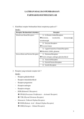
  • 1. 1 LATIHAN SOAL DAN PEMBAHASAN FARMAKOLOGIMOLEKULAR 1. Klasifikasi reseptor berdasarkan lokasi reseptornya pada sel ? Jawab : Reseptor Berdasarkan Lokasinya Reseptor Membrane Bound Receptors 1) G-Protein-Linked Receptors ➔Serotonin, muskarinik, domaninergik, noradrenergik 2) Enzyme Receptors ➔Tyrosine kinase 3) Ligand-Gated Ion Channel Receptors ➔Nikotinik, GABA,glutamat Intracellularand NuclearReceptors 1) Hormone Receptors ➔Tiroid, estrogen, androgen glukokortikoid 2) Autocoid Receptors 3) Growth Factors Receptors 4) Insulin Receptors 2. Reseptor yang termasuk reseptor inti ? Jawab : - Reseptor glukokortikoid - Reseptor mineralokortikoid - Reseptor progesteron - Reseptor androgen - Reseptor estrogen - RXR (Retinoid X Receptors) ➔ PPAR (Peroxisom Proliferators – Activated Receptor) ➔ T3R (Thyroid Hormone Receptor) - Monomeric Tethered Orphan Receptors ➔ ROR (Retinoic Acid – Related Orphan Receptor) ➔ ERR (Estrogen – Related Receptor)
  • 2. 2 3. Mekanisme aksi glucocorticoid receptor (GC) ? Jawab : a) Aktivasi genomik ➔Membutuhkan aktivasi gen-gen tertentu. Mekanismenya yaitu kortisol masuk ke dalam membran sel lalu berikatan dengan reseptor glukokortikoid yang terlebih dahulu distabilkan oleh HSP (Heat Shock Protein). Setelah reseptor glukokortikoid berikatan dengan kortisol, akan melepaskan HSp dan menduduki glukokortikoid. Respon elemen dalam untaian DNA dalam inti kemudian mRNA ditranskripsi lalu ditranslasi dan diterjemahkan membentuk protein antiinflamasi yang menghambat inflamasi. Contoh : TGF- β, IL – 10, lipocortin. b) Aktivasi non genomik ➔Kortisol masuk ke dalam membran sel lalu berikatan dengan reseptor glukokortikoid. Sebelum kortisol diikat oleh reseptor glukokortikoid, distabilkan oleh HSP. Setelah glukokortikoid berikatan dengan kortisol, HSP akan dilepaskan dan akan aktivasi protein antiinflamasi yang menghambat inflamasi. 4. Apa saja yang termasuk ligan alamiah dan ligan sintetis glucocorticoid receptor (GC)? Jawab : - Ligan alamiah = kortisol, hidrokortison - Ligan sintetis = deksametason, prednison, metil prednison, betametason 5. Bagaimanakah mekanisme molekular ikatan prednison dan reseptor GC menimbulkan efek antiinflamasi ? Jawab : Prednison adalah senyawa sintetis berupa pro-drug dari prednisolon yang memiliki aktivitas antiinflamasi dan imunomodulasi. Mekanisme kerjanya yaitu setelah prednison melekat ke reseptor membran sel dan memasuki sel, prednison memasuki nukleus lalu berikatan dan aktivasi reseptor nukleus (inti). Prednison di dalam darah akan berubah menjadi bentuk aktif (prednisolon) lalu menghambat migrasi sel polimorfonuklear (PMN) sehingga dapat inhibisi infiltrasi leukosit, supresi respon imun humoral, dan mengurangi sekresi IL – 6 sehingga inflamasi tidak terjadi. 6. Bagaimanakah mekanisme aksi genomik pada kompleks ligan dan reseptor GC ?
  • 3. 3 Jawab : Kompleks ligan dan reseptor GC, masuk ke inti dan mengikat GRE (Glucocorticoid Response Elements) sehingga menyebabkan transrepresi gen pro inflamasi. GRE sebagai suatu ‘landasan’ untuk reseptor glukokortikoid akan meregulas transkripsi gen untuk menurunkan produksi sitokin pro inflamasi dan meningkatkan protein antiinflamasi. 7. Bagaimanakah mekanisme molekular kortikosteroid memiliki efek samping osteoporosis ? Jawab : *referensi dan jurnal terlampir a) Efek Langsung b) Efek tidak Langsung ➔Osteoporosis akibat terapi glukokortikoid juga dapat terjadi karena hypogonadism, menurunkannya aktivitas fisik, menurunnya produksi growth hormone, insulin-like growth factor 1 (IGF 1) dan IGF 1 binding protein (IGF-BP) serta meningkatnya kehilangan kalsium di ginjal dan intestinal. 8. Apakah keterkaitan antara reseptor GC dengan HSP ?
  • 4. 4 Jawab : HSP (Heat Shock Protein) menstabilkan bentuk molekul GC → selama tidak mengikat ligan, reseptor GC dalam keadaan inaktif. 9. Bagaimanakah mekanisme kerja kompleks GC dan steroid dalam mengurangi inflamasi? Jawab : Kompleks GC dansteroid dapat upregulates (meningkatkan eskpresi dari gen-gen yang dikendalikan oleh lipocortin (annexin). Kortisol berikatan dengan reseptor GC menghasilkan protein annexin 1 yang dapat menghambat aktivasi enzim c PLA 2α sehingga fosfolipid tidak dapat diubah menjadi asam arachidonate → tidak dapat produksi prostaglandin → inhibisi inflamasi. Kortisol berikatan dengan reseptor GCinhibisi transkripsi NF-kB sehingga tidakterjadi inflamasi → tidak terbentuk prostaglandin oleh enzim COX-2 → inhibisi inflamasi Kortisol berikatan dengan glukokortikoid → aktivasi MAPK fosfatase 1 →inhibisi terbentuknya MAPKs → inhibisi aktivasi enzim c PL 2α yang dapat mengubah fosfolipid menjadi arachidonate → prostaglandin tidak terbentuk → inflamasi berkurang. 10. Apakah yang dimaksud dengan NF-kB, AP-1, Jun Fos, dan MAPK ? Jawab : a) NF-kB ➔Nuclear factor kappa light chain enhancer of activated β cells ➔Faktor transkripsi yang bertanggung jawab padaproses inflamasi ; berupa protein kompleks yang mengendalikan transkripsi DNA, produksi sitokin, dan pertahanan sel. b) AP-1 ➔Activated Protein 1 ➔Faktor transkripsi yang mengatur ekspresi gen sebagai respons terhadap stimulus meliputi sitokin, growth factor, stres, dan infeksi mikroorganisme dengan mengendalikan proses selular seperti diferensiasi, proliferasi, dan apoptosis.
  • 5. 5 c) Jun Fos ➔Onkogen sebagai informasi genetik yang bertanggung jawab dalam induksi tumor oleh virus sarcoma murine FBJ dan virus 17 sarkoma avian yang diderivat dari gen selular normal (c-fos). d) MAPK ➔Mitogen Activated Protein Kinase ➔Enzim yang teraktivasi sebagai respon selular terhadap berbagai jenis hormon faktor pertumbuhan, dan katalisis reaksi fosforilasi terhadap jenis protein tertentu. Referensi : 1. Ayroldi, Emira et al. Mechanismes of the Antiinflammatory Effects of Glucocorticoids : Genomic and Non-Genomic Interference with MAPK Signalling Pathways. The FASEB Journal. 2. Curran, Tom. Fos and Jun : Oncogenic Transcription Factors. Tohoku J Exp Med.1992. 3. Compston, Juliet. Glucocorticoid – Induced Osteoporosis : An Update. Springer Endocrine. 2018. 4. Lawrence, Toby. The Nuclear Factor NF-kB Pathway in Inflammation. Cold Spring Harb Perspect Biol. 2009. 5. Schijvens et al. Pharmacology and Pharmacogenetics of Prednisone and Prednisolone in Patients with Nephrotic Syndrome. Pediatric Nephrology. 2018. 6. Zenz, Rainer et al. Activator Protein 1 (Fos/Jun) Functions in Inflammatory Bone and Skin Disease. Arthritis Research and Therapy. BMC Journal. 2008.
  • 6. Endocrine https://doi.org/10.1007/s12020-018-1588-2 REVIEW Glucocorticoid-induced osteoporosis: an update Juliet Compston 1 Received: 27 December 2017 / Accepted: 22 March 2018 © The Author(s) 2018 Abstract Glucocorticoid-induced osteoporosis is the most common secondary cause of osteoporosis and the resulting fractures cause significant morbidity. Following initiation of oral glucocorticoids, rapid bone loss occurs, and fracture risk increases within a few months in a dose-dependent manner. These adverse effects are due to inhibition of bone formation accompanied by an early but transient increase in bone resorption. Multiple mechanisms underlie these changes in bone remodeling; direct effects include upregulation of PPARγR2, increased expression of sclerostin and increased RANKL/OPG ratio, whilst hypogonadism, altered renal and intestinal calcium handling, and reduced production of insulin-like growth factor 1 also contribute. Fracture risk assessment should be performed as soon as possible after glucocorticoids are initiated and bone protective therapy started promptly in individuals at high-risk, with calcium and vitamin D supplements where appropriate. Oral bisphosphonates are currently regarded as first line options on the grounds of their low cost. However, teriparatide has been shown to be superior in its effects on BMD and vertebral fracture risk in glucocorticoid-treated individuals with osteoporosis and should be considered as an alternative first line option in high-risk patients. Key words Glucocorticoids ● Bone density ● Fracture ● Bisphosphonates ● Teriparatide Introduction Glucocorticoids are used in the treatment of a wide range of diseases and it is estimated that 1–2% of the population is receiving long-term glucocorticoid therapy [1–4]. The adverse skeletal effects of glucocorticoid excess were first described over 80 years ago, and today glucocorticoid- induced osteoporosis is the most common secondary cause of osteoporosis. Research into the epidemiology, patho- physiology and clinical management of glucocorticoid- induced osteoporosis has produced substantial advances, yet the condition remains under-recognized and under-treated. This review focuses on recent developments in the field and their implications for clinical practice. Epidemiology Continuous oral glucocorticoid therapy is associated with rapid bone loss and an increase in fracture risk that is seen within 3–6 months of initiation and is dose-related. This time course can be explained, at least in part, by the higher doses of glucocorticoids and greater disease activity in the early stages of treatment. Fracture risk remains elevated for the duration of glucocorticoid therapy, but declines after its withdrawal, although whether it reverts to baseline values is unclear [5–8]. Vertebral fractures are particularly char- acteristic of glucocorticoid-induced osteoporosis, although the risk of non-vertebral fractures, including hip fractures, is also increased. Recent studies have confirmed the predilection for ver- tebral fracture, the dose-related increase in fracture risk, and decline in fracture risk with longer duration or dis- continuation of glucocorticoid therapy. Amiche et al con- ducted a Bayesian meta-analysis of fracture risk associated with oral glucocorticoid use, drawing on data from the control groups of clinical trials [9]. In individuals who had initiated glucocorticoid therapy in the last 6 months, the annual incidence of vertebral fracture was 5.1% [95% credible intervals (CrI) 2.8–8.2] and of non-vertebral fracture, 2.5% [95% CrI 1.2–4.2]. For those with duration * Juliet Compston jec1001@cam.ac.uk 1 Department of Medicine, Cambridge Biomedical Campus, Cambridge CB2 0SL, UK 1234567890();,: 1234567890();,:
  • 7. of glucocorticoid use ≥6 months, the corresponding figures were 3.2% [95% CrI 1.8–5.0] and 3.0% [95% CrI 0.8–5.0]. Using a large administrative database, Balasubramanian et al investigated the effects of initiating systemic (oral or injected) glucocorticoid therapy on fracture risk in patients with new-onset rheumatoid arthritis (mean age 49 years) [10]. Fracture incidence rates were 5 to 9/1000 person years at doses of <15 mg/day, 16 (11, 22.6) at doses ≥15 mg/day and 13.4 (10.7, 16.7) at cumulative doses ≥5400 mg. At 60-182 days after discontinuation of gluco- corticoid therapy, fracture risk was 29% lower than in those with ongoing use and by 12 months was similar to non- glucocorticoid users. The effects of inhaled or intravenous glucocorticoids on fracture risk are less well documented. There is some evi- dence that high doses of inhaled glucocorticoids may be associated with increased fracture risk, although concurrent use of oral glucocorticoids is often a confounding factor [11–14]. In a large case control study from Denmark, no increase in fracture risk was found in association with other forms of topical steroids [15]. Variation in the severity of adverse effects of gluco- corticoids, including bone loss, is well recognized but poorly understood. Pre-receptor modulation of glucocorti- coid activity by 11 beta-hydroxysteroid dehydrogenase (11βHSD) enzymes, which interconvert inactive and active cortisone/cortisol, may contribute to this variability through the effects of pro-inflammatory cytokines [16, 17] and genetic polymorphisms in the glucocorticoid receptor gene [18]. Pathophysiology Direct effects on bone Glucocorticoid-induced osteoporosis is characterized by decreased bone formation, with an additional early but transient increase in bone resorption (Fig. 1). The initial increase in remodeling rate is accompanied by reduced bone formation at the level of the individual bone multicellular unit (BMU), and this combination of increased bone turn- over and a negative remodeling balance results in rapid bone loss [19–23]. Subsequently, the decrease in bone formation, both at tissue and BMU level, predominates leading to a low turnover state. Direct effects of gluco- corticoids on bone formation are mediated largely through upregulation of peroxisome proliferator-activated receptor gamma receptor 2 (PPARγ2) [24] and effects on the Wnt/β- catenin signaling pathway [25, 26]. The former mechanism favors the differentiation of pluripotent precursor cells to adipocytes in preference to osteoblasts, resulting in decreased numbers of osteoblasts. Increased expression of sclerostin, which binds to the co-receptors for frizzled, Lrp4 and Lrp5, results in inhibition of Wnt sigalling leading to reduced differentiation of osteoblast precursors to mature osteoblasts and increased osteoblast and osteocyte apopto- sis. The importance of sclerostin in mediating the effects of glucocorticoids on bone formation is emphasized by the demonstration, in mice with sclerostin deficiency, that bone integrity is maintained in the presence of glucocorticoid excess [27]. Furthermore, in a mouse model of glucocorticoid-induced osteoporosis, treatment with an antibody to sclerostin prevented the reduction in bone mass and strength [28]. Glucocorticoids also have direct effects on bone resorp- tion, increasing the production of macrophage colony sti- mulating factor (M-CSF) and RANKL and decreasing production of osteoprotegerin (OPG) by osteoblastic cells and osteocytes, resulting in an increase both in the number and activity of osteoclasts [29, 30]. This effect diminishes with time, possibly as a result of the reduction in number of osteoblasts and osteocytes. Finally, there is some evidence from animal models that glucocorticoids affect osteocyte morphology and mineralization [31]. Indirect effects on bone Other mechanisms that may contribute to glucocorticoid- induced bone loss through indirect effects on bone include hypogonadism, reduced physical activity, increased renal and intestinal losses of calcium, and reduced production of growth hormone, insulin-like growth factor 1 (IGF1) and IGF1 binding protein (IGF-BP) [32]. In addition, the underlying diseases for which glucocorticoid therapy is administered are often associated with increased inflam- mation, which contributes to bone loss through increased production of pro-inflammatory, pro-resorptive cytokines. Whilst glucocorticoids suppress inflammation and hence should mitigate the adverse effects of inflammation, disease relapse despite therapy is associated with episodes of increased bone resorption. Finally, glucocorticoid excess has adverse effects on muscle mass and function, leading to myopathy and increased risk of falls [33]. Changes in BMD and bone microarchitecture Increased rates of bone loss in the hip, spine, and radius are well documented in individuals treated with glucocorti- coids. Data obtained from assessment of bone micro- architecture using high-resolution peripheral computed tomography (HRpQCT) are sparse. In a cross-sectional study of 30 postmenopausal women who had received oral glucocorticoids for longer than 3 months, despite similar Endocrine
  • 8. areal BMD values to 60 control subjects, significantly lower total, cortical and trabecular volumetric BMD, thinner cor- tices, increased trabecular separation and reduced trabecular number were reported in the radius and tibia; whole bone stiffness, assessed using finite element analysis, was also significantly reduced in comparison to the controls [34]. Although the patients and controls were generally well matched, however, bisphosphonate use was significantly more common in the former (100% vs. 8.6%), so definite attribution of the observed differences to glucocorticoid therapy cannot be made. Trabecular bone score (TBS) provides an indirect index of trabecular bone architecture that can be obtained from DXA images of the lumbar spine, and has predictive value for fracture independent of BMD [35]. In 64 post- menopausal women who had taken prednisolone in a dose of ≥5 mg daily for >3 months, TBS was significantly lower than in a group of non-glucocorticoid treated controls, although lumbar spine BMD T-scores were not significantly different [36]. Similar findings have been reported in 416 individuals on long-term glucocorticoids (≥5 mg daily for 3 months), the decrease in TBS being most marked in men and in individuals with fracture [37]. These findings indi- cate that glucocorticoids have adverse effects on spine bone microarchitecture that are independent of BMD and which may contribute to increased fracture risk. Fracture risk assessment in individuals treated with glucocorticoid-induced osteoporosis There is evidence from a number of studies that fracture occurs at a higher BMD in individuals receiving gluco- corticoids than in non-glucocorticoid treated people [38–40]. This independent effect of glucocorticoid therapy on fracture risk may be due to several factors, including increased falls risk and alterations in bone quality that are not captured by BMD measurements. Glucocorticoid therapy is included as a risk factor in the FRAX fracture prediction algorithm as a dichotomous variable. “Yes” is entered in the questionnaire if there is current exposure to oral glucocorticoids or past exposure for ≥ 3 months at a dose of 5 mg/day or more of prednisolone or equivalent. Using data from the UK General Research Practice Database, Kanis et al have provided adjustments that can be incorporated into the FRAX calculations to adjust for different doses of glucocorticoids (Table 1) [41]. For daily doses of over 7.5 mg daily of prednisolone or equivalent, greater upward adjustment of fracture prob- ability may be required. It should be noted that the duration of glucocorticoid therapy and cumulative dose are not accommodated within the FRAX algorithm. In addition, the use of total hip BMD in FRAX may result in under- estimation of fracture risk in patients with differentially low Fig. 1 Direct effects of glucocorticoids on bone Endocrine
  • 9. spine BMD, although a correction for this discordance has been proposed [42, 43]. A final caveat is that the response to treatment in glucocorticoid-treated individuals selected on the basis of FRAX-derived fracture probability has not been documented. Assessment of fracture risk using FRAX is recom- mended in several guidelines for the management of glucocorticoid-induced osteoporosis, including the National Osteoporosis Guideline Group (NOGG) guidance [44, 45], the updated recommendations produced by the American College of Rheumatology (ACR) [46] and guidelines pub- lished by the International Osteoporosis Foundation (IOF) and European Calcified Tissues Society (ECTS) [47, 48]. However, FRAX can only be used in people age 40 years and over; in children and young adults, fracture risk assessment should be performed using BMD measurement, together with consideration of other risk factors, particularly previous fracture. Management of glucocorticoid-induced osteoporosis Under-treatment of glucocorticoid-induced osteoporosis is widely recognized [49, 50]. In a population-based study of adults age ≥20 years, rates of BMD testing and prescription of bone protective medication between 1998 and 2008 were studied in individuals prescribed systemic glucocorticoids for 90 days or longer [51]. Overall, in the first six months after initiation of glucocorticoid therapy only 6% had BMD testing, 22% received therapy and 25% had both interven- tions. Under-treatment was greatest in younger people and men, and primary care physicians had lower prescription rates than rheumatologists. Similar results have been reported more recently using information from a national public health-insurance database in France, with only 8% undergoing BMD testing and prescription of calcium ± vitamin D and bisphosphonates in 18 and 12% respectively [52]. In a large cohort from Canada of men and women aged 66 years or over who were initiating long-term glu- cocorticoid therapy, Amiche et al reported that only 13% were prescribed bone protective therapy [53]. The problem of under-treatment is compounded by poor persistence with bisphosphonate therapy, particularly in younger people, those with co-morbidities, and those in whom BMD mea- surements have not been made [54]. General measures A number of life-style measures may mitigate the harmful skeletal effects of glucocorticoids, although the evidence base for this approach is weak and requires extrapolation from studies in non-glucocorticoid treated individuals. Falls risk should be assessed and preventive measures instituted when appropriate. Exercise, tailored to the individual patient, and good nutrition with adequate dietary calcium intake should be advocated with avoidance of smoking and alcohol abuse. Maintenance of an adequate vitamin D status should also be advised. Attention should be paid to keeping the dose of gluco- corticoids to a minimum, with the use of steroid-sparing drugs such as methotrexate or azathioprine or alternative routes of administration (for example inhaled or topical) where appropriate. Non-steroidal therapies should be used when possible to maintain remission, once achieved. However, it is also important to maintain suppression of the underlying disease, since this will prevent the adverse skeletal effects of inflammation and other effects of increased disease activity. Studies of the effects of calcium and/or vitamin D sup- plementation on BMD in patients taking glucocorticoids have produced conflicting results [55, 56]. However, cal- cium and vitamin D supplements have been included in most trials of bone protective therapy and should therefore be used as an adjunct to treatment. Pharmacological interventions Regulatory approval of drugs to reduce fracture risk in individuals taking glucocorticoids has been based on the demonstration of similar changes in BMD to those observed in postmenopausal osteoporosis and fracture has been a secondary outcome. For this reason, evidence for anti-fracture efficacy of bone protective agents in glucocorticoid-induced osteoporosis is less robust than that for postmenopausal osteoporosis. In addition, there is inevitable heterogeneity in glucocorticoid-treated trial populations, with respect to age, underlying disease, co-morbidities and co-medications, dose and duration of glucocorticoid therapy and the timing of bone protective therapy. Furthermore, the duration of most treatment stu- dies has been relatively short and this, combined with smaller trial populations, reduces the strength of the safety database. Table 1 Adjustment of FRAX-derived fracture probability according to dose of glucocorticoids. Data from ref. [40] Daily dose of prednisolone (mg) Average adjustment for major osteoporotic fracture probability Average adjustment for hip fracture probability <2.5 −20% −35% 2.5–7.5 None None ≥7.5a +15% +20a a For high doses of prednisolone, greater upward adjustment of fracture risk may be appropriate Endocrine
  • 10. Bisphosphonates Bisphosphonates are the most commonly used drugs in the management of glucocorticoid-induced osteoporosis. Oral alendronate (5 or 10 mg daily or 70 mg once weekly) and risedronate (5 mg daily or 35 mg once weekly), and intra- venous zoledronic acid (5 mg once yearly by intravenous infusion) are all approved for this indication. All have been shown to have beneficial effects on lumbar spine and hip BMD in people treated with glucocorticoids [57–64] and for alendronate and risedronate, there is also evidence from safety or post hoc analyses for a reduction in the rate of vertebral fractures [62, 63]. In the pivotal study of zole- dronic acid, a non-inferiority BMD comparison with rise- dronate, the fracture rate was too low to assess anti-fracture efficacy [64]. The number of non-vertebral and hip fractures has been insufficient in individual trials to assess an impact of bisphosphonates. However, data from cohort studies provide some evidence for efficacy at these sites. In an observational cohort study of women aged >65 years taking alendronate or risedronate, Thomas et al. studied the baseline incidence of clinical fractures in the first three months after starting glu- cocorticoid therapy and the fracture incidence in the fol- lowing 12 months [65]. Compared to the baseline incidence, both clinical vertebral and non-vertebral fracture incidence were significantly lower. Using the same database, Overton et al studied the effects of bone protective therapy (bispho- sphonates in 95.5%, denosumab or teriparatide in the remaining 4.5%) in a large cohort of new glucocorticoid users [66]. Treatment within the first 90 days of gluco- corticoid use was associated with a significant reduction in clinical fractures (including vertebral) of 48% at one year and 32% at three years when compared with non-use. Finally, in three matched cohorts derived from healthcare administrative data from Ontario, Canada, Amiche et al. reported that in individuals initiating long-term glucocorti- coids, therapy within the first six months with alendronate or risedronate was associated with a decrease in incident hip fracture (alendronate 0.49 (0.34, 0.69), risedronate 0.58 (0.36, 0.90) [53]. The results confirmed a reduction in ver- tebral fracture risk with etidronate, alendronate and rise- dronate, but no decrease in risk of forearm or humerus fractures for any bisphosphonate. The analysis was limited to oral bisphosphonates and zoledronic acid was not con- sidered. Overall, therefore, these studies would be consistent with a beneficial effect of bisphosphonates both on vertebral and non-vertebral fracture, including hip fracture. The safety profile of bisphosphonates in glucocorticoid- induced osteoporosis has been less well studied than in postmenopausal osteoporosis because of the small number of participants included and shorter duration of the trials. Because of co-morbidities and co-medications, people taking glucocorticoids may be more susceptible to side- effects, for example upper gastrointestinal disorders, and glucocorticoid therapy is a documented risk factor for osteonecrosis of the jaw [67] and probably also for atypical femoral fractures [68, 69]. Bisphosphonates should be used with caution in premenopausal women with child bearing potential, because of the ability of these drugs to cross the placenta. Teriparatide The predominant role of reduced bone formation in glucocorticoid-induced osteoporosis provides a rationale for the use of anabolic agents in its treatment. In an active- comparator controlled, randomized, double blind study the effects of 18 months treatment with subcutaneous teripara- tide, 20 µg/day, or oral alendronate 10 mg/day, were com- pared in 428 men and women with glucocorticoid-induced osteoporosis [70]. Teriparatide therapy resulted in sig- nificantly greater increases in spine and hip BMD and this was seen in both premenopausal and postmenopausal women and in men [71]. The magnitude of increase in BMD was somewhat less than that seen in non- glucocorticoid treated postmenopausal women in another study [72], possibly as a result of the opposing actions of intermittent PTH and glucocorticoids on osteoblastogenesis, and osteoblast and osteocyte apoptosis [73–75]. Although fracture was not a primary end-point of the study, significantly fewer new vertebral fractures occurred in the patients treated with teriparatide when compared to those treated with alendronate (0.6% vs. 6.1%; p = 0.004). The incidence of non-vertebral fractures was similar in the two treatment groups. Results after 36 months of treatment demonstrated a continued increase in spine and hip BMD in the teriparatide treated group, with superiority over alen- dronate at the 24 and 36 month time points [76]. A lower incidence of new vertebral fractures was also seen in the teriparatide group at 36 months (1.7% vs 7.7%, p = 0.007), with a similar incidence of non-vertebral fractures in the two groups. Interestingly, measurements of TBS in a sub- population of this study demonstrated a significant increase after 36 months in teriparatide treated patients, but no sig- nificant change in those treated with alendronate [77]. Whilst the long duration of this study is unique amongst treatment trials for glucocorticoid-induced osteoporosis, it should be noted that the participant discontinuation rate at 36 months was 44%. With regard to safety, increased pre- dose serum calcium levels were significantly more common in the teriparatide than alendronate treated group (21% vs. 7%), but no other concerns were identified In a comparison of the effects of teriparatide and rise- dronate in men treated with oral glucocorticoids for ≥3 months, significantly greater increases in lumbar spine Endocrine
  • 11. trabecular volumetric BMD were demonstrated in the teriparatide-treated group at 18 months. In addition, ter- iparatide was associated with significantly greater improvements in bone strength and stiffness at T12, mea- sured using HRQCT-based finite element analysis [78]. These data, together with the results of the study reviewed above, provide a rationale for the use of teriparatide as a first line option in some patients, particularly those at high risk of vertebral fracture. Teriparatide is contraindicated in pregnant and lactating women. Women of childbearing potential should use effective methods of contraception while on teriparatide treatment. Denosumab At present, denosumab is not approved for use in glucocorticoid-induced osteoporosis, although this is cur- rently under consideration by some regulatory agencies. In a Phase 3 randomized double blind active controlled trial of adults taking ≥7.5 mg prednisone or equivalent daily, treatment with denosumab, 60 mg once every 6 months by subcutaneous injection, was associated with a significantly greater increase in lumbar spine BMD compared to rise- dronate 5 mg daily over a 12 months treatment period [79]. This effect was seen both in patients initiating glucocorti- coids and in those established on long-term therapy, regardless of age, race, baseline BMD T-score, initial glu- cocorticoid dose and menopausal status. To date the study has only been reported in abstract form. Guidelines for the management of glucocorticoid- induced osteoporosis National guidelines for the management of glucocorticoid- induced osteoporosis have been produced by a number of countries and updated guidelines have recently been published from the US and UK. Most guidelines address users of long-term (3 months) oral glucocorticoids, and although the daily threshold dose varies it has generally been between 5 and 7.5 mg daily of prednisolone or equivalent. Treatment thresholds also vary; T-scores are used in some and generally recognize the higher BMD at which fracture occurs in glucocorticoid-treated indivi- duals. The recent guidelines from the UK and US emphasize the importance of starting bone protective therapy early in high-risk individuals, because of the rapidity with which bone loss and increased fracture risk occur. The recently updated ACR guideline defines three cate- gories of fracture risk; high, moderate and low. In adults aged ≥40 years, previous osteoporotic fracture, hip or spine BMD T-score ≤ −2.5, or 10 year fracture probability of ≥20% (major osteoporotic fracture) or ≥3% (hip fracture) are the criteria for high risk [45]. Moderate and low risk are defined solely on the basis of FRAX-derived fracture probability (10–19% and >1 to ≤3% respectively for mod- erate risk, <10% and ≤1% respectively for low risk). In adults <40 years old, the criterion for high risk is a previous osteoporotic fracture, whereas moderate and low risk are defined on the basis of BMD. Oral bisphosphonates are recommended as the first line option for individuals at moderate or high risk, with intravenous bisphosphonates, teriparatide, or denosumab in patients who are contra- indicated or intolerant to oral bisphosphonates. NOGG, in its 2017 update, takes a similar approach to risk assessment with the use of dose-adjusted FRAX, but uses the UK NOGG intervention thresholds as the basis for making treatment decisions and does not include denosumab as a treatment option [44, 45]. These recent guidelines emphasize the importance of early intervention in high-risk individuals, advocate main- tenance of adequate calcium intake and normal vitamin D status, and stress the limited evidence base to support some recommendations, particularly in younger adults. Compar- ison of the cost-effectiveness of different treatments is problematic because of the lack of fracture data, particularly hip fracture, in clinical trials. The choice of oral bispho- sphonates as a first line option in both sets of guidelines is based on their lower cost compared to either zoledronic acid or teriparatide; nevertheless, in view of the superiority of teriparatide vs. alendronate for BMD and vertebral fracture outcomes, it has been suggested that teriparatide should be considered as an alternative first line option in those at highest risk [80]. Conclusions There have been significant advances in our understanding of the mechanisms by which glucocorticoids affect bone and increase fracture risk. The clinical management of glucocorticoid-induced osteoporosis, however, remains suboptimal despite the development of more sophisticated approaches to fracture risk assessment and the availability of several effective treatment options. Whilst bispho- sphonates are currently the most widely used bone protec- tive drugs in individuals taking glucocorticoids, the use of teriparatide and of denosumab (if approved) as first-line options in some patients merits further investigation. Finally, the potential for sclerostin inhibitors to prevent or reverse adverse skeletal effects of glucocorticoids provides an exciting prospect for future research. Endocrine
  • 12. Compliance with ethical standards Conflict of interest Prof Compston has received advisory and speaking fees from Gilead and speaking fees from Amgen and UCB. Open Access This article is distributed under the terms of the Creative Commons Attribution 4.0 International License (http://crea tivecommons.org/licenses/by/4.0/), which permits unrestricted use, distribution, and reproduction in any medium, provided you give appropriate credit to the original author(s) and the source, provide a link to the Creative Commons license, and indicate if changes were made. References 1. T.P. van Staa, H.G. Leufkens, L. Abenhaim, S. Begaud, B. Zhang, C. Cooper, Use of oral corticosteroids in the United Kingdom. Q Med J. 93, 105–111 (2000) 2. L. Fardet, I. Petersen, I. Nazareth, Prevalence of long-term oral glucocorticoid prescriptions in the UK over the past 20 years. Rheumatology 50(11), 1982–1990 (2011) 3. R.A. Overman, J.Y. Yeh, C.L. Deal, Prevalence of oral gluco- corticoid usage in the United States: a general population per- spective. Arthritis Care Res. 65(2), 294–298 (2013) 4. S. Silverman, J. Curtis, K. Saag, J. Flahive, J. Adachi, F. Anderson, R. Chapurlat, C. Cooper, A. Diez-Perez, S. Greenspan, F. Hooven, A. Le Croix, L. March, J.C. Netelenbos, J. Nieves, J. Pfeilschifter, M. Rossini, C. Roux, E. Siris, N. Watts, J. Comp- ston, International management of bone health in glucocorticoid- exposed individuals in the observational GLOW study. Osteo- poros. Int. 26(1), 419–420 (2015) 5. T. van Staa, H.G.M. Leufkens, L. Abenhaim, B. Zhang, C. Cooper, Use of oral corticosteroids and risk of fractures. J. Bone Miner. Res. 15, 933–1000 (2000) 6. F. De Vries, M. Bracke, H.G. Leufkens, J.W. Lammers, C. Cooper, T.P. Van Staa, Fracture risk with intermittent high-dose glucocorticoid therapy. Arthritis Rheum. 56, 206–214 (2007) 7. T.P. van Staa, H.G.M. Leufkens, L. Abenhaim, B. Zhang, C. Cooper, Oral corticosteroids and fracture risk: relationship to daily and cumulative doses. Rheumatology 39, 1383–1389 (2000) 8. T.P. van Staa, H.G.M. Leufkens, C. Cooper, A meta-analysis of the epidemiology of corticosteroid-induced osteoporosis. Osteo- poros. Int. 13, 777–787 (2002) 9. M.A. Amiche, J.M. Albaum, M. Tadrous, P. Pechlivanoglou, L.E. Lévesque, J.D. Adachi, S.M. Cadarette, Fracture risk in oral glucocorticoid users: a Bayesian meta-regression leveraging con- trol arms of osteoporosis clinical trials. Osteoporos. Int. 27(5), 1709–1718 (2016) 10. A. Balasubramanian, S.W. Wade, R.A. Adler, C.J.F. Lin, M. Maricic, C.D. O’Malley, K. Saag, J.R. Curtis, Glucocorticoid exposure and fracture risk in patients with new-onset rheumatoid arthritis. Osteoporos. Int. 27(11), 3239–3249 (2016) 11. J.H. Toogood, J.C. Baskerville, A.E. Markov, A.B. Hodsman, L.J. Fraher, B. Jennings, R.G. Haddad, D. Drost, Bone mineral density and the risk of fractures in patients receiving long-term inhaled steroid therapy for asthma. J. Allergy Clin. Immunol. 96, 157–166 (1995) 12. C.A. Wong, L.J. Walsh, C.J. Smith, A.F. Wisniewski, S.A. Lewis, R. Hubbard, S. Cawte, D.J. Green, M. Pringle, A.E. Tattersfield, Inhaled corticosteroid use and bone mineral density in patients with asthma. Lancet 355, 1399–1403 (2000) 13. T.P. Van Staa, H.G. Leufkens, C. Cooper, Use of inhaled corti- costeroids and risk of fractures. J. Bone Miner. Res. 16, 581–588 (2001) 14. T.P. Van Staa, N. Bishop, H.G. Leufkens, C. Cooper, Are inhaled corticosteroids associated with an increased risk of fracture in children? Osteoporos. Int. 15, 785–791 (2004) 15. P. Vestergaard, L. Rejnmark, L. Mosekilde, Fracture risk asso- ciated with systemic and topical corticosteroids. J. Intern. Med. 257(4), 374–84 (2005) 16. M.S. Cooper, A. Blumsohn, P.E. Goddard, W.A. Bartlett, C.H. Shackleton, R. Eastell, M. Hewison, P.M. Stewart, 11 beta- hydroxysteroid dehydrogenase type 1 activity predicts the effects of glucocorticoids on bone. J. Clin. Endocrinol. Metab. 83, 3874–3877 (2003) 17. M.S. Cooper, I. Bujalska, E. Rabbitt, E.A. Walker, R. Bland, M.C. Sheppard, M. Hewison, Stewart PM.Modulation of 11-beta hydroxysteroid dehydrogenase enzymes by proinflammatory cytokines in osteoblasts: an autocrine switch from glucocorticoid inactivation to activation. J. Bone Miner. Res. 16, 1037–1044 (2001) 18. H. Russcher, P. Smit, E.L.T. van den Akker, E.F. van Rossum, A. O. Brinkmann, F.H. de Jong, S.W. Lamberts, J.W. Koper, Two polymorphisms in the glucocorticoid receptor gene directly affect glucocorticoid-regulated gene expression. J. Clin. Endocrinol. Metab. 90, 5804–5810 (2005) 19. C. Bresssot, P.J. Meunier, M.C. Chapuy, E. Lejeune, C. Edouard, A.J. Darby, Histomorphometric profile, pathophysiology and reversibility of corticosteroid-induced osteoporosis. Metab. Bone Dis. Rel. Res. 1, 303–311 (1979) 20. D.W. Dempster, M.A. Arlot, P.J. Meunier, Mean wall thickness and formation periods of trabecular bone packets in corticosteroid-induced osteoporosis. Calcif. Tissue Int. 35, 410–417 (1983) 21. D.W. Dempster, Bone histomorphometry in glucocorticoid- induced osteoporosis. J. Bone Miner. Res. 4, 137–147 (1989) 22. D. Chappard, E. Legrand, M.F. Basle, P. Fromont, J.L. Racineux, A. Rebel, M. Audran, Altered trabecular architecture induced by corticosteroids: a bone histomorphometric study. J. Bone Miner. Res. 11, 676–685 (1996) 23. L. Dalle Carbonare, M.E. Arlot, P.M. Chavassieux, J.P. Roux, N.R. Portero, P.J. Meunier, Comparison of trabecular bone architecture and remodeling in glucocorticoid-induced and postmenopausal osteoporosis. J. Bone Miner. Res. 16, 97–103 (2001) 24. Z. Wu, N.L.R. Bucher, S.R. Farmer, Induction of peroxisome proliferator-activated receptor γ during the conversion of 3T3 fibroblasts into adipocytes is mediated by C/EBPh, C/EBPy, and glucocorticoids. Mol. Cell. Biol. 16, 4128–4136 (1996) 25. R.S. Weinstein, R.L. Jilka, A.M. Parfitt, S.C. Manolagas, Inhibi- tion of osteoblastogenesis and promotion of apoptosis of osteo- blasts and osteocytes by glucocorticoids—potential mechanisms of their deleterious effects on bone. J. Clin. Invest. 102, 274–282 (1998) 26. K. Ohnaka, M. Tanabe, H. Kawate, H. Nawata, R. Takayanagi, Glucocorticoid suppresses the canonical Wnt signal in cultured human osteoblasts. Biochem. Biophys. Res. Commun. 329, 177–181 (2005) 27. A.Y. Sato, M. Cregor, J. Delgado-Calle, K.W. Condon, M.R. Allen, M. Peacock, L.I. Plotkin, T. Bellido, Protection from glucocorticoid-induced osteoporosis by anti-catabolic signaling in the absence of sost/sclerostin. J. Bone Miner. Res. 31, 1791–1802 (2016) 28. W. Yao, W. Dai, L. Jiang, E.Y. Lay, Z. Zhong, R.O. Ritchie, X. Li, H. Ke, N.E. Lane, Sclerostin-antibody treatment of glucocorticoid-induced osteoporosis maintained bone mass and strength. Osteoporos. Int. 27(1), 283–294 (2016) Endocrine
  • 13. 29. C. Swanson, M. Lorentzon, H.H. Conaway, U.H. Lerner, Glu- cocorticoid regulation of osteoclast differentiation and expression of receptor activator of nuclear factor-kappaB (NF-kappaB) ligand, osteoprotegerin, and receptor activator of NF-kappaB in mouse calvarial bones. Endocrinology 147(7), 3613–3622 (2006) 30. L.C. Hofbauer, F. Gori, B.L. Riggs, D.L. Lacey, C.R. Dunstan, T. C. Spelsberg, S. Khosla, Stimulation of osteoprotegerin ligand and inhibition of osteoprotegerin production by glucocorticoids in human osteoblasts: potential paracrine mechanisms of glucocorticoid-induced osteoporosis. Endocrinology 140, 4382–4389 (1999) 31. N.E. Lane, W. Yao, M. Balooch, R.K. Nalla, G. Balooch, S. Habelitz, J.H. Kinney, L.F. Bonewald, Glucocorticoid-treated mice have localized changes in trabecular bone material properties and osteocyte lacunar size that are not observed in placebo-treated or oestrogen-deficient mice. J. Bone Miner. Res. 21, 466–476 (2006) 32. G. Mazziotti, A.M. Formenti, R.A. Adler, J.P. Bilezikian, A. Grossman, E. Sbardella, S. Minisola, A. Giustina, Glucocorticoid- induced osteoporosis: pathophysiological role of GH/IGF-I and PTH/VITAMIN D axes, treatment options and guidelines. Endo- crine 54(3), 603–611 (2016) 33. A.Y. Sato, D. Richardson, M. Cregor, H.M. Davis, E.D. Au, K. McAndrews, T.A. Zimmers, J.M. Organ, M. Peacock, L.I. Plot- kin, T. Bellido, Glucocorticoids induce bone and muscle atrophy by tissue-specific mechanisms upstream of E3 ubiquitin ligases. Endocrinology 158(3), 664–677 (2017) 34. S. Sutter, K.K. Nishiyama, A. Kepley, B. Zhou, J. Wang, D.J. McMahon, X.E. Guo, E.M. Stein, Abnormalities in cortical bone, trabecular plates, and stiffness in postmenopausal women treated with glucocorticoids. J. Clin. Endocrinol. Metab. 99(11), 4231–40 (2014) 35. E.V. McCloskey, A. Odén, N.C. Harvey, W.D. Leslie, D. Hans, H. Johansson, R. Barkmann, S. Boutroy, J. Brown, R. Chapurlat, P.J. Elders, Y. Fujita, C.C. Glüer, D. Goltzman, M. Iki, M. Karlsson, A. Kindmark, M. Kotowicz, N. Kurumatani, T. Kwok, O. Lamy, J. Leung, K. Lippuner, Ö. Ljunggren, M. Lorentzon, D. Mellström, T. Merlijn, L. Oei, C. Ohlsson, J.A. Pasco, F. Riva- deneira, B. Rosengren, E. Sornay-Rendu, P. Szulc, J. Tamaki, J.A. Kanis, A meta-analysis of trabecular bone score in fracture risk prediction and its relationship to FRAX. J. Bone Miner. Res. 31 (5), 940–948 (2016). 36. M.A. Paggiosi, N.F. Peel, R. Eastell, The impact of glucocorticoid therapy on trabecular bone score in older women. Osteoporos. Int. 26(6), 1773–1780 (2015) 37. K.G. Saag, D. Agnusdei, D. Hans, L.A. Kohlmeier, K.D. Krohn, E.S. Leib, E.J. MacLaughlin, J. Alam, C. Simonelli, K.A. Taylor, R. Marcus, Trabecular bone score in patients with chronic glu- cocorticoid therapy-induced osteoporosis treated With alendronate or teriparatide. Arthritis Rheumatol. 68(9), 2122–2128 (2016) 38. M. Luengo, C. Picado, L. Del Rio, N. Guañabens, J.M. Mon- tserrat, J. Setoain, Vertebral fractures in steroid dependent asthma and involutional osteoporosis: a comparative study. Thorax 46 (11), 803–806 (1991) 39. T.P. van Staa, R.F. Laan, I.P. Barton, S. Cohen, D.M. Reid, C. Cooper, Bone density threshold and other predictors of vertebral fracture in patients receiving oral glucocorticoid therapy. Arthritis Rheum. 48, 3224–3229 (2003) 40. J.A. Kanis, H. Johansson, A. Oden, O. Johnell, C. de Laet, L.J. Melton III, A. Tenenhouse, J. Reeve, A.J. Silman, H.A. Pols, J.A. Eisman, E.V. McCloskey, D. Mellstrom, A meta-analysis of prior corticosteroid use and fracture risk. J. Bone Miner. Res. 19, 893–899 (2004) 41. J.A. Kanis, H. Johansson, A. Oden, E.V. McCloskey, Guidance for the adjustment of FRAX according to the dose of gluco- corticoids. Osteoporos. Int. 22(3), 809–816 (2011) 42. W.D. Leslie, L.M. Lix, H. Johansson, A. Oden, E. McCloskey, J. A. Kanis, Spine-hip discordance and fracture risk assessment: a physician-friendly FRAX enhancement. Osteoporos. Int. 22(3), 839–847 (2011) 43. H. Johansson, J.A. Kanis, A. Odén, W.D. Leslie, S. Fujiwara, C. C. Glüer, H. Kroger, A.Z. LaCroix, E. Lau, L.J. Melton 3rd, J.A. Eisman, T.W. O’Neill, D. Goltzman, D.M. Reid, E. McCloskey, Impact of femoral neck and lumbar spine BMD discordances on FRAX probabilities in women: a meta-analysis of international cohorts. Calcif. Tissue Int. 95(5), 428–435 (2014) 44. J. Compston, A. Cooper, C. Cooper, N. Gittoes, C. Gregson, N. Harvey, S. Hope, J.A. Kanis, E.V. McCloskey, K.E.S. Poole, D. M. Reid, P. Selby, F. Thompson, A. Thurston, N. Vine; National Osteoporosis Guideline Group (NOGG), UK clinical guideline for the prevention and treatment of osteoporosis. Arch. Osteoporos. 12(1), 43 (2017) 45. National Osteoporosis Guideline Group: clinical guideline for the prevention and treatment of osteoporosis. https://www.sheffield. ac.uk/NOGG/ (2017) 46. L. Buckley, G. Guyatt, H.A. Fink, M. Cannon, J. Grossman, K.E. Hansen, M.B. Humphrey, N.E. Lane, M. Magrey, M. Miller, L. Morrison, M. Rao, A. Byun Robinson, S. Saha, S. Wolver, R.R. Bannuru, E. Vaysbrot, M. Osani, M. Turgunbaev, A.S. Miller, T. McAlindon, American college of rheumatology guideline for the prevention and treatment of glucocorticoid-induced osteoporosis. Arthritis Care Res. 69(8), 1095–1110 (2017) 47. S. Lekamwasam, J.D. Adachi, D. Agnusdei, J. Bilezikian, S. Boonen, F. Borgström, C. Cooper, A. Diez Perez, R. Eastell, L.C. Hofbauer, J.A. Kanis, B.L. Langdahl, O. Lesnyak, R. Lorenc, E. McCloskey, O.D. Messina, N. Napoli, B. Obermayer-Pietsch, S. H. Ralston, P.N. Sambrook, S. Silverman, M. Sosa, J. Stepan, G. Suppan, D.A. Wahl, J.E. Compston; Joint IOF-ECTS GIO Guidelines Working Group, A framework for the development of guidelines for the management of glucocorticoid-induced osteo- porosis. Osteoporos. Int. 23(9), 2257–2276 (2012) 48. S. Lekamwasam, J.D. Adachi, D. Agnusdei, J. Bilezikian, S. Boonen, F. Borgström, C. Cooper, A.D. Perez, R. Eastell, L.C. Hofbauer, J.A. Kanis, B.L. Langdahl, O. Lesnyak, R. Lorenc, E. McCloskey, O.D. Messina, N. Napoli, B. Obermayer-Pietsch, S. H. Ralston, P.N. Sambrook, S. Silverman, M. Sosa, J. Stepan, G. Suppan, D.A. Wahl, J.E. Compston; Joint IOF–ECTS GIO Guidelines Working Group, An appendix to the 2012 IOF-ECTS guidelines for the management of glucocorticoid-induced osteo- porosis. Arch. Osteoporos. 7, 25–30 (2012) 49. J.R. Curtis, A.O. Westfall, J.J. Allison, A. Becker, L. Casebeer, A. Freeman, C.M. Spettell, N.W. Weissman, S. Wilke, K.G. Saag, Longitudinal patterns in the prevention of osteoporosis in glucocorticoid-treated patients. Arthritis Rheum. 52, 2485–2494 (2005) 50. A.C. Feldstein, P.J. Elmer, G.A. Nichols, M. Herson, Practice patterns in patients at risk for glucocorticoid-induced osteoporosis. Osteoporos. Int. 16, 2168–2174 (2005) 51. S.R. Majumdar, L.M. Lix, S.N. Morin, M. Yogendran, C.J. Metge, W.D. Leslie, The disconnect between better quality of glucocorticoid-induced osteoporosis preventive care and better outcomes: a population-based cohort study. J. Rheumatol. 40(10), 1736–1741 (2013) 52. S. Trijau, G. de Lamotte, V. Pradel, F. Natali, V. Allaria-Lapierre, H. Coudert, T. Pham, V. Sciortino, P. Lafforgue, Osteoporosis prevention among chronic glucocorticoid users: results from a public health insurance database. RMD Open 2, e000249 (2016) 53. M.A. Amiche, L.E. Lévesque, T. Gomes, J.D. Adachi, S.M. Cadarette, Effectiveness of oral bisphosphonates in reducing fracture risk among oral glucocorticoid users: three matched cohort analyses. J. Bone Miner. Res. 33, 419–429 (2017). https:// doi.org/10.1002/jbmr.3318. Endocrine
  • 14. 54. J.R. Curtis, A.O. Westfall, J.J. Allison, A. Freeman, K.G. Saag, Channeling and adherence with alendronate and risedronate among chronic glucocorticoid users. Osteoporos. Int. 17(8), 1268–1274 (2006) 55. L.M. Buckley, E.S. Leib, K.S. Cartularo, P.M. Vacek, S.M. Cooper, Calcium and vitamin D3 supplementation prevents bone loss in the spine secondary to low-dose corticosteroids in patients with rheumatoid arthritis. A randomized, double-blind, placebo- controlled trial. Ann. Intern. Med. 125(12), 961–968 (1996) 56. J.D. Adachi, W.G. Bensen, F. Bianchi, A. Cividino, S. Pillersdorf, R.J. Sebaldt, P. Tugwell, M. Gordon, M. Steele, C. Webber, C.H. Goldsmith, Vitamin D and calcium in the prevention of corti- costeroid induced osteoporosis: a 3 year followup. J. Rheumatol. 23(6), 995–1000 (1996) 57. K.G. Saag, R. Emkey, T.J. Schnitzer, J.P. Brown, F. Hawkins, S. Goemaere, G. Thamsborg, U.A. Liberman, P.D. Delmas, M.P. Malice, M. Czachur, A.G. Daifotis, Alendronate for the preven- tion and treatment of glucocorticoid induced osteoporosis. Glucocorticoid-Induced Osteoporosis Intervention Study Group. N. Engl. J. Med. 339, 292–299 (1998) 58. J.D. Adachi, K.G. Saag, P.D. Delmas, U.A. Liberman, R.D. Emkey, E. Seeman, N.E. Lane, J.M. Kaufman, P.E. Poubelle, F. Hawkins, R. Correa-Rotter, C.J. Menkes, J.A. Rodriguez-Portales, T.J. Schnitzer, J.A. Block, J. Wing, H.H. McIlwain, R. Westho- vens, J. Brown, J.A. Melo-Gomes, B.L. Gruber, M.J. Yanover, M. O. Leite, K.G. Siminoski, M.C. Nevitt, J.T. Sharp, M.P. Malice, T. Dumortier, M. Czachur, W. Carofano, A. Daifotis, Two-year effects of alendronate on bone mineral density and vertebral fracture in patients receiving glucocorticoids: a randomized, double-blind, placebo-controlled extension trial. Arthritis Rheum. 44, 202–211 (2001) 59. S.A. Stoch, K.G. Saag, M. Greenwald, A.I. Sebba, S. Cohen, N. Verbruggen, H. Giezek, J. West, T.J. Schnitzer, Once-weekly oral alendronate 70 mg in patients with glucocorticoid-induced bone loss: a 12-month randomized, placebo-controlled clinical trial. J. Rheumatol. 36, 1705–1714 (2009) 60. S. Cohen, R.M. Levy, M. Keller, E. Boling, R.D. Emkey, M. Greenwald, T.M. Zizic, S. Wallach, K.L. Sewell, B.P. Lukert, D. W. Axelrod, A.A. Chines, Risedronate therapy prevents corticosteroid-induced bone loss: a twelve-month, multicenter, randomized, double-blind, placebo-controlled, parallel group study. Arthritis Rheum. 42, 2309–2318 (1999) 61. D.M. Reid, R.A. Hughes, R.F. Laan, N.A. Sacco-Gibson, D.H. Wenderoth, S. Adami, R.A. Eusebio, J.P. Devogelaer, Efficacy and safety of daily risedronate in the treatment of corticosteroid- induced osteoporosis in men and women: a randomized trial. European Corticosteroid-Induced Osteoporosis Treatment Study. J. Bone Miner. Res. 15, 1006–1013 (2000) 62. S. Wallach, S. Cohen, D.M. Reid, R.A. Hughes, D.J. Hosking, R. F. Laan, S.M. Doherty, M. Maricic, C. Rosen, J. Brown, I. Barton, A.A. Chines, Effects of risedronate treatment on bone density and vertebral fracture in patients on corticosteroid therapy. Calcif. Tissue Int. 67, 277–285 (2000) 63. D.M. Reid, S. Adami, J.P. Devogelaer, A.A. Chines, Risedronate increases bone density and reduces vertebral fracture risk within one year in men on corticosteroid therapy. Calcif. Tissue Int. 69, 242–247 (2001) 64. D.M. Reid, J.P. Devogelaer, K. Saag, C. Roux, C.S. Lau, J.Y. Reginster, P. Papanastasiou, A. Ferreira, F. Hartl, T. Fashola, P. Mesenbrink, P.N. Sambrook, HORIZON investigators. Zole- dronic acid and risedronate in the prevention and treatment of glucocorticoid-induced osteoporosis (HORIZON): a multicentre, double-blind, double-dummy, randomised controlled trial. Lancet 373, 1253–1263 (2009) 65. T. Thomas, S. Horlait, J.D. Ringe, A. Abelson, D.T. Gold, P. Atlan, J.L. Lange, Oral bisphosphonates reduce the risk of clinical fractures in glucocorticoid-induced osteoporosis in clinical prac- tice. Osteoporos. Int. 24(1), 263–269 (2013) 66. R.A. Overman, M.L. Gourlay, C.L. Deal, J.F. Farley, M.A. Brookhart, J.B. Layton, Fracture rate associated with quality metric-based anti-osteoporosis treatment in glucocorticoid- induced osteoporosis. Osteoporos. Int. 26(5), 1515–1524 (2015) 67. A.A. Khan, A. Morrison, D.A. Hanley, D. Felsenberg, L.K. McCauley, F. O’Ryan, I.R. Reid, S.L. Ruggiero, A. Taguchi, S. Tetradis, N.B. Watts, M.L. Brandi, E. Peters, T. Guise, R. Eastell, A.M. Cheung, S.N. Morin, B. Masri, C. Cooper, S.L. Morgan, B. Obermayer-Pietsch, B.L. Langdahl, R. Al Dabagh, K.S. Davison, D.L. Kendler, G.K. Sándor, R.G. Josse, M. Bhandari, M. El Rabbany, D.D. Pierroz, R. Sulimani, D.P. Saunders, J.P. Brown, J. Compston, International Task Force on Osteonecrosis of the Jaw. Diagnosis and management of osteonecrosis of the jaw: a sys- tematic review and international consensus. J. Bone Miner. Res. 30(1), 3–23 (2015) 68. E. Shane, D. Burr, B. Abrahamsen, R.A. Adler, T.D. Brown, A.M. Cheung, F. Cosman, J.R. Curtis, R. Dell, D.W. Dempster, P.R. Ebeling, T.A. Einhorn, H.K. Genant, P. Geusens, K. Klaushofer, J.M. Lane, F. McKiernan, R. McKinney, A. Ng, J. Nieves, R. O’Keefe, S. Papapoulos, T.S. Howe, M.C. van der Meulen, R.S. Weinstein, M.P. Whyte, Atypical subtrochanteric and diaphyseal femoral fractures: second report of a task force of the American Society for Bone and Mineral Research. J. Bone Miner. Res. 29 (1), 1–23 (2014) 69. J.H. Koh, J.P. Myong, J. Yoo, Y.W. Lim, J. Lee, S.K. Kwok, S.H. Park, J.H. Ju, Predisposing factors associated with atypical femur fracture among postmenopausal Korean women receiving bisphosphonate therapy: 8 years’ experience in a single center. Osteoporos. Int. 28(11), 3251–3259 (2017) 70. K.G. Saag, E. Shane, S. Boonen, F. Marín, D.W. Donley, K.A. Taylor, G.P. Dalsky, R. Marcus, Teriparatide or alendronate in glucocorticoid-induced osteoporosis. N. Engl. J. Med. 357, 2028–2039 (2007) 71. B.L. Langdahl, F. Marin, E. Shane, H. Dobnig, J.R. Zanchetta, M. Maricic, K. Krohn, K. See, M.R. Warner, Teriparatide versus alendronate for treating glucocorticoid-induced osteoporosis: an analysis by gender and menopausal status. Osteoporos. Int. 20 (12), 2095–2104 (2009) 72. R.M. Neer, C.D. Arnaud, J.R. Zanchetta, R. Prince, G.A. Gaich, J. Y. Reginster, A.B. Hodsman, E.F. Eriksen, S. Ish-Shalom, H.K. Genant, O. Wang, B.H. Mitlak, Effect of parathyroid hormone (1- 34) on fractures and bone mineral density in postmenopausal women with osteoporosis. N. Engl. J. Med. 344, 1434–1441 (2001) 73. H. Oxlund, G. Ortoft, J.S. Thomsen, C.C. Danielsen, C. Ejersted, T.T. Andreassen, The anabolic effect of PTH on bone is atte- nuated by simultaneous glucocorticoid treatment. Bone 39, 244–252 (2006) 74. S. Nishida, A. Yamaguchi, T. Tanizawa, N. Endo, T. Mashiba, Y. Uchiyama, T. Suda, S. Yoshiki, H.E. Takahashi, Increased bone formation by intermittent parathyroid hormone administration is due to the stimulation of proliferation and differentiation of osteoprogenitor cells in bone marrow. Bone 15, 717–723 (1994) 75. R.L. Jilka, R.S. Weinstein, T. Bellido, P. Roberson, A.M. Parfitt, S.C. Manolagas, Increased bone formation by prevention of osteoblast apoptosis by parathyroid hormone. J. Clin. Invest. 104, 439–446 (1999) 76. K.G. Saag, J.R. Zanchetta, J.P. Devogelaer, R.A. Adler, R. Eastell, K. See, J.H. Krege, K. Krohn, M.R. Warner, Effects of teriparatide versus alendronate for treating glucocorticoid-induced osteoporosis: thirty-six-month results of a randomized, double- blind, controlled trial. Arthritis Rheum. 60, 3346–3355 (2009) 77. K.G. Saag, D. Agnusdei, D. Hans, L.A. Kohlmeier, K.D. Krohn, E.S. Leib, E.J. MacLaughlin, J. Alam, C. Simonelli, K.A. Taylor, Endocrine
  • 15. R. Marcus, Trabecular bone score in patients with chronic glu- cocorticoid therapy-induced osteoporosis treated with alendronate or teriparatide. Arthritis Rheumatol. 68(9), 2122–2128 (2016) 78. C.C. Glüer, F. Marin, J.D. Ringe, F. Hawkins, R. Möricke, N. Papaioannu, P. Farahmand, S. Minisola, G. Martínez, J.M. Nolla, C. Niedhart, N. Guañabens, R. Nuti, E. Martín-Mola, F. Thoma- sius, G. Kapetanos, J. Peña, C. Graeff, H. Petto, B. Sanz, A. Reisinger, P.K. Zysset, Comparative effects of teriparatide and risedronate in glucocorticoid-induced osteoporosis in men: 18- month results of the EuroGIOPs trial. J. Bone Miner. Res. 28(6), 1355–68 (2013) 79. K. Saag, N. Pannacciulli, P. Geusens, J. Adachi, E. Lespessailles, J. Malouf-Serra, O. Messina, A. Wang, R.B. Wagman, W.F. Lems, Effect of denosumab compared with risedronate on per- centage change in lumbar spine BMD at 12 months in subgroups of glucocorticoid-treated individuals. American Society of Bone and Mineral Research Annual Meeting Abstracts FR0301 S109 (2017). 80. M. Maricic, C. Deal, R. Dore, A. Laster, Comment on 2017 American college of rheumatology guideline for the prevention and treatment of glucocorticoid-induced osteoporosis. Arthritis Care Res. (2017) https://doi.org/10.1002/acr.23424. Endocrine
  • 16. ORIGINAL RESEARCH Management of Glucocorticoid-Induced Osteoporosis R. Rizzoli • J. D. Adachi • C. Cooper • W. Dere • J. P. Devogelaer • A. Diez-Perez • J. A. Kanis • A. Laslop • B. Mitlak • S. Papapoulos • S. Ralston • S. Reiter • G. Werhya • J. Y. Reginster Received: 22 February 2012 / Accepted: 29 May 2012 / Published online: 10 August 2012 Ó Springer Science+Business Media, LLC 2012 Abstract This review summarizes the available evidence-based data that form the basis for therapeutic intervention and covers the current status of glucocorti- coid-induced osteoporosis (GIOP) management, regulatory requirements, and risk-assessment options. Glucocorticoids are known to cause bone loss and fractures, yet many patients receiving or initiating glucocorticoid therapy are not appropriately evaluated and treated. An European Society for Clinical and Economic Aspects of Osteoporosis and Osteoarthritis workshop was convened to discuss GIOP management and to provide a report by a panel of experts. An expert panel reviewed the available studies that discussed approved therapeutic agents, focusing on ran- domized and controlled clinical trials reporting on bone mineral density and/or fracture risk of at least 48 weeks’ duration. There is no evidence that GIOP and postmeno- pausal osteoporosis respond differently to treatments. The FRAX algorithm can be adjusted according to glucocorti- coid dose. Available antiosteoporotic therapies such as bisphosphonates and teriparatide are efficacious in GIOP management. Several other agents approved for the treat- ment of postmenopausal osteoporosis may become avail- able for GIOP. It is advised to stop antiosteoporotic treatment after glucocorticoid cessation, unless the patient remains at increased risk of fracture. Calcium and vitamin D supplementation as an osteoporosis-prevention measure is less effective than specific antiosteoporotic treatment. Fracture end-point studies and additional studies investi- gating specific subpopulations (pediatric, premenopausal, or elderly patients) would strengthen the evidence base and facilitate the development of intervention thresholds and treatment guidelines. J. A. has received consultant/speaker fees from Amgen, Eli Lilly, GlaxoSmithKline, Merck, Novartis, Pfizer, Procter & Gamble, Roche, Sanofi Aventis, and Warner Chilcott. B. M. is an employee and owns stock in Eli Lilly. S. P. has received consultant/speaker fees from Amgen, Merck, Novartis, GlaxoSmithKline, Eli Lilly, and Roche. J.-Y. R. has received consulting and lecture fees, has been on paid advisory boards, and/or has received grant support from Servier, Novartis, Negma, Lilly, Wyeth, Amgen, GlaxoSmithKline, Roche, Merckle, Nycomed, NPS, Theramex, UCB, Merck Sharp and Dohme, Rottapharm, IBSA, Genevrier, Teijin, Teva, Ebewee Pharma, Zodiac, Analis, Novo-Nordisk, and Bristol Myers Squibb. R. R. has received lecture fees and/or has been on advisory boards for Amgen, Novartis, Servier, Roche, Nycomed, and Danone. S. R. has received consultant/ speaker fees from Novartis, Merck, and Eli Lilly. W. D. owns stock in Amgen and Eli Lilly. All other authors have stated that they have no conflict of interest. R. Rizzoli (&) Service of Bone Diseases, Faculty of Medicine, Geneva University Hospitals, 1211 Geneva 14, Switzerland e-mail: rene.rizzoli@unige.ch J. D. Adachi Division of Rheumatology, Department of Medicine, McMaster University, Hamilton, Ontario, Canada C. Cooper MRC Lifecourse Epidemiology Unit, University of Southampton, Southampton General Hospital, Southampton, UK C. Cooper NIHR Musculoskeletal Biomedical Research Unit, Institute of Musculoskeletal Sciences, University of Oxford, Oxford, UK W. Dere Amgen, Uxbridge, UK J. P. Devogelaer Arthritis Unit UCL5390, Université Catholique de Louvain, 1200 Brussels, Belgium 123 Calcif Tissue Int (2012) 91:225–243 DOI 10.1007/s00223-012-9630-5
  • 17. Keywords Glucocorticoid FRAX Bone therapy Osteoporosis Fracture Glucocorticoids are widely used to suppress inflammation and to treat various immune-mediated diseases, such as rheumatic diseases (rheumatoid arthritis, polymyalgia rheumatica, systemic lupus erythematosus, vasculitis), lung diseases, inflammatory bowel disease, chronic liver dis- ease, and skin diseases, or in organ transplant recipients. Glucocorticoid use is associated with deleterious effects on bone (Fig. 1), leading to glucocorticoid-induced osteopo- rosis (GIOP), a common cause of secondary osteoporosis. Long-term glucocorticoid therapy causes osteoporotic fractures in *30–50 % of treated adult patients [reviewed in 1]. A particular characteristic of GIOP is early rapid bone loss [2]. Early intervention with bone protective therapy is therefore important in individuals at increased risk of fracture [3]. Compared with postmenopausal osteoporosis, fewer studies have investigated treatment efficacy in GIOP patients [4], despite the fact that *1.4–1.7 % of women in the United Kingdom above the age of 55 receive gluco- corticoid therapy [5, 6]. Of note, glucocorticoid use varies markedly in different countries. The global longitudinal study of osteoporosis in women (GLOW) observational study reported higher use of glucocorticoids in women above the age of 55 years (2.7–4.6 %), but sampling was weighted toward older and less healthy subjects [7]. Although glucocorticoids are known to cause bone loss and fractures, many patients receiving or starting long-term glucocorticoid therapy are not appropriately evaluated and treated, in terms of bone health and fracture prevention [8]. This review summarizes the available evidence-based data that form the basis for therapeutic intervention and cover the current status of GIOP management, regulatory requirements, and options for risk assessment. Methods A workshop was convened to identify areas of consensus among a panel of experts (the authors), with the objective of discussing the available data for the evaluation and manage- ment of GIOP. The studies included in this position paper reflect those discussed by the panel during the workshop. The studies discussed approved therapeutic agents, focusing on randomized controlled clinical trials (RCTs) of human subjects reported in English. The emphasis was on studies with information on bone mineral density (BMD) and/or fracture, with a minimum duration of 48 weeks. Relevant meta-analyses were also described. In cases where RCT data were not available as supportive evidence, qualitative literature reviews and expert-identified articles were used to summarize the existing evidence. Epidemiology Epidemiological data on GIOP mostly relate to oral glu- cocorticoid therapy given continuously for 3–6 months or longer. In 2000, *0.9 % of the total adult population in the United Kingdom used oral glucocorticoids (2.5–7.5 mg prednisolone or equivalent) at any one time. Of these, an estimated 22 % took higher doses long term ([6 months) [6]. A pharmacies database study of glucocorticoid pre- scriptions in Iceland collected over a 2-year period, A. Diez-Perez Department of Internal Medicine, Hospital del Mar-IMIM, Universitat Autònoma de Barcelona, Barcelona, Spain A. Diez-Perez RETICEF, Instituto Carlos III, Barcelona, Spain J. A. Kanis Centre for Metabolic Bone Diseases (WHO Collaborating Centre), University of Sheffield Medical School, Sheffield, UK A. Laslop AGES PharmMed, Vienna, Austria B. Mitlak Lilly Research Labs, Eli Lilly and Company, Indianapolis, IN, USA S. Papapoulos Department of Endocrinology and Metabolic Diseases, Leiden University Medical Center, Leiden, The Netherlands S. Ralston School of Molecular and Clinical Medicine and Arthritis Research, Molecular Medicine Centre, Western General Hospital, University of Edinburgh, Edinburgh, UK S. Reiter Federal Institute for Drugs and Medical Devices (BfArM), Bonn, Germany G. Werhya Department of Endocrinology, CHU Nancy, Vandoeuvre, France J. Y. Reginster Head Bone and Cartilage Metabolism Unit, CHU Centre-Ville, Liège, Belgium 226 R. Rizzoli et al.: Management of GIOP 123
  • 18. combined with information obtained from medical records, estimated that 26 % of patients on long-term glucocorti- coid therapy ([6 months) develop osteoporosis [9]. Fracture rates increase with age regardless of gluco- corticoid use in both men and women, but there is an additional dose-dependent increase in risk for people tak- ing higher doses of oral glucocorticoids [10, 11]. Daily doses of C5 mg prednisolone have been shown to increase fracture risk by about 20 %, rising to 60 % in individuals on C20 mg/day [2, 12]. In a meta-analysis of *42,000 subjects from seven cohort studies across different coun- tries, current and past glucocorticoid use was an important predictor of fracture risk, independent of prior fracture and BMD. Relative risk of any fracture ranged from 1.98 at the age of 50 years to 1.66 at the age of 85 years. Relative risk ranged 2.63–1.71 and 2.48–4.42 for osteoporotic and hip fractures, respectively [13]. Cumulative glucocorticoid dose correlates strongly with loss of BMD [2]. A correlation has also been established between daily dose and fracture risk, which increases rapidly (within 3–6 months of initiating oral glucocorticoid therapy) and independently of underlying disease, age, and gender [14]. The underlying disease being treated often contributes to loss of bone mass and increased fracture risk [15]. Factors such as general health status, age, sex, body mass index (BMI), fracture history (personal and family), diet, exercise, smoking, alcohol consumption, and meno- pausal status must also be considered when evaluating fracture risk [12]. Nature of Glucocorticoid Therapy Glucocorticoid doses and treatment schedules vary con- siderably among patients, complicating the quantification of fracture risk. Daily prednisolone doses as low as 2.5 mg have been associated with increased fracture risk [12]. However, the effects on bone of low-dose oral glucocor- ticoids, as well as of inhaled glucocorticoids and inter- mittent dosing, are not yet fully established [16]. An increased risk of vertebral fracture has been reported in patients on inhaled glucocorticoid therapy, although this effect was less pronounced than with systemic glucocorti- coid therapy [16]. Inhaled low-dose glucocorticoids are not associated with increased fracture risk [17], whereas inhaled beclomethasone above 800 lg/day was associated with a small increase in fracture risk [18]. The metabolic impact of inhaled corticosteroids is reviewed elsewhere [19]. An increased risk of fracture has been reported in children on inhaled glucocorticoid therapy, but after adjusting for asthma severity, the risk disappeared. Thus, it is likely that the increase in fracture risk was related to the underlying disease as well [20]. Few studies have documented the effect of intermittent (pulse) therapy on bone metabolism. Early studies con- cluded that bone metabolism was not seriously affected during glucocorticoid pulse therapy but that hypercalciuria was consistently reported [21–23]. Although a significant increase is initially seen in serum calcium, parathyroid hormone (PTH), and 1,25-dihydroxyvitamin D, these Fig. 1 Effects of glucocorticoids on bone metabolism (adapted from [29, 35, 121]). Dkk-1 dickkopf- 1, OPG osteoprotegerin, RANKL receptor activator of nuclear factor jB ligand, BMP bone morphogenetic protein, GSK3b glycogen synthase kinase 3b, PPAR peroxisome proliferator– activated receptor, Runx2 runt-related protein 2, AP-1 activator protein 1 R. Rizzoli et al.: Management of GIOP 227 123
  • 19. values typically return to baseline levels in subsequent pulses. Rather than an actual increase in PTH levels, a greater sensitivity of bone cells to PTH induced by glu- cocorticoids has been advocated [24]. Contrary to expec- tations, the use of long-term, intermittent, high-dose glucocorticoids in children, after adjusting for BMI, has been linked with an increase in whole-body bone mineral content and maintenance of the bone mineral content of the lumbar spine [25]. Typically, however, there is an increased fracture risk associated with intermittent gluco- corticoid use, with higher doses and more frequent use leading to greater risk [17]. Oral pulse therapy increases vertebral fracture risk similarly to inhaled glucocorticoids, but these patients are less likely to fracture than those on continuous therapy [26, 27]. Although withdrawal of glucocorticoids leads to a reduction in fracture risk, it is not clear whether the risk returns to baseline values [4, 27, 28]. In one case–con- trolled study, it took [1 year for fracture risk to return to the general population level [29]. There is some evidence that BMD loss is partially reversible upon cessation of glucocorticoid therapy [4, 10, 30]. This raises the question of when to discontinue antiosteoporotic treatment after cessation of glucocorticoid use as there is no current consensus. Pathophysiology Reduced bone formation is the key process in GIOP and probably the main difference between GIOP and post- menopausal osteoporosis. Even at low doses, glucocorti- coids have been shown to rapidly and significantly suppress several indices of osteoblast activity, including serum propeptide of type I N-terminal procollagen (P1NP), pro- peptide of type I C-terminal procollagen (P1CP), and osteocalcin [31]. Glucocorticoid-induced apoptosis of osteoblasts and osteocytes has long been recognized in the pathophysiology of GIOP [1, 32, 33], but it is only in the past few years that our understanding of the molecular mechanisms involved has extended to a more in-depth process (Fig. 1). It has been suggested that osteocyte–canalicular network interruption, could trigger apoptosis in the immediately surrounding cells and activate bone remodeling [34]. Recent studies have identified activation of caspase-3 as an important trigger for osteoblast and osteocyte apoptosis. Osteoblast loss has also been linked with activation of glycogen synthase kinase 3b (GSK3b), a serine/threonine- specific protein kinase involved in the Wnt signaling pathway [35]. In bone metabolism, the Wnt signaling pathway plays an important role in osteoblastogenesis. This pathway is negatively regulated by dickkopf-1 (Dkk-1) and sclerostin. Glucocorticoids upregulate expression of these inhibitors [36, 37], thereby suppressing the binding of Wnt to low- density lipoprotein receptor–related proteins 5 and 6 (LPR5/-6). This leads to a reduced ability to stabilize b-catenin and to the inhibition of bone formation by blocking the transcription of target genes [35]. Glucocorticoids also stimulate osteoblast precursor cells in bone marrow (bone marrow stromal cells) to differen- tiate toward adipogenesis instead of osteoblastogenesis. High-dose glucocorticoids may initiate this preferential shift through the repression of activator protein 1 (AP-1), which is further implicated in the regulation of genomic anti-inflammatory activity [35]. Other important processes include upregulation of expression of peroxisome prolif- erator–activated receptor c2 (PPARc2) and repression of runt-related protein 2 (Runx2) [35]. In contrast to osteo- blastogenesis, osteoclastogenesis is rather promoted by glucocorticoid use. Osteoclast apoptosis is suppressed during glucocorticoid therapy, through both inhibition of osteoprotegerin (OPG) and increased expression of recep- tor activator for nuclear factor-jB ligand (RANKL). The Wnt signaling pathway is a candidate mechanism for glu- cocorticoid-mediated suppression of OPG. Direct effects of glucocorticoids on osteoclasts have been shown in vitro to result in a reduced capacity for bone resorption [35]. In addition to their direct effects on osteoblasts, osteo- clasts, and osteocytes, glucocorticoids exert indirect effects on bone by reducing intestinal calcium absorption and increasing renal calcium clearance and by affecting gona- dal hormones and the neuromuscular system (as seen by an increased propensity for falls in glucocorticoid-treated patients) [1]. Overall, these effects on bone metabolism contribute to early and rapid bone loss and increased fracture risk [1, 29, 38, 39]. Furthermore, continuous exposure to glucocorticoids has been shown to increase expression of genes that inhibit bone mineralization (Dmp- 1, Phex). Glucocorticoid transrepression of two important matrix proteins, osteocalcin and type 1 collagen, may have an additional effect on bone mineralization [35]. Comparisons of the pathophysiology of GIOP and postmenopausal osteoporosis may have some parallels with comparisons of osteoporosis in men vs. women. Although the pathophysiology of osteoporosis differs between the sexes, there are similarities in the relationship of BMD to fracture and treatment response. In the context of GIOP, however, additional factors, such as age and comorbidity profiles and the fact that glucocorticoids have a negative effect on muscle strength, may obscure such parallels. Patients with GIOP fracture at a lower BMD value than those with postmenopausal osteoporosis [2]. A potential biological basis for the focus on older age as an indepen- dent risk factor was highlighted in studies indicating that 228 R. Rizzoli et al.: Management of GIOP 123
  • 20. the effects of glucocorticoids on bone are dependent on autocrine actions of 11b-hydroxysteroid dehydrogenase type 1 (11b-HSD1), the expression of which facilitates local synthesis of active glucocorticoids in osteoblasts, affecting their proliferation and differentiation. Age-spe- cific variation in osteoblastic 11b-HSD1 was characterized using primary cultures of human osteoblasts. 11b-HSD1 reductase activity was detected in all osteoblast cultures, which correlated positively with age. Moreover, gluco- corticoid treatment caused a time- and dose-dependent increase in 11b-HSD1 activity compared with controls. Thus, activation of glucocorticoids at an autocrine level within bone may have a role in the age-related decrease in bone formation and increased GIOP risk [40]. Intervention Thresholds In GIOP, the terms ‘‘prevention’’ and ‘‘treatment’’ distin- guish between the initiation of antiosteoporosis interven- tion at the start of glucocorticoid therapy and after C3 months of glucocorticoid therapy. Ideally, however, the timing of antiosteoporotic intervention in GIOP would be based on absolute fracture risk [41]. The World Health Organization (WHO) developed the FRAXÒ computer- based fracture risk-assessment tool (http://www.shef. ac.uk/FRAX) to calculate 10-year fracture probability from clinical risk factors with or without BMD testing [42, 43]. This risk-assessment tool provides estimates of frac- ture probability in individuals receiving glucocorticoid doses (2.5–7.5 mg/day), whereas the algorithm can be adjusted according to glucocorticoid dose defined in terms of prednisolone-equivalent doses [44]. For low-dose exposure (2.5 mg/day prednisolone equivalent) the probability of a major fracture may need revising down- ward, depending on age, whereas with high doses ([7.5 mg/day) probabilities can be revised upward [44, 45]. Various guidelines for GIOP stress the importance of initiating antiosteoporosis prophylaxis in patients receiving chronic glucocorticoid therapy [46, 47]. The most recent (2010) recommendations from the American College of Rheumatology (ACR) are stratified by glucocorticoid dose and fracture risk based on FRAX calculations. They rec- ommend that low- and medium-risk patients (FRAX 10 and 10–20 % probability of a major fracture, respectively) should be treated if their glucocorticoid dose is C7.5 mg/ day. High-risk patients (FRAX[20 %) should be treated if they receive glucocorticoids at any dose for[1 month or if they are on C5 mg/day prednisolone equivalent even for 1 month [46]. The 10 % threshold is lower than the generally accepted 20 % threshold for postmenopausal osteoporosis in use in North America. Further recommendations from the ACR include avoiding routine osteoporosis prophylaxis in young women of childbearing potential who are due to receive gluco- corticoids for 3 months. In premenopausal women who will receive glucocorticoids for C3 months, it is assumed that the fracture risk is high enough to recommend treat- ment with alendronate, risedronate, zoledronic acid, or teriparatide to prevent GIOP [46]. A joint Guideline Working Group of the International Osteoporosis Foundation (IOF) and European Calcified Tissue Society (ECTS) recently published a framework for the development of national guidelines for the management of GIOP [47]. The IOF–ECTS recommends that, in post- menopausal women and men aged C50 years exposed to C3 months of oral glucocorticoids, a decision should be made on whether to consider treatment directly or to assess risk with adjusted FRAX (with or without BMD testing). This decision should be based on history of fracture, age (C70 years), and glucocorticoid dose (C7.5 mg/day); intervention thresholds will depend on the country. In premenopausal women and men aged 50 years exposed to C3 months of oral glucocorticoids, treatment should be considered in patients with prior fracture. Treatment deci- sions in individuals with no prior fracture should be based on clinical judgment. In addition, it is recommended that all individuals receiving glucocorticoid therapy should be counseled as to the risks of treatment. General measures (e.g., good nutrition and regular weight-bearing exercise) should be taken, and patients should be monitored as appropriate (Appendix) [47]. Regulatory Requirements Current European guidelines for the evaluation of new medicinal products in the treatment of primary osteoporo- sis do not include any requirements for GIOP [48, 49]. However, work is in progress to provide guidelines for this group of patients, including pediatric subjects. The simi- larities with respect to the pathophysiology of bone loss between GIOP and postmenopausal osteoporosis, including increased bone resorption (at least initially in GIOP) and reduced bone formation, form the basis of the claim for a BMD end point in GIOP as being sufficient (reviewed in [41]). In line with WHO guidelines [50], patients starting glucocorticoid therapy and/or those already on glucocorti- coid therapy may be included in phase III clinical trials. Stratification by duration of glucocorticoid use may be recommended. Phase III trials in GIOP should aim to demonstrate a minimum of 2-year superior antifracture efficacy (vertebral and/or nonvertebral fractures) vs. pla- cebo in a randomized, double-blind study. For the R. Rizzoli et al.: Management of GIOP 229 123
  • 21. treatment of GIOP in postmenopausal women, bridging studies may be performed with drugs (same formulation, dose, and route of administration) already approved in this patient population, provided that the trial in postmeno- pausal osteoporosis lasts C1 year, that spine BMD will be the primary end point, and that the comparator is an approved and established treatment for GIOP. Bridging studies are not considered to be adequate or appropriate in premenopausal women. Due to uncertainties regarding the exact effect of a specific therapy on facture rates in men and the double BMD-based extrapolation (from postmenopausal women to men without GIOP, then to men with GIOP), three-armed studies of new chemical entities in men with primary osteoporosis vs. new chemical entities in men with GIOP versus an active comparator in GIOP are recommended. Osteoporosis Treatment in GIOP Although GIOP and postmenopausal osteoporosis are pathophysiologically different, approved treatments are similar in both groups of patients, with some geographical variations. For instance, vitamin D metabolites and analogs are used in GIOP, but neither 1a-hydroxyvitamin D3 (alfacalcidol) nor 1,25-dihydroxyvitamin D3 (calcitriol) is approved for this indication in Europe or North America [28, 51]. There is no convincing evidence that GIOP and postmenopausal osteoporosis respond differently to current treatments. However, some are restricted to the indication of postmenopausal osteoporosis [selective estrogen recep- tor modulators (SERMs), tibolone, calcitonin, and stron- tium ranelate in Europe]. Treatment of GIOP in Different Population Subgroups The evidence base in GIOP is limited, and information on glucocorticoid response and safety is incomplete for some subpopulations, especially premenopausal women, pediat- ric subjects, and older adults. To date, few fractures have been recorded in premenopausal women enrolled in RCTs of bisphosphonates and teriparatide (these women have higher baseline BMD values) [52–55]. Initiation of bis- phosphonates is recommended in premenopausal patients on high-dose steroids with evidence of a prevalent fracture (Appendix). The 2010 ACR guidelines recommend newer therapies such as zoledronic acid and teriparatide, along with alendronate and risedronate, for the treatment of GIOP in women of childbearing potential with a prior fragility fracture. Estrogen replacement is no longer endorsed in these women [46]. This panel believes that bone protective therapy may be indicated in premenopausal women and younger men, e.g., in individuals with a history of fracture or those receiving high glucocorticoid doses. Women of childbearing potential should be under appropriate contra- ception before introducing bisphosphonates at relatively high doses because of potential deleterious effects on the fetus. Pediatricians tend to treat children with glucocorticoids because of low bone mass [56]. Reported fracture rates in children with GIOP are very similar to those seen in postmenopausal women [57]. Age must be taken into consideration when evaluating fracture rates as baseline fracture risk in pediatric disease is high. Peak fracture rate in children occurs between the ages of 8 and 15 years, independent of the use of glucocorticoids. Conservative measures (Appendix) may be inadequate in the pediatric population, giving rise to fragility fractures; and the growing skeleton may be especially vulnerable to adverse glucocorticoid effects on bone formation. Safety concerns have been raised about the use of bisphosphonates in children, and their efficacy in pediatric populations remains unclear [58]. Further studies are needed to differentiate between population subgroups, particularly adolescents and chil- dren, for whom there are insufficient data to provide evi- dence-based guidelines for GIOP prevention and treatment. In general, however, the lowest possible effective gluco- corticoid dose should be used for a limited period. Alter- native approaches such as calcium and vitamin D supplementation, exercise, and balanced diet should also be considered (Appendix) [59]. If BMD decreases, or even remains stable in a growing individual, bisphosphonate treatment may be recommended. Bisphosphonates should also be considered when higher doses of glucocorticoids are likely to be used long term or in patients with a history of fracture [60]. Preliminary studies in children with GIOP have suggested a benefit of bisphosphonate intervention during periods of high bone turnover [61], but the phar- macokinetics of alendronate in different age groups have yet to be described in larger subsets of patients [62]. A Cochrane review of bisphosphonate treatment (including oral alendronate, clodronate, and intravenous [iv] pamidr- onate) in 281 children 0–18 years with GIOP concluded that further evaluation of bisphosphonates among children with secondary osteoporosis is justified but did not support bisphosphonates as standard therapy [63]. These drugs were well tolerated with short-term use (B3 years). Calcium and Vitamin D Supplementation Intestinal calcium absorption is decreased in GIOP, for reasons that are not fully understood, making the pre- scription of calcium and vitamin D supplementation a 230 R. Rizzoli et al.: Management of GIOP 123
  • 22. logical approach [64]. Vitamin D increases intestinal cal- cium absorption and may have a role in the maintenance of muscular strength. Two meta-analyses demonstrated a clinically and statistically significant prevention of bone loss at the lumbar spine and forearm with vitamin D and calcium in glucocorticoid-treated patients, with vitamin D plus calcium being superior to calcium alone or no therapy in GIOP management. Prophylactic treatment with vitamin D plus calcium can therefore be recommended as a mini- mum preventive strategy for GIOP in patients receiving long-term glucocorticoids (Appendix) [65, 66]. Studies investigating the efficacy of calcium and vita- min D supplementation in glucocorticoid-treated patients fall into three categories: prevention studies, treatment studies, and studies on both prevention and treatment (Table 1). Prevention studies are those that enroll patients within 3–4 months of starting glucocorticoid therapy and are mostly designed to maintain BMD. Treatment studies are those that enroll patients who have been on gluco- corticoid therapy for [3–4 months, in whom an inter- vention is expected to increase BMD. In general, prevention studies used higher glucocorticoid doses and evaluated patients at greater risk, whereas treatment studies used lower doses; baseline T-Scores were similar in both study types. Despite having an effect on BMD, calcium and vitamin D supplementation as a preventive measure in osteoporosis is much less effective than specific antiosteoporotic treatments, regardless of the subpopulation under study (postmenopausal and pre- menopausal women, or men) [52–55]. Some studies have shown that bone loss can be attenu- ated by using more polar vitamin D derivatives. Supple- mentation with calcium and active vitamin D metabolites (alfacalcidol or calcitriol) blunts the decline in lumbar spine and femoral neck BMD in patients on glucocorticoid therapy compared with untreated patients [68, 69]. How- ever, a study in patients with asthma reported ineffective inhibition of both lumbar spine and femoral neck BMD decreases with calcitriol supplementation [70]. To date, only one head-to-head comparator study of vitamin D supplementation (alfacalcidol) versus bisphosphonates (alendronate) has been conducted. However, the study was not double-blinded [71]. This study is discussed further in the bisphosphonates section. In a study of 103 patients starting long-term glucocorticoid therapy who were ran- domized to receive calcium with or without calcitriol and/ or calcitonin supplementation for 1 year, a greater degree of bone loss from the lumbar spine occurred in patients treated with calcium alone than in patients treated with calcium plus calcitriol with or without calcitonin (mean rate of change –4.3, –0.2, and –1.3 %, respectively; p = 0.0035). Treatment did not have a significant effect on bone loss at the femoral neck and distal radius [68]. A meta-analysis showed that, compared with no treat- ment or calcium alone, supplementation with active vita- min D derivatives resulted in a higher BMD increase and more effective reduction of vertebral fracture risk. Com- pared with bisphosphonates, however, active vitamin D treatment is not as potent at increasing BMD [71, 72]. Overall, the incidence of vertebral fractures in glucocorti- coid-treated patients receiving calcium and vitamin D from 48 weeks to 2 years, according to quantitative morpho- metric analyses, ranged from 3.7 [53] to 18.0 % [52, 54, 73, 74] and was consistently lower in patients who received antiosteoporosis treatment (Table 1). Bisphosphonates in the Treatment of GIOP Bisphosphonates are the current standard of care for GIOP. Etidronate A 12-month, double-blind, multicenter study in 141 patients initiating glucocorticoid therapy randomized patients to receive etidronate (400 mg/day for 14 days, every 3 months) or placebo [67]. All patients received calcium 500 mg/day. The demographic distribution was characteristic of GIOP trials, with men accounting for one- third of participants, postmenopausal women for half, and the remaining being premenopausal women; mean age was around 60 years. As this was a prevention study rather than a treatment study, the primary end point was maintenance of BMD at 1 year. Significant maintenance of lumbar spine BMD was seen in the etidronate group compared with a loss of BMD in the placebo group (p 0.05). When these data were broken down into subgroups, the difference between etidronate-treated and placebo-treated patients remained statistically significant in premenopausal (p = 0.015) and postmenopausal (p = 0.001) women, but not in men. The difference in femoral neck BMD was not statistically significant (Table 1) [67]. This study included a population with a high rate of prevalent vertebral fracture (49 vs. 45 % in the placebo- and etidronate-treated groups, respectively). A fair number of participants had rheumatoid arthritis and were older, which may explain higher baseline fracture prevalence compared with other studies. Fewer new vertebral fractures were reported in the etidronate group (8.8 %) than the placebo group (15.4 %) (Table 1). In addition, no patients in the etidronate group had multiple fractures compared with 9.2 % of patients in the placebo group [67]. Etidronate therapy reduced the proportion of postmenopausal women with new vertebral fractures by 85 % compared with pla- cebo (7/32 vs. 1/31, p = 0.05). Etidronate-treated post- menopausal women also had significantly fewer vertebral R. Rizzoli et al.: Management of GIOP 231 123
  • 23. Table 1 Effects of approved treatments on BMD and incident vertebral fractures in selected GIOP RCTs Study drug [Reference] Comparator n Design Duration % BMD change Vertebral fractures Spine Femoral neck Comparator Study drug Comparator Study drug Comparator (%) Study drug (%) Etidronate [67] Placebo (Ca) 141 Prevention 12 months -3.2 ?0.61*** -1.6 ?0.19 10/63 (15) 5/54 (9) Alendronate [53] Placebo (Ca and vit D) 447 Both 48 weeks -0.4 ?2.9* -1.2 ?1.0** 8/135 (5.9) 8/268 (2.9) Alendronate [73] Placebo (Ca and vit D) 447 Both 24 months -0.8 ?3.9* -2.9 ?0.6** 4/59 (6.8) 1/143 (0.7)*** Alendronate [71] Placebo (Ca and alfacalcidol) 201 Treatment 18 months -1.9 ?2.1* -2.0 ?1.4 13/101 (12.9) 3/99 (3.0) Risedronate [52] Placebo (Ca and vit D) 224 Prevention 12 months -2.8 ?0.6*** -3.1 ?0.8* 9/52 (17.3) 3/53 (5.7) Risedronate [74] Placebo (Ca and vit D) 290 Treatment 12 months ?0.4 ?2.9* -0.3 ?1.8** 9/60 (15) 3/60 (5) Risedronate a [54] Placebo (Ca and vit D) 518 Both 12 months -1.0 ?1.9* -1.5 ?1.3* 18/111 (16.2) 6/111 (5.4)** Zoledronic acid [87] Risedronate (Ca and vit D) 545 Treatment 12 months ?2.7 ?4.1   ?1.5 ?0.4* 5/833 (0.6) 3/833 (0.4) 288 Prevention ?2.0 ?2.6   ?1.3 -0.03* Teriparatide [91] Alendronate 428 Treatment 18 months ?3.4 ?7.2* – – 10/165 (6) 1/171 (0.6)** Teriparatide [92] Alendronate 428 Treatment 36 months ?5.3 ?11.0* ?3.4 ?6.3* 13/169 (7.7) 3/173 (1.7)** Teriparatide [94] Estrogen 51 Treatment 12 months ?1.3 ?11.1* ?1.2 ?2.9 1/18 (5.5) 0/26 (0.0) * p B 0.001, ** p B 0.01, *** p B 0.05,   p B 0.0001 a Combination of two studies BMD bone mineral density, Ca calcium, Vit D, vitamin D 232 R. Rizzoli et al.: Management of GIOP 123
  • 24. fractures per patient (p = 0.04). No significant difference in the rate of new vertebral fractures was observed among male patients, and no new vertebral fractures occurred in either group in premenopausal women. A number of smaller studies have evaluated etidronate in GIOP, including two other prevention trials [75, 76] and two treatment studies [77, 78]. Overall, the prevention studies reported BMD maintenance, whereas the treatment studies reported increases in BMD. Alendronate A combined analysis of data from two double-blind RCTs investigated the use of alendronate in the treatment of 477 men and women aged 18–80 years with GIOP [53]. Par- ticipants were randomized to 48-week treatment with alendronate or placebo, stratified into ‘‘prevention’’ and ‘‘treatment’’ arms according to prior glucocorticoid expo- sure. One hundred and sixty patients entered the prevention arm. Two treatment arms were established according to duration of glucocorticoid therapy: 99 patients had 4–12 months of glucocorticoid therapy, and 218 were treated for [12 months. All patients received background calcium (800–1,000 mg/day) and vitamin D (250–500 IU/ day) supplementation. At baseline, 43 % of patients had lumbar spine BMD within 1 standard deviation (SD) below the peak value for sex-matched healthy young adults; 32 % had osteoporosis, as defined by a lumbar spine T-Score–2 SD. Loss of lumbar spine BMD was noted in the placebo group, but this was not statistically significant compared with alendronate-treated patients overall. Loss of femoral neck BMD was also noted in the placebo group, whereas a slight improvement was seen in the alendronate group (p 0.01) (Table 1) [53]. When considering the prevention versus treatment groups separately, patients in the prevention arm had a lumbar spine BMD response in favor of alendronate that was highest in patients on the 10-mg dose. A gain in lumbar spine BMD from baseline to week 48 was reported in all four subgroups (men, premenopausal and postmen- opausal women, and estrogen-treated postmenopausal women) [53]. At 1 year, the combined analysis showed a 38 % reduction in lumbar spine BMD in the placebo group (not statistically significant). Few new vertebral fractures occurred during the study, and the incidence of morpho- metrically defined vertebral fractures in the alendronate groups (combined 5 and 10 mg) was not significantly lower than that in the placebo group. The majority of new ver- tebral fractures occurred in postmenopausal women (5/134 in the alendronate groups vs. 4/53 in the placebo group, p = 0.05) [53]. Compared with the etidronate trial discussed above [66], patients in the alendronate study were, on average, younger and received a lower dose of glucocorticoids. They were also less likely to have a prevalent vertebral fracture. Other demographic characteristics were similar between the two studies. Both trials found no differences in male patients, and no new vertebral fractures occurred in premenopausal women [53]. The selected patient populations did not allow for further comparison of fracture risk between the two studies. A 2-year extension of the alendronate trial revealed that the effect of 10 mg alendronate on lumbar spine BMD plateaued after *1 year [73]. However, upon taking the large number of dropouts into consideration, the extension study showed a progressive decline in femoral neck BMD and indicated that, after longer follow-up, femoral neck BMD may decline more than spine BMD. Overall, there were fewer patients with new vertebral fractures in the alendronate group compared with the placebo group (0.7 vs. 6.8 %, p = 0.026). No new vertebral fractures occurred in patients treated with any dose of alendronate during the second year [73]. In most studies on GIOP, the increase in lumbar spine BMD was smaller than the reported effect in postmeno- pausal women [53, 71, 73, 79–81]; it was similar in one study [82] and greater in another (small) study [83]. These comparisons should, however, be interpreted with caution as the treatment duration and the comparator arms in these studies were not consistent with those used in the post- menopausal osteoporosis studies. The pooled estimate of the relative risk for vertebral fracture for alendronate C5 mg in postmenopausal osteo- porosis was 0.52 (95 % CI 0.43–0.65) [79], and similar values for vertebral fracture risk in GIOP patients treated with alendronate have been published [73, 79, 80]. In three of the alendronate trials, the reported effect on vertebral fracture reduction in GIOP patients was lower than that in postmenopausal women [71, 73, 83]. Again, this compar- ison must be interpreted with caution as none of the dif- ferences were statistically significant. The studies were small and underpowered, and each used different doses and treatment durations and included patients with different underlying diseases. Risedronate Two key risedronate trials (one prevention study, one treatment study) have been carried out in the context of GIOP (Table 1) [52, 74, 84]. The prevention study was a 1-year RCT in 228 patients (151 women, 77 men, mean age *59 years) initiating glucocorticoid therapy treated with risedronate (2.5 or 5.0 mg) or placebo, with maintenance of lumbar spine BMD as a primary outcome. All patients received calcium (500 mg/day), and vitamin D supplementation (up to 500 IU/day) was recommended for patients whose baseline R. Rizzoli et al.: Management of GIOP 233 123