Downloaded 1,163 times
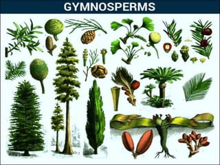

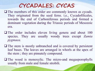
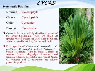

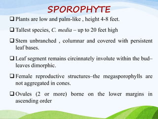
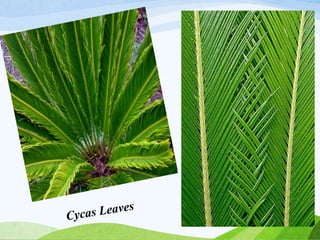
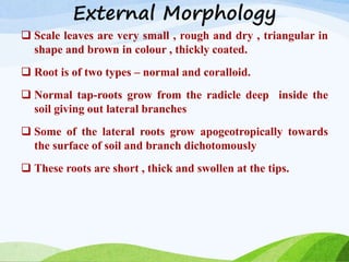
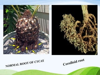


The document discusses the morphology, anatomy, and reproductive structures of gymnosperms. It focuses on Cycas, describing its external morphology such as its unbranched stem covered in persistent leaf bases and pinnately compound leaves. Internally, it notes Cycas has monoxylic wood and coralloid roots that form a symbiotic relationship with cyanobacteria. It also details the structures and development of male and female reproductive organs in Cycas, which are dioecious and wind pollinated. Cycas reproduces sexually through seeds and vegetatively through bulbils.


![Cordaitales - Yudhvir Singh Checked[1].pptx gymnosperms](https://cdn.slidesharecdn.com/ss_thumbnails/cordaitales-yudhvirsinghchecked1-250516114347-93347ab7-thumbnail.jpg?width=640&height=640&fit=bounds)


