This document summarizes key details about the fern genus Pteris. It describes the systematic position of Pteris within the plant kingdom, common Indian species, and global occurrence/distribution. The morphology, anatomy, and reproductive structures of the sporophyte (fern plant) are then explained in detail, covering the rhizome, fronds, leaflets, and roots. Key anatomical features include dictyostele stele in the rhizome, vascular bundles with endodermis and pericycle, and hypostomatous leaflets. Reproduction occurs vegetatively from the rhizome as well as sexually from spores.
The "Telome theory" of Walter Zimmermann (1930, 1952) is the most accepted theory that is based on fossil record and synthesizes the major steps in the evolution of vascular plants.
It describes how the primitive type of vascular plants developed from Rhynia like plants.
This is a detailed presentation on Morphology, anatomy and reproduction of Marchantia spp. with high quality pics and eye capturing transitions and animations
Pteridophytes are vascular plants and have leaves (known as fronds), roots and sometimes true stems, and tree ferns have full trunks. Examples include ferns, horsetails and club-mosses. Fronds in the largest species of ferns can reach some six metres in length!
Many ferns from tropical rain forests are epiphytes, which means they only grow on other plant species; their water comes from the damp air or from rainfall running down branches and tree trunks. There are also some purely aquatic ferns such as water fern or water velvet (Salvinia molesta) and mosquito ferns (Azolla species).
Pteridophytes do not have seeds or flowers either, instead they also reproduce via spores.
There are around 13,000 species of Pteridophytes.
Gnetum: A Powerpoint Presentation on Gymnospemsshivduraigaran
The Gymnosperms are a group of seed-producing plants (spermatophytes) that includes conifers (Pinophyta), cycads, Ginkgo, and gnetophytes. The term "gymnosperm" comes from the Greek composite word γυμνόσπερμος (γυμνός gymnos, "naked" and σπέρμα sperma, "seed"), meaning "naked seeds". The name is based on the unenclosed condition of their seeds (called ovules in their unfertilized state). The non-encased condition of their seeds stands in contrast to the seeds and ovules of flowering plants (angiosperms), which are enclosed within an ovary. Gymnosperm seeds develop either on the surface of scales or leaves, which are often modified to form cones, or solitary as in Yew, Torreya, Ginkgo.
The gymnosperms and angiosperms together compose the spermatophytes or seed plants. The gymnosperms are divided into six phyla. Organisms that belong to the Cycadophyta, Ginkgophyta, Gnetophyta, and Pinophyta (also known as Coniferophyta) phyla are still in existence while those in the Pteridospermales and Cordaitales phyla are now extinct.
By far the largest group of living gymnosperms are the conifers (pines, cypresses, and relatives), followed by cycads, gnetophytes (Gnetum, Ephedra and Welwitschia), and Ginkgo biloba (a single living species). Roots in some genera have fungal association with roots in the form of micorrhiza(Pinus), while in some others(Cycas) small specialised roots called coralloid roots are associated with nitrogen fixing cyanobacteria.
Gnetum is a genus of gymnosperms, the sole genus in the family Gnetaceae and order Gnetales. They are tropical evergreen trees, shrubs and lianas. Unlike other gymnosperms, they possess vessel elements in the xylem. Some species have been proposed to have been the first plants to be insect-pollinated as their fossils occur in association with extinct pollinating scorpion flies. Molecular phylogenies based on nuclear and plastid sequences from most of the species indicate hybridization among some of the Southeast Asian species. Fossil-calibrated molecular-clocks suggest that the Gnetum lineages now found in Africa, South America and Southeast Asia are the result of ancient long-distance dispersal across seawater
This presentation explores a brief idea about the structural and functional attributes of nucleotides, the structure and function of genetic materials along with the impact of UV rays and pH upon them.
Slide 1: Title Slide
Extrachromosomal Inheritance
Slide 2: Introduction to Extrachromosomal Inheritance
Definition: Extrachromosomal inheritance refers to the transmission of genetic material that is not found within the nucleus.
Key Components: Involves genes located in mitochondria, chloroplasts, and plasmids.
Slide 3: Mitochondrial Inheritance
Mitochondria: Organelles responsible for energy production.
Mitochondrial DNA (mtDNA): Circular DNA molecule found in mitochondria.
Inheritance Pattern: Maternally inherited, meaning it is passed from mothers to all their offspring.
Diseases: Examples include Leber’s hereditary optic neuropathy (LHON) and mitochondrial myopathy.
Slide 4: Chloroplast Inheritance
Chloroplasts: Organelles responsible for photosynthesis in plants.
Chloroplast DNA (cpDNA): Circular DNA molecule found in chloroplasts.
Inheritance Pattern: Often maternally inherited in most plants, but can vary in some species.
Examples: Variegation in plants, where leaf color patterns are determined by chloroplast DNA.
Slide 5: Plasmid Inheritance
Plasmids: Small, circular DNA molecules found in bacteria and some eukaryotes.
Features: Can carry antibiotic resistance genes and can be transferred between cells through processes like conjugation.
Significance: Important in biotechnology for gene cloning and genetic engineering.
Slide 6: Mechanisms of Extrachromosomal Inheritance
Non-Mendelian Patterns: Do not follow Mendel’s laws of inheritance.
Cytoplasmic Segregation: During cell division, organelles like mitochondria and chloroplasts are randomly distributed to daughter cells.
Heteroplasmy: Presence of more than one type of organellar genome within a cell, leading to variation in expression.
Slide 7: Examples of Extrachromosomal Inheritance
Four O’clock Plant (Mirabilis jalapa): Shows variegated leaves due to different cpDNA in leaf cells.
Petite Mutants in Yeast: Result from mutations in mitochondrial DNA affecting respiration.
Slide 8: Importance of Extrachromosomal Inheritance
Evolution: Provides insight into the evolution of eukaryotic cells.
Medicine: Understanding mitochondrial inheritance helps in diagnosing and treating mitochondrial diseases.
Agriculture: Chloroplast inheritance can be used in plant breeding and genetic modification.
Slide 9: Recent Research and Advances
Gene Editing: Techniques like CRISPR-Cas9 are being used to edit mitochondrial and chloroplast DNA.
Therapies: Development of mitochondrial replacement therapy (MRT) for preventing mitochondrial diseases.
Slide 10: Conclusion
Summary: Extrachromosomal inheritance involves the transmission of genetic material outside the nucleus and plays a crucial role in genetics, medicine, and biotechnology.
Future Directions: Continued research and technological advancements hold promise for new treatments and applications.
Slide 11: Questions and Discussion
Invite Audience: Open the floor for any questions or further discussion on the topic.
Multi-source connectivity as the driver of solar wind variability in the heli...Sérgio Sacani
The ambient solar wind that flls the heliosphere originates from multiple
sources in the solar corona and is highly structured. It is often described
as high-speed, relatively homogeneous, plasma streams from coronal
holes and slow-speed, highly variable, streams whose source regions are
under debate. A key goal of ESA/NASA’s Solar Orbiter mission is to identify
solar wind sources and understand what drives the complexity seen in the
heliosphere. By combining magnetic feld modelling and spectroscopic
techniques with high-resolution observations and measurements, we show
that the solar wind variability detected in situ by Solar Orbiter in March
2022 is driven by spatio-temporal changes in the magnetic connectivity to
multiple sources in the solar atmosphere. The magnetic feld footpoints
connected to the spacecraft moved from the boundaries of a coronal hole
to one active region (12961) and then across to another region (12957). This
is refected in the in situ measurements, which show the transition from fast
to highly Alfvénic then to slow solar wind that is disrupted by the arrival of
a coronal mass ejection. Our results describe solar wind variability at 0.5 au
but are applicable to near-Earth observatories.
What is greenhouse gasses and how many gasses are there to affect the Earth.moosaasad1975
What are greenhouse gasses how they affect the earth and its environment what is the future of the environment and earth how the weather and the climate effects.
Deep Behavioral Phenotyping in Systems Neuroscience for Functional Atlasing a...Ana Luísa Pinho
Functional Magnetic Resonance Imaging (fMRI) provides means to characterize brain activations in response to behavior. However, cognitive neuroscience has been limited to group-level effects referring to the performance of specific tasks. To obtain the functional profile of elementary cognitive mechanisms, the combination of brain responses to many tasks is required. Yet, to date, both structural atlases and parcellation-based activations do not fully account for cognitive function and still present several limitations. Further, they do not adapt overall to individual characteristics. In this talk, I will give an account of deep-behavioral phenotyping strategies, namely data-driven methods in large task-fMRI datasets, to optimize functional brain-data collection and improve inference of effects-of-interest related to mental processes. Key to this approach is the employment of fast multi-functional paradigms rich on features that can be well parametrized and, consequently, facilitate the creation of psycho-physiological constructs to be modelled with imaging data. Particular emphasis will be given to music stimuli when studying high-order cognitive mechanisms, due to their ecological nature and quality to enable complex behavior compounded by discrete entities. I will also discuss how deep-behavioral phenotyping and individualized models applied to neuroimaging data can better account for the subject-specific organization of domain-general cognitive systems in the human brain. Finally, the accumulation of functional brain signatures brings the possibility to clarify relationships among tasks and create a univocal link between brain systems and mental functions through: (1) the development of ontologies proposing an organization of cognitive processes; and (2) brain-network taxonomies describing functional specialization. To this end, tools to improve commensurability in cognitive science are necessary, such as public repositories, ontology-based platforms and automated meta-analysis tools. I will thus discuss some brain-atlasing resources currently under development, and their applicability in cognitive as well as clinical neuroscience.
Comparing Evolved Extractive Text Summary Scores of Bidirectional Encoder Rep...University of Maribor
Slides from:
11th International Conference on Electrical, Electronics and Computer Engineering (IcETRAN), Niš, 3-6 June 2024
Track: Artificial Intelligence
https://www.etran.rs/2024/en/home-english/
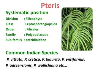
1. Systematic position
Division : Filicophyta
Class : Leptosporangiopsida
Order : Filicales
Family : Polypodiaceae
Sub-family : pteridioideae
Common Indian Species
P. vittata, P. cretica, P. biaurita, P. ensiformis,
P. adscensionis, P. wallichiana etc...
Pteris
2. Occurrence of distribution
lCosmopolitan fern being distributed in almost
all area.
lIt prefers tropical and subtropical climates.
lPlants usually grow in well drained places or in
crevices of rocks.
lThey are very common along the slopes of hills
and can be seen at 1200 metre above sea level.
lThere are aboubt 250-280 species reported for
the genus.
3.
4. SPOROPHYTE
Morphology of Sporophyte
lSporophyte is differentiated into root, stem and
leaves.
lPrimary root is soon replaced by the
adventitious roots. Roots are slender, black, wiry
and arise from ventral side of rhizome or all over
the surface.
lStem is rhizomatous, underground, branched,
perennial and covered by brown scales. Some
species have persistant leaf bases on rhizome.
5. lLeaves- Macrophyllous, unipinnate or imparipinnate
(P. vittata) or Bipinnate (P. biaurita). Arise
acropetally on the rhizome. Develped leaves are
called fronds.
lPetiole base is covered with brown scales and
sometimes with ramenta
lRachis has several sessile, lanceolate leaflets
arranged in pairs except the terminal one.
lLeaflet is rough ,has a midrib from which the lateral
veins with dichotomous branching arise. Venation is
open dichotomous venation.
lYounger leaves shows circinate vernation.
6. Anatomy of sporophyte
1) Anatomy of Rhizome
lRhizome is differentiated in to epidermis,cortex
and stele
lEpidermis-single layered with quadrangular
cells,covered by cuticle.
lCortex-multi layered,differentiated in to
Sclerenchymatous hypodermis and inner broad
parenchymatous region. parenchymatous region
has root and leaf traces.
7. lStele-In P. vittata it is dictyostele with a ring of
vascular strand (meristele).
lMeristele is embedded in the parenchymatous
ground tissue.
lEach meristele is elliptical with single layered
endodermis having casparian strips in its radial
walls.
l1-2 layered thin walled pericycle is present below
the epidermis and surrounding the phloem.
lPhloem has only sieve cells and phloem
parenchyma. It completely surrounds the xylem.
8. lXylem is present at the centre of meristele. It
shows central protoxylem surrounded on
either side by metaxylem.
lIt consists of tracheids and xylem parenchyma.
9. 2) Anatomy of Petiole or Rachis
lIt is differentiated in to epidermis,ground
tissue and vascular bundle.
lEpidermis-single layered with narrow
quadrangular cells coverd by thick cuticle.
lSome epidermal cells give rise uniseriate
bicellular hairs called as ramenta.
lGround tissue-It has multilayered
sclerenchymatous hypodermis followed by
parenchymatous tissue in which the vascular
bundle is embedded.
10. lVascular bundle-It is 'V' or 'U' shaped.
lIt resembles meristele and has single layered
endodermis with casparian strips.
lPericycle is 1 or 2 layered and
parenchymatous.
lXylem is at the centre with mesarch condition
surrounded by phloem.
11. 3) Anatomy of leaflet
lIt has epidermis, mesophyll and vascular bundle.
lEpidermis - single layered present on both upper
and lower surfaces. It has stomata only on the
lower epidermis(Hypostomatous condition).
lMesophyll - either homogenous or differentiated in
to upper pallisade and lower spongy with broader
intercellular spaces.
lHypodermal region of mid rib has
sclerenchymatous strips in both abaxial and adaxial
sides.
12. lMid rib has single concentric, amphicribal
vascular bundles surrounded by single
layered pericycle and endodermis.
lVascular strands are embedded in the
mesophyll.
13. 4) Anatomy of Root
lIt is differentiated in to epidermis ,cortex and
stele.
lEpidermi-single layered with thin walled cells.A
few cells form root hairs.
lCortex -multilayered and differentiated in to
parenchymatous outer cortex
,sclerenchymatous inner cortex and single
layered endodermis with casparian thickenings.
lStele -it has single layered thin walled
pericycle,central plate like exarch and diarch
xylem surrounded on either side by phloem.