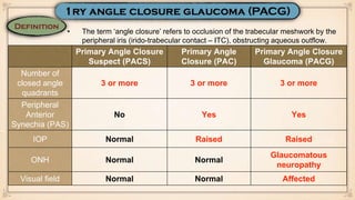
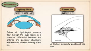
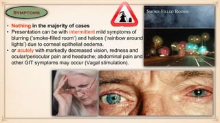
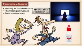
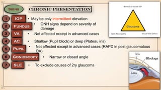
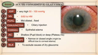
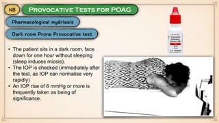
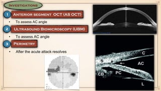
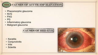
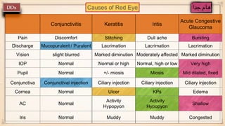
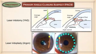
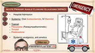
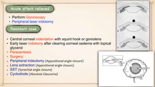
Primary Angle Closure
Suspect (PACS)
Primary Angle
Closure (PAC)
Primary Angle Closure
Glaucoma (PACG)
Number of
closed angle
quadrants
3 or more 3 or more 3 or more
Peripheral
Anterior
Synechia (PAS)
No Yes Yes
IOP Normal Raised Raised
ONH Normal Normal
Glaucomatous
neuropathy
Visual field Normal Normal Affected
• The term ‘angle closure’ refers to occlusion of the trabecular meshwork by the
peripheral iris (irido-trabecular contact – ITC), obstructing aqueous outflow.
Failure of physiological aqueous
flow through the pupil leads to a
pressure differential between the
anterior and posterior chambers,
with resultant anterior bowing of the
iris.
A thicker, anteriorly positioned iris
root
• More common in old age
• More common in Asian individuals
• More common in Hyperopic individuals with
short axial length
• More common in females
• Nothing in the majority of cases
• Presentation can be with intermittent mild symptoms of
blurring (‘smoke-filled room’) and haloes (‘rainbow around
lights’) due to corneal epithelial oedema.
• or acutely with markedly decreased vision, redness and
ocular/periocular pain and headache; abdominal pain and
other GIT symptoms may occur (Vagal stimulation).
• Watching TV in darkened room
• Pharmacological mydriasis
• Acute emotional stress
• May be only intermittent elevation
• ONH signs depend on severity of
damage
• Not affected except in advanced cases
• Not affected except in advanced cases (RAPD in post glaucomatous
OA)
• Narrow or closed angle
• To exclude causes of 2ry glaucoma
• Shallow (Pupil block) or deep (Plateau iris)
• very high 50 - 100 mmHg
• Epithelial edema
• 6/60 to HM
• Mid dilated , fixed
• Narrow or closed angle (might be
difficult due to corneal edema)
• To exclude causes of 2ry glaucoma
• Ciliary injection
• Shallow (Pupil block) or deep (Plateau iris)
• The patient sits in a dark room, face
down for one hour without sleeping
(sleep induces miosis).
• The IOP is checked (immediately after
the test, as IOP can normalise very
rapidly)
• An IOP rise of 8 mmHg or more is
frequently taken as being of
significance.
• To assess AC angle
• After the acute attack resolves
• Phacomorphic glaucoma
• NVG
• PXG
• PG
• Inflammatory glaucoma
• Malignant glaucoma
• Keratitis
• Conjunctivitis
• Iritis
• Scleritis
Causes of Red Eye
Conjunctivitis Keratitis Iritis
Acute Congestive
Glaucoma
Pain Discomfort Stitching Dull ache Bursting
Discharge Mucopurulent / Purulent Lacrimation Lacrimation Lacrimation
Vision slight blurred Marked diminution Moderately affected Marked diminution
IOP Normal Normal or high Normal, high or low Very high
Pupil Normal +/- miosis Miosis Mid dilated, fixed
Conjunctiva Conjunctival injection Ciliary injection Ciliary injection Ciliary injection
Cornea Normal Ulcer KPs Edema
AC Normal
Activity
Hypopyon
Activity
Hypopyon
Shallow
Iris Normal Muddy Muddy Congested
Laser iridotomy (YAG)
Laser iridoplasty (Argon)
• Systemic: Oral Acetazolamide, IV Mannitol
• Topical:
Pilocarpine (Parasympathomimetic)
Timolol
Prednisolone
• Hospital Admission
• Systemic analgesics, anti emetics
• Perform Gonioscopy
• Peripheral laser iridotomy
• Central corneal indentation with squint hook or goniolens
• Early laser iridotomy after clearing corneal oedema with topical
glycerol
• Paracentesis
• Surgery:
Peripheral iridectomy (Appositional angle closure)
Lens extraction (Appositional angle closure)
SST (Synechial angle closure)
Cyclodiode (Absolute Glaucoma)

Primary Angle Closure Glaucoma

  • 1.
    Primary Angle Closure Suspect(PACS) Primary Angle Closure (PAC) Primary Angle Closure Glaucoma (PACG) Number of closed angle quadrants 3 or more 3 or more 3 or more Peripheral Anterior Synechia (PAS) No Yes Yes IOP Normal Raised Raised ONH Normal Normal Glaucomatous neuropathy Visual field Normal Normal Affected • The term ‘angle closure’ refers to occlusion of the trabecular meshwork by the peripheral iris (irido-trabecular contact – ITC), obstructing aqueous outflow.
  • 2.
    Failure of physiologicalaqueous flow through the pupil leads to a pressure differential between the anterior and posterior chambers, with resultant anterior bowing of the iris. A thicker, anteriorly positioned iris root
  • 3.
    • More commonin old age • More common in Asian individuals • More common in Hyperopic individuals with short axial length • More common in females
  • 4.
    • Nothing inthe majority of cases • Presentation can be with intermittent mild symptoms of blurring (‘smoke-filled room’) and haloes (‘rainbow around lights’) due to corneal epithelial oedema. • or acutely with markedly decreased vision, redness and ocular/periocular pain and headache; abdominal pain and other GIT symptoms may occur (Vagal stimulation).
  • 5.
    • Watching TVin darkened room • Pharmacological mydriasis • Acute emotional stress
  • 6.
    • May beonly intermittent elevation • ONH signs depend on severity of damage • Not affected except in advanced cases • Not affected except in advanced cases (RAPD in post glaucomatous OA) • Narrow or closed angle • To exclude causes of 2ry glaucoma • Shallow (Pupil block) or deep (Plateau iris)
  • 7.
    • very high50 - 100 mmHg • Epithelial edema • 6/60 to HM • Mid dilated , fixed • Narrow or closed angle (might be difficult due to corneal edema) • To exclude causes of 2ry glaucoma • Ciliary injection • Shallow (Pupil block) or deep (Plateau iris)
  • 8.
    • The patientsits in a dark room, face down for one hour without sleeping (sleep induces miosis). • The IOP is checked (immediately after the test, as IOP can normalise very rapidly) • An IOP rise of 8 mmHg or more is frequently taken as being of significance.
  • 9.
    • To assessAC angle • To assess AC angle • After the acute attack resolves
  • 10.
    • Phacomorphic glaucoma •NVG • PXG • PG • Inflammatory glaucoma • Malignant glaucoma • Keratitis • Conjunctivitis • Iritis • Scleritis
  • 11.
    Causes of RedEye Conjunctivitis Keratitis Iritis Acute Congestive Glaucoma Pain Discomfort Stitching Dull ache Bursting Discharge Mucopurulent / Purulent Lacrimation Lacrimation Lacrimation Vision slight blurred Marked diminution Moderately affected Marked diminution IOP Normal Normal or high Normal, high or low Very high Pupil Normal +/- miosis Miosis Mid dilated, fixed Conjunctiva Conjunctival injection Ciliary injection Ciliary injection Ciliary injection Cornea Normal Ulcer KPs Edema AC Normal Activity Hypopyon Activity Hypopyon Shallow Iris Normal Muddy Muddy Congested
  • 12.
    Laser iridotomy (YAG) Laseriridoplasty (Argon)
  • 13.
    • Systemic: OralAcetazolamide, IV Mannitol • Topical: Pilocarpine (Parasympathomimetic) Timolol Prednisolone • Hospital Admission • Systemic analgesics, anti emetics
  • 14.
    • Perform Gonioscopy •Peripheral laser iridotomy • Central corneal indentation with squint hook or goniolens • Early laser iridotomy after clearing corneal oedema with topical glycerol • Paracentesis • Surgery: Peripheral iridectomy (Appositional angle closure) Lens extraction (Appositional angle closure) SST (Synechial angle closure) Cyclodiode (Absolute Glaucoma)