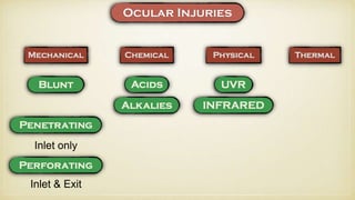
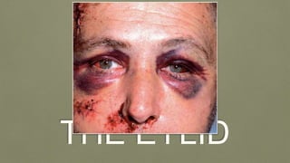
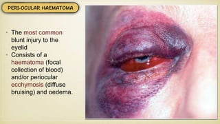
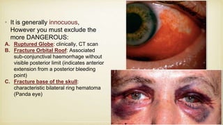
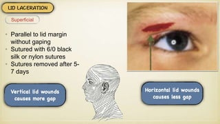
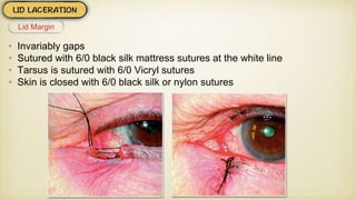
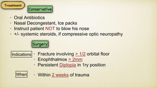
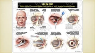
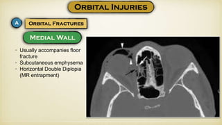
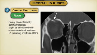
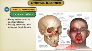
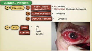
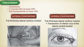
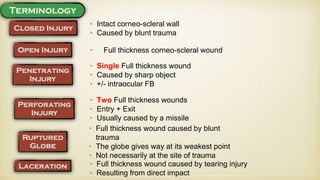
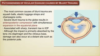
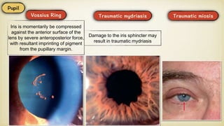
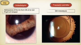
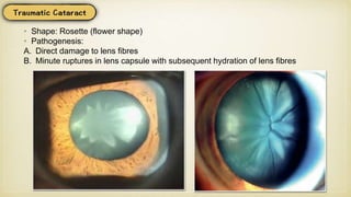
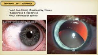
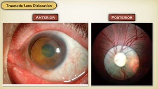
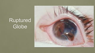
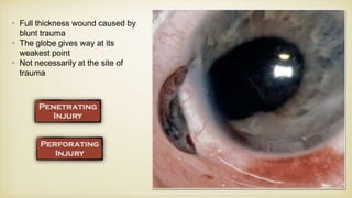
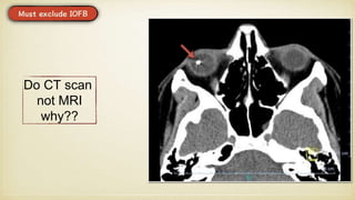
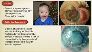
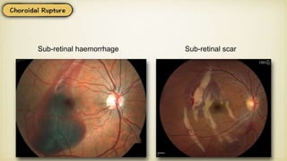
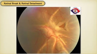
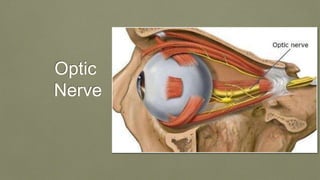
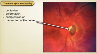
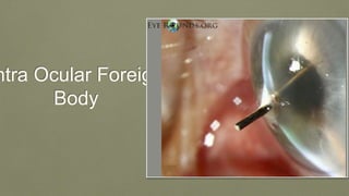
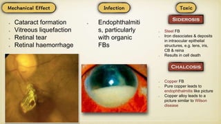
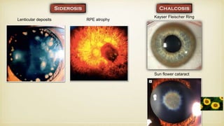
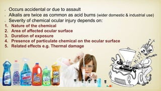
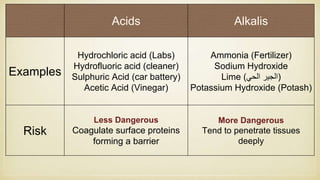
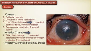
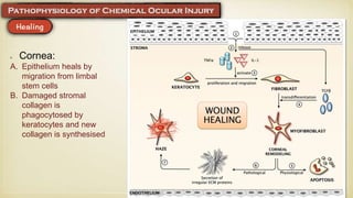
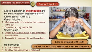
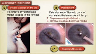
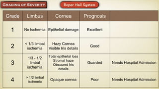
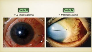
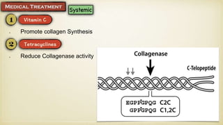
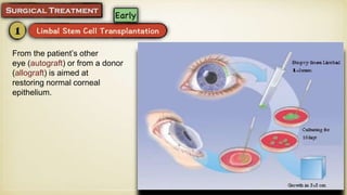
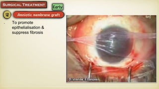
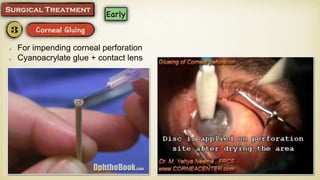
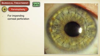
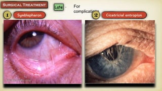
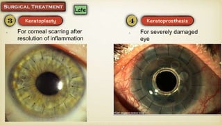
Ocular
Mohamed Abdelzaher MD, FRCS
might be
a part of
Poly
trauma
Inlet only
Inlet & Exit
THE EYLID
• The most common
blunt injury to the
eyelid
• Consists of a
haematoma (focal
collection of blood)
and/or periocular
ecchymosis (diffuse
bruising) and oedema.
• It is generally innocuous,
However you must exclude the
more DANGEROUS:
A. Ruptured Globe: clinically, CT scan
B. Fracture Orbital Roof: Associated
sub-conjunctival haemorrhage without
visible posterior limit (indicates anterior
extension from a posterior bleeding
point)
C. Fracture base of the skull:
characteristic bilateral ring hematoma
(Panda eye)
• Cold fomentation in 1st 24 hours then
warm fomentation
• Anti oedema drugs, e.g. alpha
chymotrypsin
• Parallel to lid margin
without gaping
• Sutured with 6/0 black
silk or nylon sutures
• Sutures removed after 5-
7 days
Superficial
• Invariably gaps
• Sutured with 6/0 black silk mattress sutures at the white line
• Tarsus is sutured with 6/0 Vicryl sutures
• Skin is closed with 6/0 black silk or nylon sutures
Lid Margin
• Should be repaired within the 1st 24 hours
• The laceration is bridged by silicone tubing, which is threaded down the lacrimal
system and tied in the nose, then the laceration is sutured
• Tube is left in-situ for 3-6 months
Canalicular Laceration
Orbit
• Caused by sudden increase in the
orbital pressure from an impacting
object that is greater in diameter
than the orbital aperture (about 5
cm), such as a fist or tennis ball,
• Lid oedema, Laceration
• Conjunctiva Chemosis
• +/-
Enophthalmos
• Double Diplopia;
Restrictive limitation in ocular motility in UP & DOWN gaze (IR entrapment)
UPDown
Orbital margin
Fingertip exploration 360°; palpation for orbital
margin fracture.
• Infra-
orbital
Anaesthe
sia
• CT Orbit: Tear drop sign
• Oral Antibiotics
• Nasal Decongestant, Ice packs
• Instruct patient NOT to blow his nose
• +/- systemic steroids, if compressive optic neuropathy
Indications • Fracture involving > 1/2 orbital floor
• Enophthalmos > 2mm
• Persistent Diplopia in 1ry position
When • Within 2 weeks of trauma
• Usually accompanies floor
fracture
• Subcutaneous emphysema
• Horizontal Double Diplopia
(MR entrapment)
• Rarely encountered by
ophthalmologists
• Might be associated with
other craniofacial fractures
• +/- pulsating proptosis (CSF)
• Rarely encountered by
ophthalmologists
• Usually associated with
extensive facial damage
• Iatrogenic; retrobulbar anaesthesia
• Trauma
• Spontaneous e.g. bleeding disorder
• Lid oedema
• Conjunctiva Chemosis, hematoma
• Proptosis
• Limitation
• Hig
h
• ONH
swelling
• Compression
• IV mannitol 20% (to lower IOP)
• Oral Acetazolamide (to lower IOP)
Full thickness lateral canthus incision Full thickness lateral canthus incision
+ Transection of inferior crus of the
lateral canthal tendon
The Eyeball
• Intact corneo-scleral wall
• Caused by blunt trauma
• Full thickness corneo-scleral wound
• Single Full thickness wound
• Caused by sharp object
• +/- intraocular FB
• Two Full thickness wounds
• Entry + Exit
• Usually caused by a missile
• Full thickness wound caused by blunt
trauma
• The globe gives way at its weakest point
• Not necessarily at the site of trauma
• Full thickness wound caused by tearing injury
• Resulting from direct impact
• The most common causes of blunt trauma are
squash balls, elastic luggage straps and
champagne corks.
• Severe blunt trauma to the globe results in
anteroposterior compression with simultaneous
expansion in the equatorial plane
• Associated with sharp, transient rise in IOP
• Although the impact is primarily absorbed by the
lens–iris diaphragm and the vitreous base,
damage can also occur at a distant site such as
the posterior pole.
Conjunctiva
• Commonly in Sulcus
Subtarsalis
• Removed under
topical anaesthesia
(Search for other
FBs)
• Causes:
A. Spontaneous; uncontrolled hypertension, bleeding disorder, Valsalva
B. Trauma
• Resolve spontaneously
Cornea
• Breach of corneal epithelium
• Stained with fluorescein
• Treatment:
1. Topical antibiotics
2. Topical lubricants
3. +/- Topical Cycloplegics
• Focal or Diffuse endothelial
dysfunction 2ry to blunt trauma
• Associated with folds in
Descemet membrane
• Resolve spontaneously
• Usually vertical
• Most commonly as a result of
birth trauma
• Symptoms: Stitching pain, lacrimation & photophobia
• Removed under topical anaesthesia (Search for other FBs)
Anterior
Chamber
• Haemorrhage in the AC
• Source: Iris root, CB face (Why?)
• The blood settles inferiorly giving a fluid level, except in total hyphema
• Obstruction of trabecular meshwork by
RBCs
• Pupil occlusion by blood clot leading to iris
bombe
Severe and prolonged elevation of IOP may cause corneal blood staining and
damage the optic nerve
1. Coagulation abnormality should be
excluded e.g. Sickle cell anaemia
2. Any current anticoagulant therapy
should be discontinued
3. Hospital admission; in case of large
hyphaema
4. Bed rest in sitting or semi upright
position
1. IOP lowering agents:
Topical; beta blockers +/- carbonic
anhydrase inhibitors (CAIs)
Systemic; hyper osmotic agents (
mannitol), &/or CAIs
2. Topical steroids: to reduce
inflammation & risk of 2ry
haemorrhage
3. Atropine: is recommended by some
authorities to reduce inflammation &
risk of 2ry haemorrhage (clear
evidence is lacking)
4. Systemic aminocaproic acid
(Kapron): especially in 2ry
haemorrhage
Indications for surgical evacuation of hyphaema:
Especially In case of Sickling haemoglobinopathies:
1. IOP > 25 mmHg for 5 days (to prevent corneal staining)
2. IOP > 60 mmHg for 2 days (to prevent optic atrophy)
3. Repeated IOP spikes > 30 mmHg daily for 2-4 days
4. Corneal staining
5. Any hyphaema failing to resolve less than 50% of AC for 8 days
• Rupture of CB face
• The resultant cleft is obscured by fibrosis and hyper pigmentation
• Resultant rise in IOP
Iris
Iris is momentarily be compressed
against the anterior surface of the
lens by severe anteroposterior force,
with resultant imprinting of pigment
from the pupillary margin.
Damage to the iris sphincter may
result in traumatic mydriasis
Dehiscence of the iris from CB at its root
Pupil is D shaped
360 iridodialysis
Lens
• Shape: Rosette (flower shape)
• Pathogenesis:
A. Direct damage to lens fibres
B. Minute ruptures in lens capsule with subsequent hydration of lens fibres
• Result from tearing of suspensory zonules
• Phacodonesis & Iridodonesis
• Result in monocular diplopia
Ruptured
Globe
• Full thickness wound caused by
blunt trauma
• The globe gives way at its
weakest point
• Not necessarily at the site of
trauma
Do CT scan
not MRI
why??
• Cover the injured eye with
sterile eye patch (Avoid any
ocular pressure)
• Refer to the hospital
• Closure of all corneo-scleral
wounds As Early As Possible
• Prolapsed uveal tissue might be
excised if necrotic or there is risk of
contamination by foreign material
• Prolapsed vitreous is cut with
vitrectomy probe
Vitreous
&
Retina
• Red reflex appears dark red
HOW??
• Retinal oedema, giving the retina a
greyish appearance
• If macular is involved, a cherry red
spot is seen at the fovea
Sub-retinal haemorrhage Sub-retinal scar
Optic
Nerve
contusion,
deformation,
compression or
transection of the nerve
ntra Ocular Foreign
Body
Cataract formation
Vitreous liquefaction
Retinal tear
Retinal haemorrhage
Endophthalmiti
s, particularly
with organic
FBs
Steel FB
Iron dissociates & deposits
in intraocular epithelial
structures, e.g. lens, iris,
CB & reina
Results in cell death
Copper FB
Pure copper leads to
endophthalmitis like picture
Copper alloy leads to a
picture similar to Wilson
disease
Lenticular deposits RPE atrophy
Kayser Fleischer Ring
Sun flower cataract
Chemical Ocular
Injuries
Occurs accidental or due to assault
Alkalis are twice as common as acid burns (wider domestic & industrial use)
Severity of chemical ocular injury depends on:
1. Nature of the chemical
2. Area of affected ocular surface
3. Duration of exposure
4. Presence of particulate chemical on the ocular surface
5. Related effects e.g. Thermal damage
Acids Alkalis
Examples
Hydrochloric acid (Labs)
Hydrofluoric acid (cleaner)
Sulphuric Acid (car battery)
Acetic Acid (Vinegar)
Ammonia (Fertilizer)
Sodium Hydroxide
Lime ( ‫الجير‬‫الحي‬ )
Potassium Hydroxide (Potash)
Risk
Less Dangerous
Coagulate surface proteins
forming a barrier
More Dangerous
Tend to penetrate tissues
deeply
Cornea:
A. Epithelial necrosis
B. Occlusion of limbal vasculature
C. Loss of limbal stem cells persistent
epithelial defect, conjunctivalization
D. Deep corneal penetration & stromal
opacity
Anterior Chamber:
1. Ciliary body damage decreased
ascorbate production required for collagen
production & corneal repair
• Hypotony & phthisis bulbs may ensues
Cornea:
A. Epithelium heals by
migration from limbal
stem cells
B. Damaged stromal
collagen is
phagocytosed by
keratocytes and new
collagen is synthesised
Speed & Efficacy of eye irrigation are
the most important prognostic factors
following chemical injury.
Ocular irrigation:
A. Minimise period of contact of the chemical
to the eye
B. Balance the conjunctival PH
What to use?
1. Sterile buffered solution e.g. Ringer lactate,
Normal saline
2. Tap water if necessary to avoid any delay
For how long??
A. 15 - 30 minutes
B. Till pH of the conjunctiva is balanced
To remove any particulate
matter trapped in the fornices
Debridement of Necrotic parts of
corneal epithelium under slit lamp:
A. To promote re-epithelialisation
B. Remove associated chemical residue
Grade Limbus Cornea Prognosis
1 No Ischemia Epithelial damage Excellent
2
< 1/3 limbal
ischemia
Hazy Cornea
Visible Iris details
Good
3
1/3 - 1/2
limbal
ischemia
Total epithelial loss
Stromal haze
Obscured Iris
details
Guarded Needs Hospital Admission
4
> 1/2 limbal
ischemia
Opaque cornea Poor Needs Hospital Admission
< 1/3 limbal ischemia > 1/2 limbal ischemia
Reduce Inflammation
Used initially 4-8 times daily, then tailed off after 7-10 days (Steroids reduce
collagen synthesis & inhibit fibroblast migration), and replaced with NSAIDs
Replace steroids (Do not affect Keratocyte function)
Improve comfort
Prophylaxis against bacterial infection
Promote collagen Synthesis
Reduce Collagenase activity
From the patient’s other
eye (autograft) or from a donor
(allograft) is aimed at
restoring normal corneal
epithelium.
To promote
epithelialisation &
suppress fibrosis
For impending corneal perforation
Cyanoacrylate glue + contact lens
For impending
corneal perforation
For
complications
For corneal scarring after
resolution of inflammation
For severely damaged
eye
Physical Ocular Injuries
Inflammation of the
cornea and conjunctiva
(PEE) due to exposure
to intense light of short
wavelength (as
ultraviolet light), as in:
A. Snow blindness
B. Arc Flash (Welding)
Physical damage of the
eye due to exposure to
intense light of long
wavelength (as Infrared
Rays), as in:
A. Iron melter
B. Glass blower
C. Solar eclipse
• Effects:
A. Cataract
B. Retinal burn (Solar eclipse macular burn)
Vision
Loss
Painful:
A. Acute
Congestive
Glaucoma
B. Ruptured Globe
Vision Loss
Painless:
A. Vitreous hemorrhage
B. Retinal vein occlusion
C. Retinal artery occlusion
D. Malingerer
Vision Loss
Ocula er

Ocula er

  • 1.
  • 2.
    might be a partof Poly trauma
  • 3.
  • 4.
  • 5.
    • The mostcommon blunt injury to the eyelid • Consists of a haematoma (focal collection of blood) and/or periocular ecchymosis (diffuse bruising) and oedema.
  • 6.
    • It isgenerally innocuous, However you must exclude the more DANGEROUS: A. Ruptured Globe: clinically, CT scan B. Fracture Orbital Roof: Associated sub-conjunctival haemorrhage without visible posterior limit (indicates anterior extension from a posterior bleeding point) C. Fracture base of the skull: characteristic bilateral ring hematoma (Panda eye)
  • 7.
    • Cold fomentationin 1st 24 hours then warm fomentation • Anti oedema drugs, e.g. alpha chymotrypsin
  • 8.
    • Parallel tolid margin without gaping • Sutured with 6/0 black silk or nylon sutures • Sutures removed after 5- 7 days Superficial
  • 9.
    • Invariably gaps •Sutured with 6/0 black silk mattress sutures at the white line • Tarsus is sutured with 6/0 Vicryl sutures • Skin is closed with 6/0 black silk or nylon sutures Lid Margin
  • 10.
    • Should berepaired within the 1st 24 hours • The laceration is bridged by silicone tubing, which is threaded down the lacrimal system and tied in the nose, then the laceration is sutured • Tube is left in-situ for 3-6 months Canalicular Laceration
  • 11.
  • 12.
    • Caused bysudden increase in the orbital pressure from an impacting object that is greater in diameter than the orbital aperture (about 5 cm), such as a fist or tennis ball,
  • 13.
    • Lid oedema,Laceration • Conjunctiva Chemosis
  • 14.
  • 15.
    • Double Diplopia; Restrictivelimitation in ocular motility in UP & DOWN gaze (IR entrapment) UPDown
  • 16.
    Orbital margin Fingertip exploration360°; palpation for orbital margin fracture. • Infra- orbital Anaesthe sia
  • 17.
    • CT Orbit:Tear drop sign
  • 18.
    • Oral Antibiotics •Nasal Decongestant, Ice packs • Instruct patient NOT to blow his nose • +/- systemic steroids, if compressive optic neuropathy Indications • Fracture involving > 1/2 orbital floor • Enophthalmos > 2mm • Persistent Diplopia in 1ry position When • Within 2 weeks of trauma
  • 20.
    • Usually accompaniesfloor fracture • Subcutaneous emphysema • Horizontal Double Diplopia (MR entrapment)
  • 21.
    • Rarely encounteredby ophthalmologists • Might be associated with other craniofacial fractures • +/- pulsating proptosis (CSF)
  • 22.
    • Rarely encounteredby ophthalmologists • Usually associated with extensive facial damage
  • 23.
    • Iatrogenic; retrobulbaranaesthesia • Trauma • Spontaneous e.g. bleeding disorder
  • 24.
    • Lid oedema •Conjunctiva Chemosis, hematoma • Proptosis • Limitation • Hig h • ONH swelling
  • 25.
    • Compression • IVmannitol 20% (to lower IOP) • Oral Acetazolamide (to lower IOP) Full thickness lateral canthus incision Full thickness lateral canthus incision + Transection of inferior crus of the lateral canthal tendon
  • 26.
  • 27.
    • Intact corneo-scleralwall • Caused by blunt trauma • Full thickness corneo-scleral wound • Single Full thickness wound • Caused by sharp object • +/- intraocular FB • Two Full thickness wounds • Entry + Exit • Usually caused by a missile • Full thickness wound caused by blunt trauma • The globe gives way at its weakest point • Not necessarily at the site of trauma • Full thickness wound caused by tearing injury • Resulting from direct impact
  • 28.
    • The mostcommon causes of blunt trauma are squash balls, elastic luggage straps and champagne corks. • Severe blunt trauma to the globe results in anteroposterior compression with simultaneous expansion in the equatorial plane • Associated with sharp, transient rise in IOP • Although the impact is primarily absorbed by the lens–iris diaphragm and the vitreous base, damage can also occur at a distant site such as the posterior pole.
  • 29.
  • 30.
    • Commonly inSulcus Subtarsalis • Removed under topical anaesthesia (Search for other FBs)
  • 31.
    • Causes: A. Spontaneous;uncontrolled hypertension, bleeding disorder, Valsalva B. Trauma • Resolve spontaneously
  • 32.
  • 33.
    • Breach ofcorneal epithelium • Stained with fluorescein • Treatment: 1. Topical antibiotics 2. Topical lubricants 3. +/- Topical Cycloplegics
  • 34.
    • Focal orDiffuse endothelial dysfunction 2ry to blunt trauma • Associated with folds in Descemet membrane • Resolve spontaneously
  • 35.
    • Usually vertical •Most commonly as a result of birth trauma
  • 36.
    • Symptoms: Stitchingpain, lacrimation & photophobia • Removed under topical anaesthesia (Search for other FBs)
  • 37.
  • 38.
    • Haemorrhage inthe AC • Source: Iris root, CB face (Why?) • The blood settles inferiorly giving a fluid level, except in total hyphema
  • 39.
    • Obstruction oftrabecular meshwork by RBCs • Pupil occlusion by blood clot leading to iris bombe Severe and prolonged elevation of IOP may cause corneal blood staining and damage the optic nerve
  • 40.
    1. Coagulation abnormalityshould be excluded e.g. Sickle cell anaemia 2. Any current anticoagulant therapy should be discontinued 3. Hospital admission; in case of large hyphaema 4. Bed rest in sitting or semi upright position
  • 41.
    1. IOP loweringagents: Topical; beta blockers +/- carbonic anhydrase inhibitors (CAIs) Systemic; hyper osmotic agents ( mannitol), &/or CAIs 2. Topical steroids: to reduce inflammation & risk of 2ry haemorrhage 3. Atropine: is recommended by some authorities to reduce inflammation & risk of 2ry haemorrhage (clear evidence is lacking) 4. Systemic aminocaproic acid (Kapron): especially in 2ry haemorrhage
  • 42.
    Indications for surgicalevacuation of hyphaema: Especially In case of Sickling haemoglobinopathies: 1. IOP > 25 mmHg for 5 days (to prevent corneal staining) 2. IOP > 60 mmHg for 2 days (to prevent optic atrophy) 3. Repeated IOP spikes > 30 mmHg daily for 2-4 days 4. Corneal staining 5. Any hyphaema failing to resolve less than 50% of AC for 8 days
  • 44.
    • Rupture ofCB face • The resultant cleft is obscured by fibrosis and hyper pigmentation • Resultant rise in IOP
  • 45.
  • 46.
    Iris is momentarilybe compressed against the anterior surface of the lens by severe anteroposterior force, with resultant imprinting of pigment from the pupillary margin. Damage to the iris sphincter may result in traumatic mydriasis
  • 47.
    Dehiscence of theiris from CB at its root Pupil is D shaped 360 iridodialysis
  • 48.
  • 49.
    • Shape: Rosette(flower shape) • Pathogenesis: A. Direct damage to lens fibres B. Minute ruptures in lens capsule with subsequent hydration of lens fibres
  • 50.
    • Result fromtearing of suspensory zonules • Phacodonesis & Iridodonesis • Result in monocular diplopia
  • 52.
  • 53.
    • Full thicknesswound caused by blunt trauma • The globe gives way at its weakest point • Not necessarily at the site of trauma
  • 54.
    Do CT scan notMRI why??
  • 55.
    • Cover theinjured eye with sterile eye patch (Avoid any ocular pressure) • Refer to the hospital • Closure of all corneo-scleral wounds As Early As Possible • Prolapsed uveal tissue might be excised if necrotic or there is risk of contamination by foreign material • Prolapsed vitreous is cut with vitrectomy probe
  • 56.
  • 58.
    • Red reflexappears dark red HOW??
  • 59.
    • Retinal oedema,giving the retina a greyish appearance • If macular is involved, a cherry red spot is seen at the fovea
  • 60.
  • 62.
  • 63.
  • 64.
  • 65.
    Cataract formation Vitreous liquefaction Retinaltear Retinal haemorrhage Endophthalmiti s, particularly with organic FBs Steel FB Iron dissociates & deposits in intraocular epithelial structures, e.g. lens, iris, CB & reina Results in cell death Copper FB Pure copper leads to endophthalmitis like picture Copper alloy leads to a picture similar to Wilson disease
  • 66.
    Lenticular deposits RPEatrophy Kayser Fleischer Ring Sun flower cataract
  • 67.
  • 68.
    Occurs accidental ordue to assault Alkalis are twice as common as acid burns (wider domestic & industrial use) Severity of chemical ocular injury depends on: 1. Nature of the chemical 2. Area of affected ocular surface 3. Duration of exposure 4. Presence of particulate chemical on the ocular surface 5. Related effects e.g. Thermal damage
  • 69.
    Acids Alkalis Examples Hydrochloric acid(Labs) Hydrofluoric acid (cleaner) Sulphuric Acid (car battery) Acetic Acid (Vinegar) Ammonia (Fertilizer) Sodium Hydroxide Lime ( ‫الجير‬‫الحي‬ ) Potassium Hydroxide (Potash) Risk Less Dangerous Coagulate surface proteins forming a barrier More Dangerous Tend to penetrate tissues deeply
  • 70.
    Cornea: A. Epithelial necrosis B.Occlusion of limbal vasculature C. Loss of limbal stem cells persistent epithelial defect, conjunctivalization D. Deep corneal penetration & stromal opacity Anterior Chamber: 1. Ciliary body damage decreased ascorbate production required for collagen production & corneal repair • Hypotony & phthisis bulbs may ensues
  • 71.
    Cornea: A. Epithelium healsby migration from limbal stem cells B. Damaged stromal collagen is phagocytosed by keratocytes and new collagen is synthesised
  • 72.
    Speed & Efficacyof eye irrigation are the most important prognostic factors following chemical injury. Ocular irrigation: A. Minimise period of contact of the chemical to the eye B. Balance the conjunctival PH What to use? 1. Sterile buffered solution e.g. Ringer lactate, Normal saline 2. Tap water if necessary to avoid any delay For how long?? A. 15 - 30 minutes B. Till pH of the conjunctiva is balanced
  • 73.
    To remove anyparticulate matter trapped in the fornices Debridement of Necrotic parts of corneal epithelium under slit lamp: A. To promote re-epithelialisation B. Remove associated chemical residue
  • 74.
    Grade Limbus CorneaPrognosis 1 No Ischemia Epithelial damage Excellent 2 < 1/3 limbal ischemia Hazy Cornea Visible Iris details Good 3 1/3 - 1/2 limbal ischemia Total epithelial loss Stromal haze Obscured Iris details Guarded Needs Hospital Admission 4 > 1/2 limbal ischemia Opaque cornea Poor Needs Hospital Admission
  • 75.
    < 1/3 limbalischemia > 1/2 limbal ischemia
  • 76.
    Reduce Inflammation Used initially4-8 times daily, then tailed off after 7-10 days (Steroids reduce collagen synthesis & inhibit fibroblast migration), and replaced with NSAIDs Replace steroids (Do not affect Keratocyte function) Improve comfort Prophylaxis against bacterial infection
  • 77.
  • 78.
    From the patient’sother eye (autograft) or from a donor (allograft) is aimed at restoring normal corneal epithelium.
  • 79.
  • 80.
    For impending cornealperforation Cyanoacrylate glue + contact lens
  • 81.
  • 82.
  • 83.
    For corneal scarringafter resolution of inflammation For severely damaged eye
  • 84.
  • 85.
    Inflammation of the corneaand conjunctiva (PEE) due to exposure to intense light of short wavelength (as ultraviolet light), as in: A. Snow blindness B. Arc Flash (Welding)
  • 86.
    Physical damage ofthe eye due to exposure to intense light of long wavelength (as Infrared Rays), as in: A. Iron melter B. Glass blower C. Solar eclipse • Effects: A. Cataract B. Retinal burn (Solar eclipse macular burn)
  • 87.
  • 88.
  • 89.
    Painless: A. Vitreous hemorrhage B.Retinal vein occlusion C. Retinal artery occlusion D. Malingerer Vision Loss