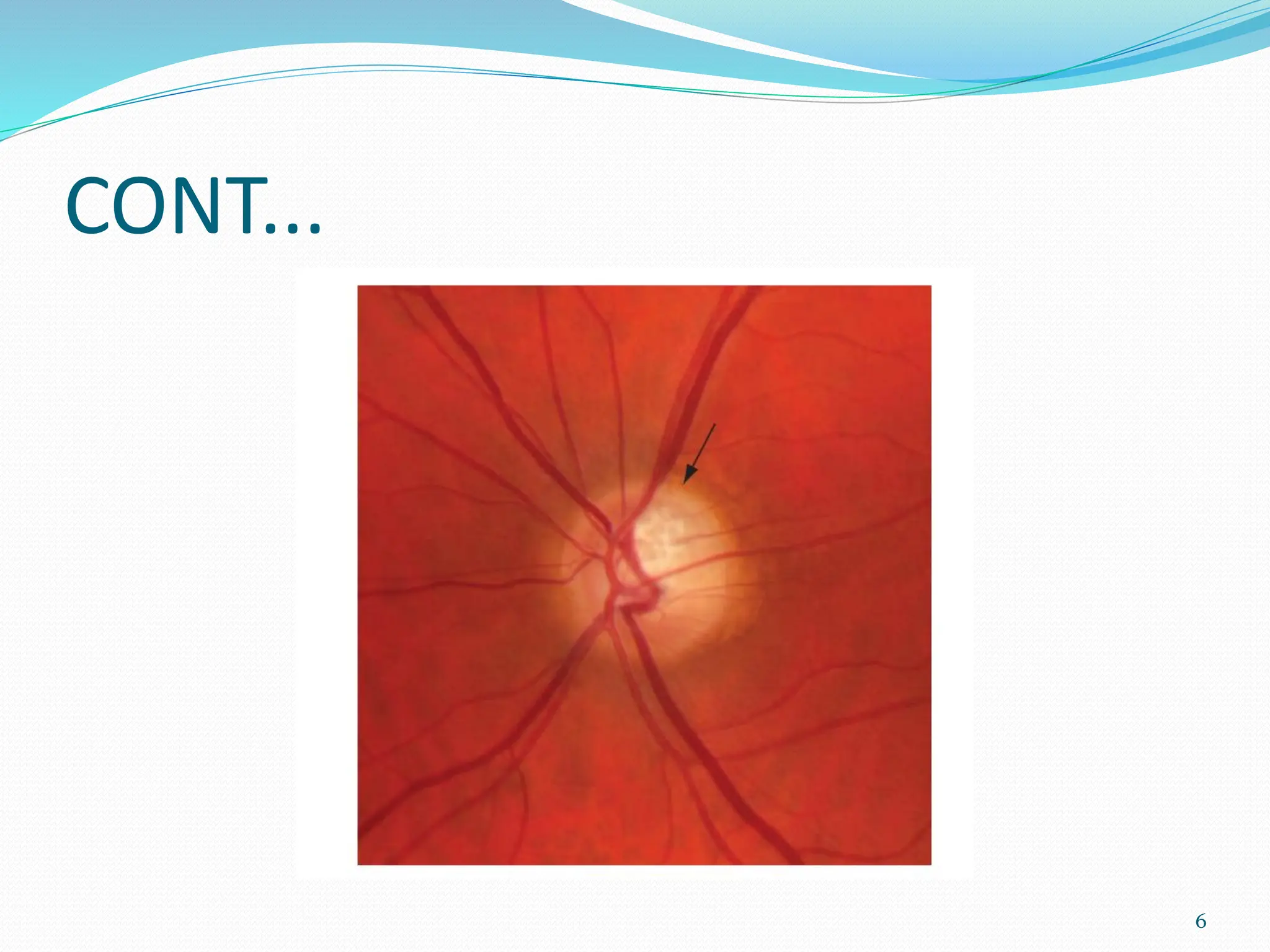
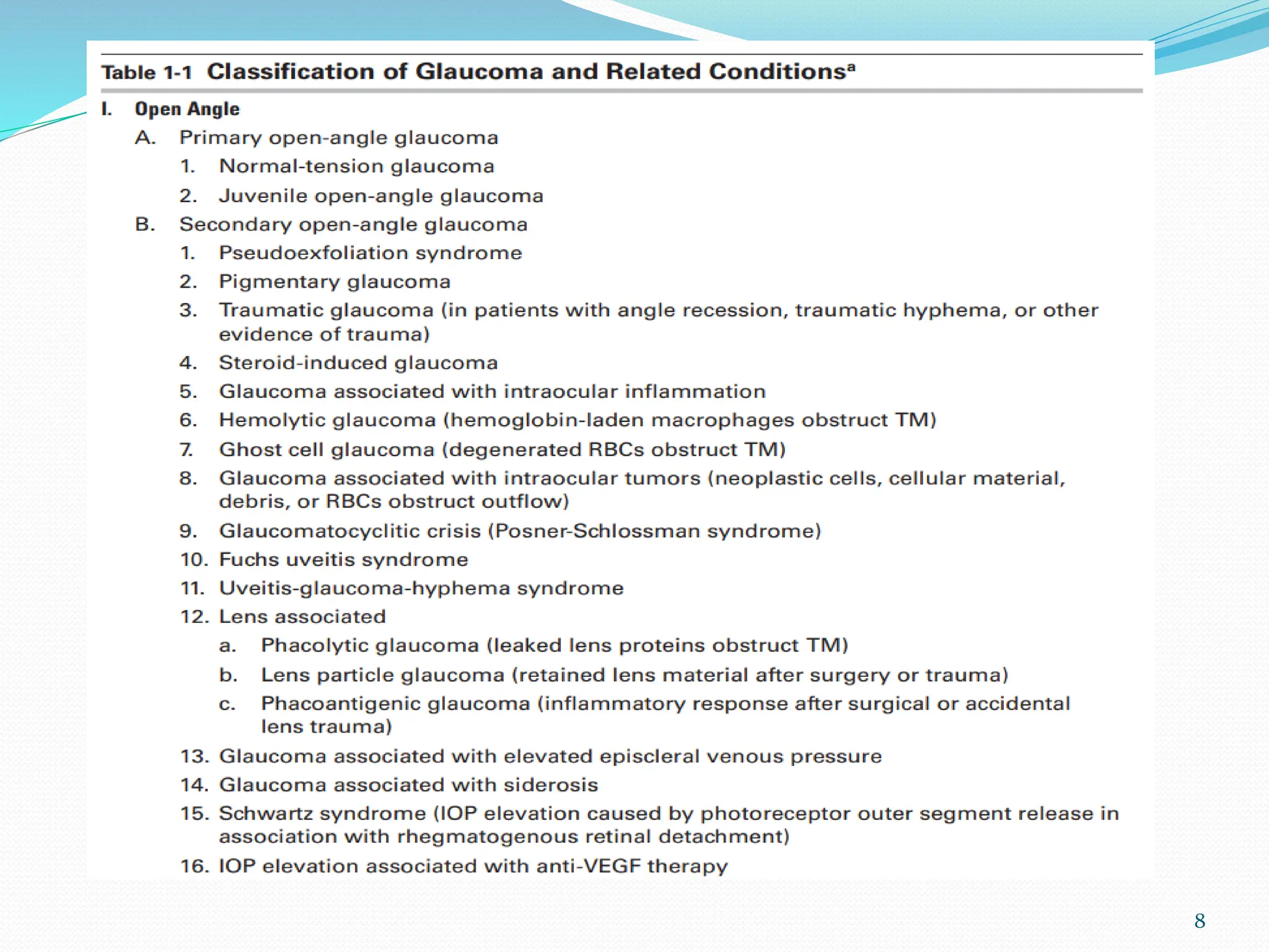
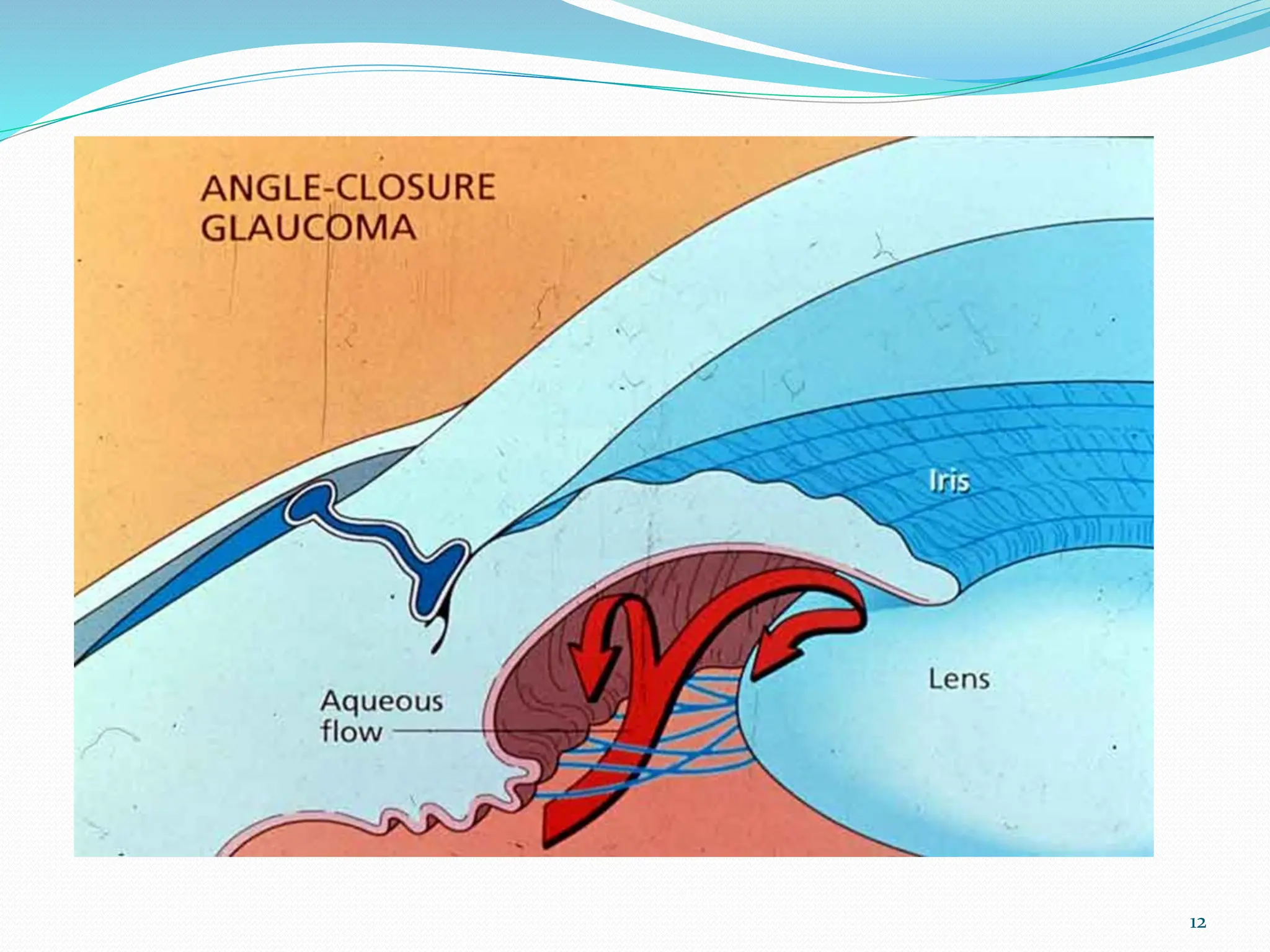
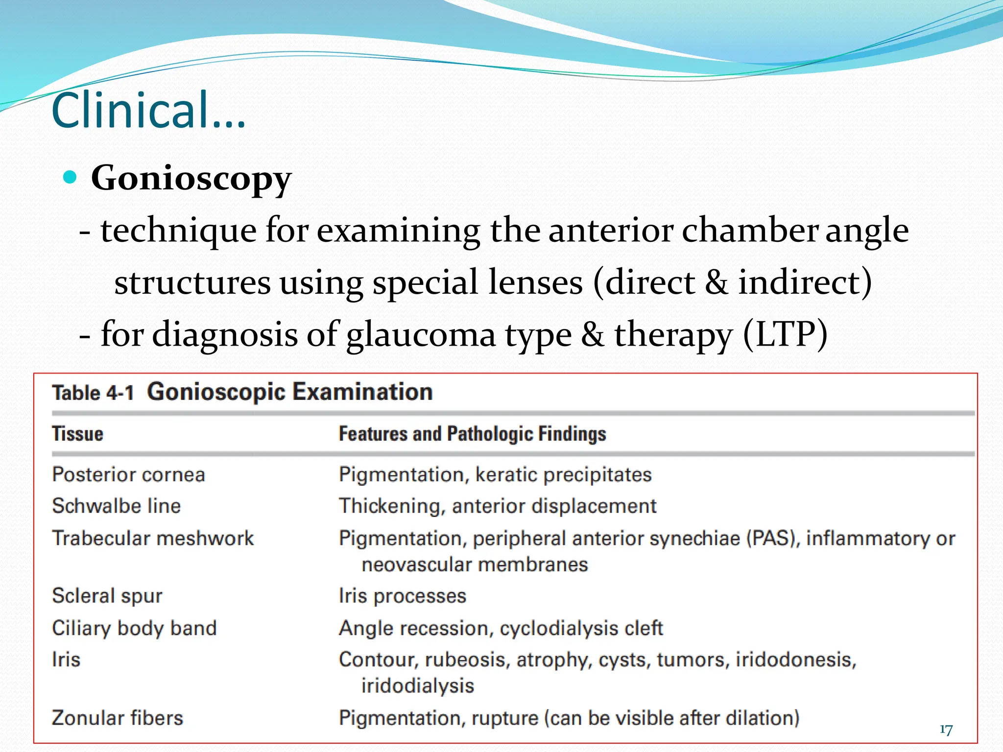
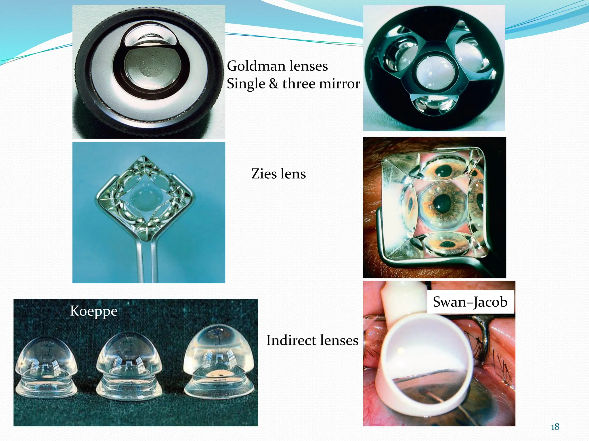
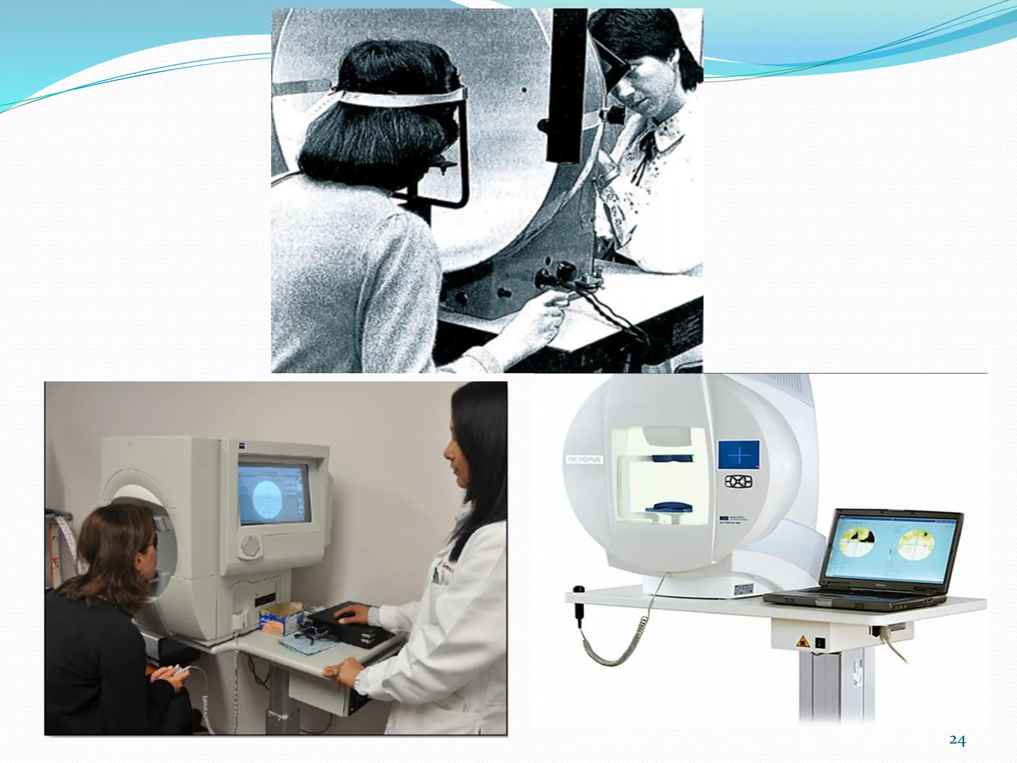
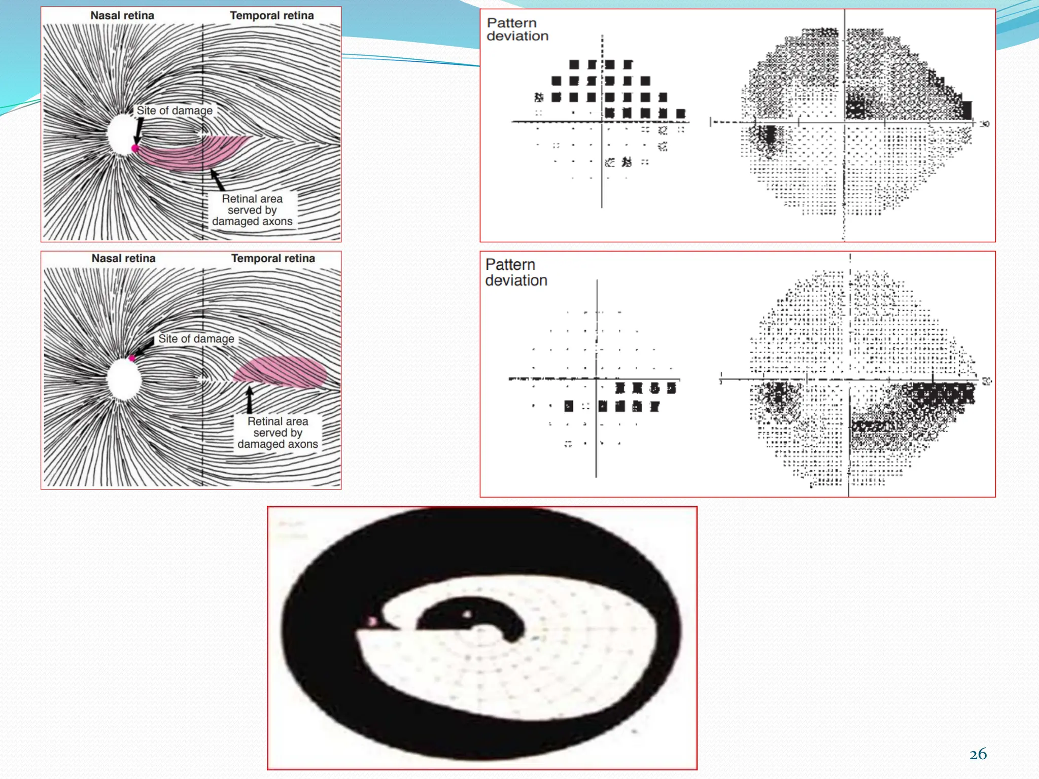
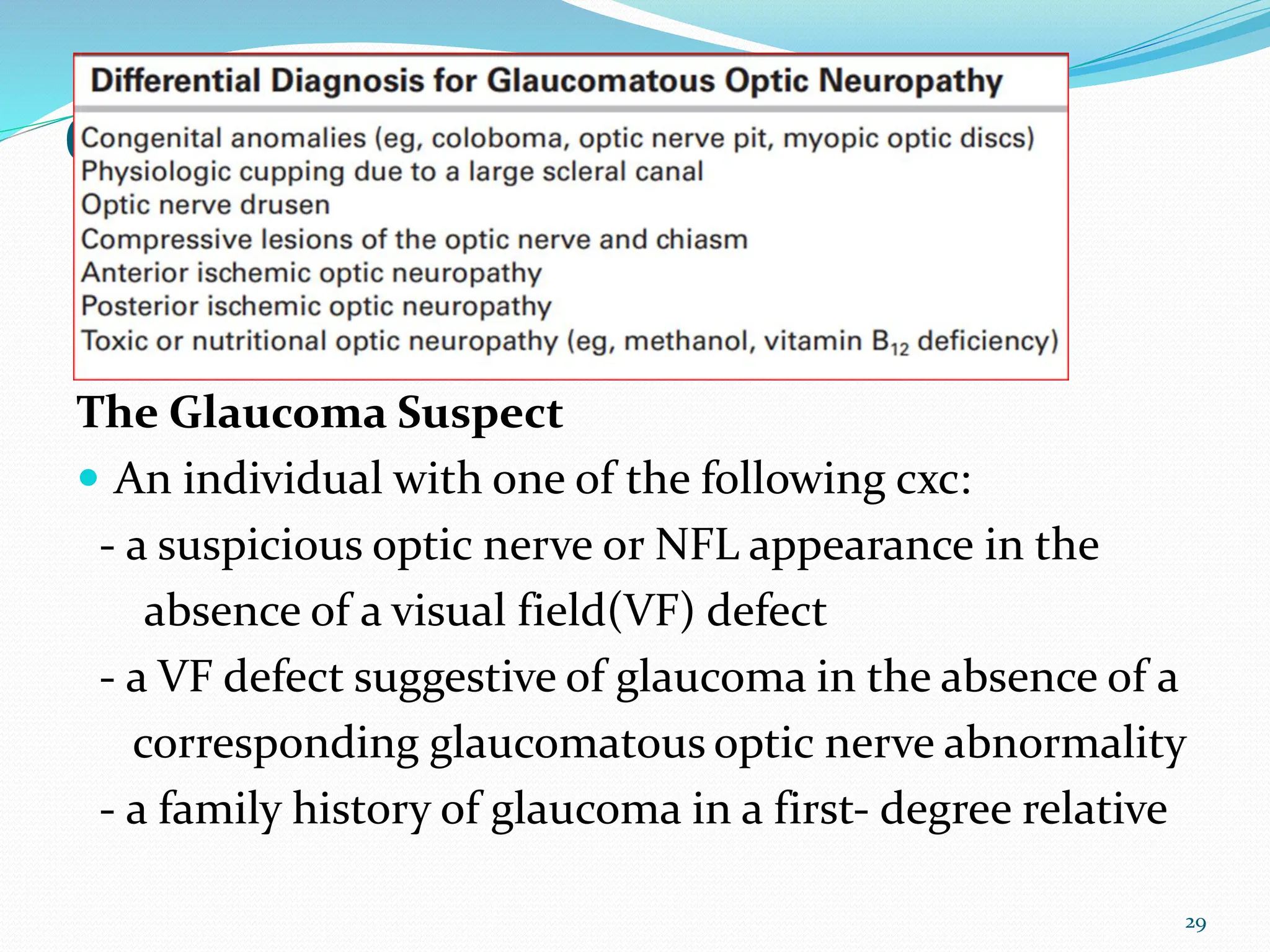
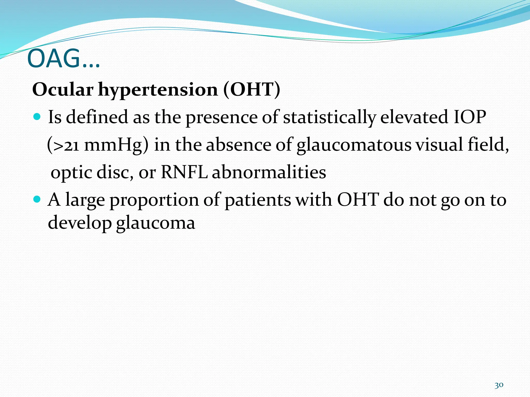
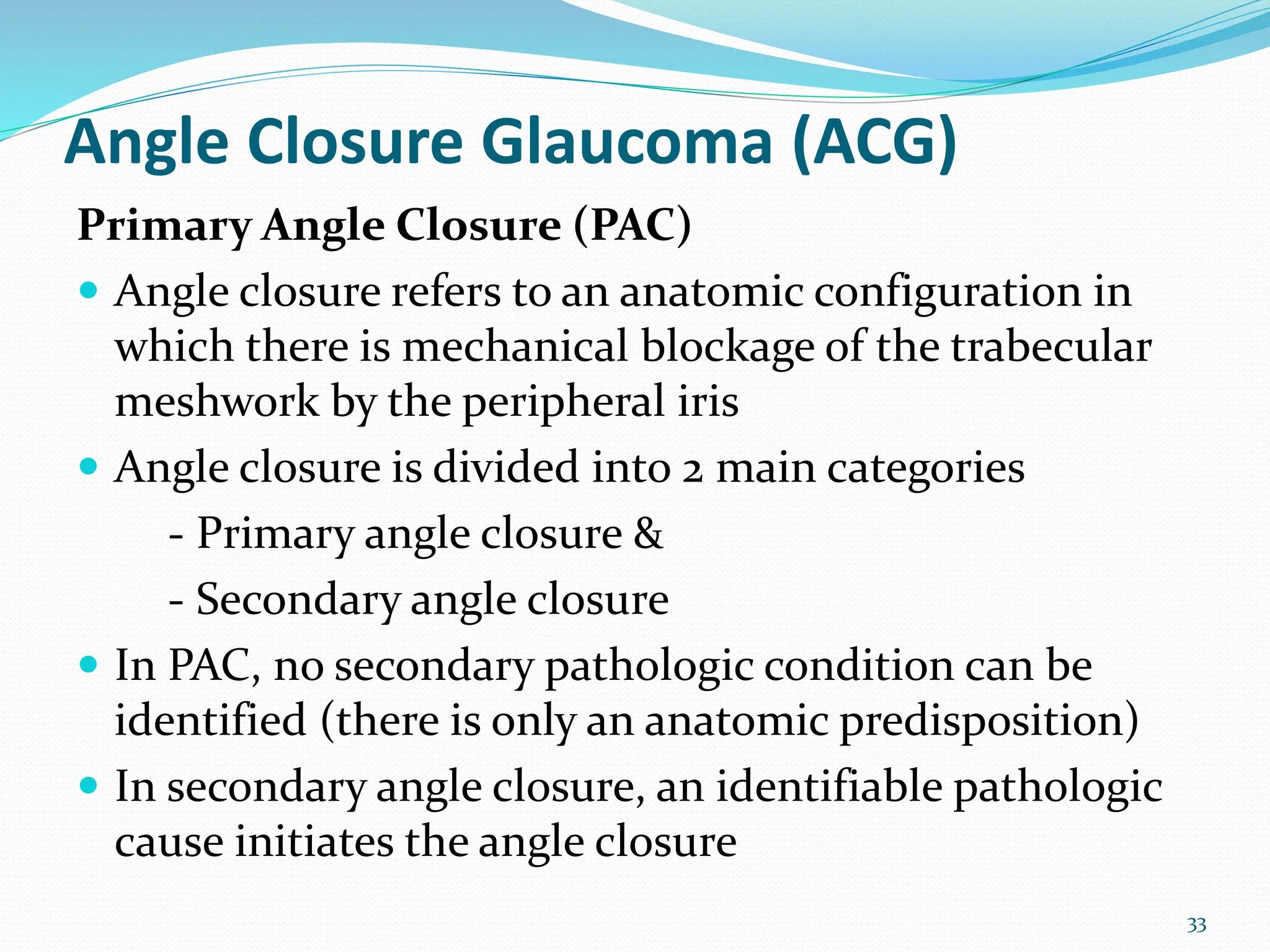
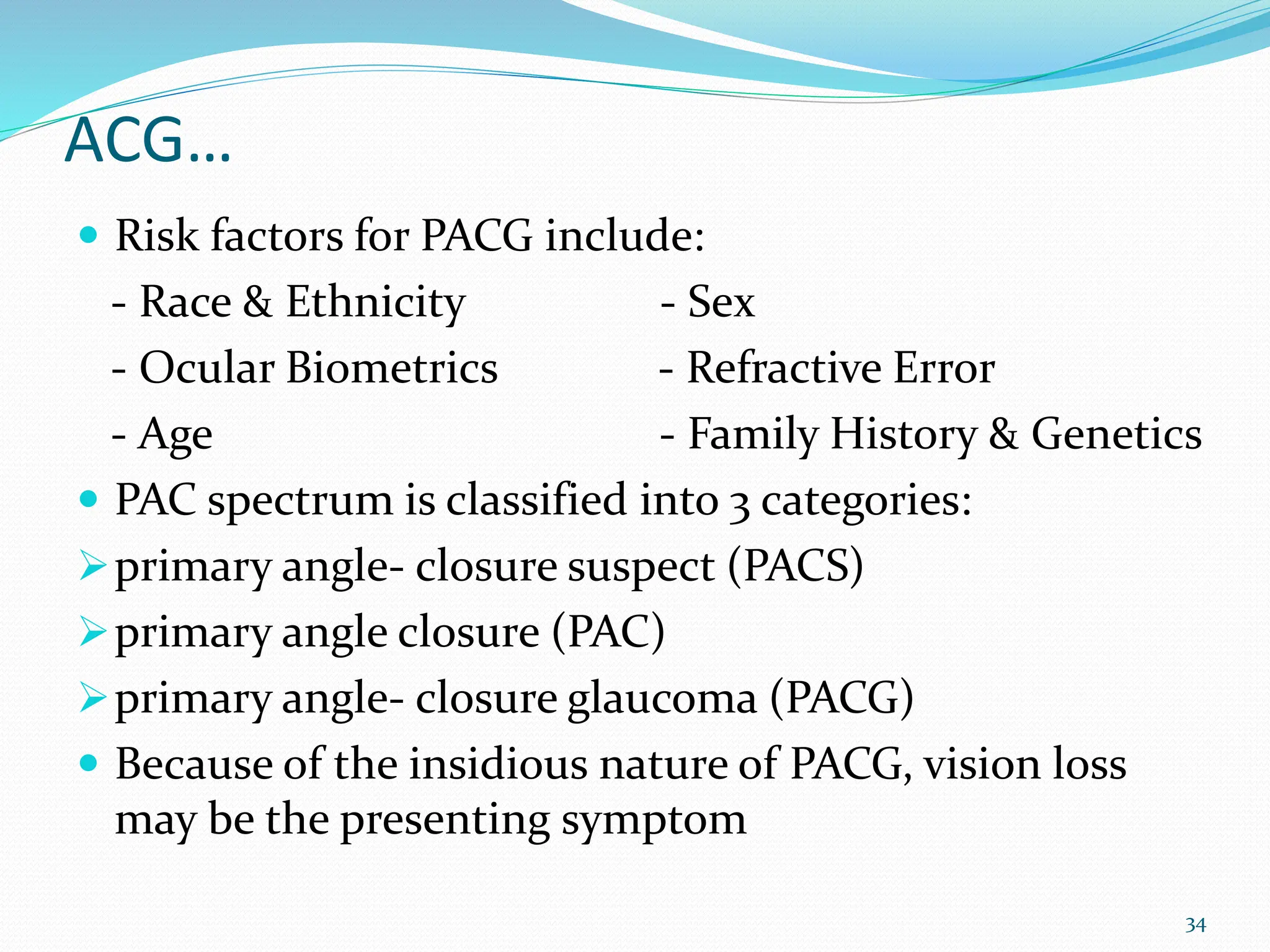
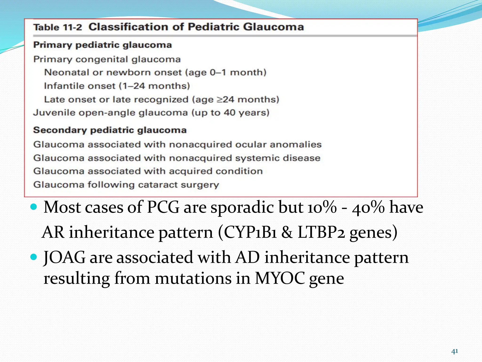
DEPARTMENT OF OPHTGALMOLOGY
Deresse Belie (MD)
OUTLINE
 INTRODUCTION
 CLASSIFICATION
OPEN ANGLE GLAUCOMA (OAG)
ANGLE CLOSURE GAUCOMA (ACG)
 PEDIATRIC GLAUCOMA
 CLINICAL EVALUATION
 CLINICAL FEATURES
(OAG, ACG, PEDIATRIC GLAUCOMA)
 MANAGEMENT
2
INTRODUCTION
Definition
 Glaucoma is a group of disorders characterized by a
progressive optic neuropathy resulting in a
characteristic appearance of the optic disc and a
specific pattern of irreversible visual field defects
 The causes of glaucoma are multifactorial and include
genetic and environmental factors
 Intraocular pressure (IOP) is the most common risk
factor but not the only risk factor for development of
glaucoma
3
Int…
 But, IOP is presently the only factor that can be
controlled to prevent progressive optic neuropathy
 Normal range of IOP in the general population is 10-21
mmHg (Average 15.5 mmHg with SD of 2.6 mmHg)
 In patients with glaucoma, the IOP at baseline -
regardless of its actual level - is too high for retinal
ganglion cell function and survival
 In most patients with glaucoma, lowering the IOP will
stop or slow visual field loss
 During patient evaluation, visual field tests and IOP
readings are assessed in addition to ON evaluation
4
Int…
 Early diagnosis and treatment is crucial in the
prevention of visual loss from glaucoma
 If untreated, it can lead to total irreversible blindness
 But in nearly all types of glaucoma, blindness is
preventable
 Glaucoma is the leading cause of irreversible blindness
worldwide
 The global prevalence of glaucoma is about 3.5% in the
population aged 40–80 years
5
CONT...
6
CLASSIFICATION
A. Based on Angle
 Open angle glaucoma
 Angle closure glaucoma
B. Based on Cause
 Primary
 Secondary
C. Pediatric glaucoma
7
8
9
Normal Aqueous flow
10
11
12
Class…
Pediatric glaucoma
Primary congenital glaucoma (PCG)
Juvenile open- angle glaucoma (JOAG)
Glaucoma associated with non acquired ocular
anomalies
Glaucoma associated with acquired conditions
Glaucoma following cataract surgery
13
CLINICAL EVALUATION
History
 The history includes :
• Demography (Age, Race & ethnicity)
• Symptoms, onset, duration, and severity
• Past ocular history (medical & surgical)
• Family history of glaucoma
• General medical history (medications & allergies)
• Social and occupational history
14
Clinical…
Examination
 Visual acuity
 IOP measurement (Tonometry)
 Slit- Lamp Biomicroscopy
 Ocular adnexal examination
- conditions associated with 2⁰ glaucoma
- for possible external effects of glaucoma therapy
 Pupillary function assessment (RAPD, drug effects)
 Anterior segment evaluation & Imaging (UBM, OCT)
 Refraction
15
Tonometry
• process of measuring intraocular pressure using a
variety of instruments
• Goldman tonometer is the gold standard
16
Clinical…
 Gonioscopy
- technique for examining the anterior chamber angle
structures using special lenses (direct & indirect)
- for diagnosis of glaucoma type & therapy (LTP)
17
18
Goldman lenses
Single & three mirror
Zies lens
Koeppe
Swan–Jacob
Indirect lenses
19
Clinical…
 Posterior Segment/Optic Nerve Head Evaluation
- Characteristic changes in the optic nerve head are the
defining feature of glaucoma
- Glaucomatous optic neuropathy is a progressive
degeneration of RGCs and their axons in association
with lamina cribrosa damage
- Examination of ONH may be performed with
-> direct ophthalmoscope
-> indirect ophthalmoscope
-> Slit-lamp biomicroscope combined with high-
magnification posterior pole lens
20
21
ONH Changes in Glaucoma
- Generalized enlargement of the cup
- Thinning, undermining & focal
notching of neuroretinal rim (NRR)
- Progressive loss of NRR tissue
- Retinal nerve fiber layer (RNFL) atrophy
- Nasal shift of retinal vessels
- Peripapillary atrophy
- Disc hemorrhag
- Asymmetry of cupping
Clinical…
- The normal ONH ranges from approximately 1.5 to
2.2 mm in diameter
- The vertical cup– disc ratio typically ranges b/n 0.1 & 0.4
- Asymmetry of the cup– disc ratio of more than 0.2 is
suspicious & significant
22
ONH, RNFL & Macula may also be evaluated
for glaucomatous changes using OCT
Clinical…
Visual field
 Perimetry is the method of visual field assessment
 Perimetry serves 2 major purposes in the management
of glaucoma:
1. identification and quantification of abnormalities
in the visual field
2. longitudinal assessment to detect glaucomatous
progression and measure rates of change
 Two major types of perimetry
Automated Static perimetry (HFA, Octopus, FDT)
Manual kinetic & static perimetry (Goldmann)
23
24
 Visual field defects in glaucoma usually run parallel to
the changes at the ONH
 These continue to progress if IOP is not controlled
& can be described as early or advanced field defects
Glaucomatous Visual Field Loss
25
Early Advanced
- Diffuse reduction /
constriction of isopters
- Paracentral scotoma
- Bjerrum / arcuate
- Nasal steps
- Temporal wedge
- Double arctuate with
peripheral extension
- Central and temporal
island of vision
26
Open- Angle Glaucoma (OAG)
Primary Open Angle Glaucoma
 Most common type of glaucoma in adults
 No clearly identifiable cause for the glaucoma
 Risk factors associated with POAG include:
- higher IOP - race
- lower ocular perfusion pressure - ethnicity
- older age - genetics
- lower CCT (thinner cornea) - ?associated
- high myopia systemic conditions
27
 POAG is typically insidious in onset, slowly
progressive, and painless
 It is usually bilateral but can be asymmetric
 AC angle is open & IOP may be elevated
 Diagnosis is based primarily on the appearance of the
ONH and on the results of visual field testing
28
Prevalence of POAG with
age and race
OAG…
The Glaucoma Suspect
 An individual with one of the following cxc:
- a suspicious optic nerve or NFL appearance in the
absence of a visual field(VF) defect
- a VF defect suggestive of glaucoma in the absence of a
corresponding glaucomatous optic nerve abnormality
- a family history of glaucoma in a first- degree relative
29
OAG…
Ocular hypertension (OHT)
 Is defined as the presence of statistically elevated IOP
(>21 mmHg) in the absence of glaucomatous visual field,
optic disc, or RNFL abnormalities
 A large proportion of patients with OHT do not go on to
develop glaucoma
30
Normal or low tension glaucoma (NTG/LTG)
 Is suggested for the typical cupping of the disc and/or
visual field defects associated with a normal or low
IOP
 Patients with “normal” IOP in clinic may experience
higher pressures outside clinic hours
 Vascular factors may have a significant role in the
development of NTG in these persons
 As in POAG, NTG is characteristically bilateral but
often asymmetric
 The visual field defects in NTG tend to be more focal,
deeper, and closer to fixation
31
OAG…
Secondary Open- Angle Glaucoma
Pseudoexfoliation Syndrome
Pigment Dispersion Syndrome
Lens- Induced Glaucoma
Intraocular Tumors
Ocular Inflammation/Uveitis
Elevated Episcleral Venous Pressure
Trauma and Surgery
Schwartz Syndrome (Schwartz- Matsuo Syndrome)
Drug induced (corticosteroids)
32
Angle Closure Glaucoma (ACG)
Primary Angle Closure (PAC)
 Angle closure refers to an anatomic configuration in
which there is mechanical blockage of the trabecular
meshwork by the peripheral iris
 Angle closure is divided into 2 main categories
- Primary angle closure &
- Secondary angle closure
 In PAC, no secondary pathologic condition can be
identified (there is only an anatomic predisposition)
 In secondary angle closure, an identifiable pathologic
cause initiates the angle closure
33
ACG…
 Risk factors for PACG include:
- Race & Ethnicity - Sex
- Ocular Biometrics - Refractive Error
- Age - Family History & Genetics
 PAC spectrum is classified into 3 categories:
primary angle- closure suspect (PACS)
primary angle closure (PAC)
primary angle- closure glaucoma (PACG)
 Because of the insidious nature of PACG, vision loss
may be the presenting symptom
34
ACG…
 Causes of angle closure
Pupillary Block
Plateau Iris and Iris- Induced Angle Closure
Drugs (either mydriatic or miotic)
 The clinical course of PACG usually resembles that of
open- angle glaucoma in its
- lack of initial symptoms,
- modest elevation of IOP,
- progressive glaucomatous optic nerve damage, and
- characteristic patterns of visual field loss
35
ACG…
Acute primary angle closure
 IOP rises rapidly as a result of relatively sudden
blockage of the trabecular meshwork by the iris
 Is an ocular emergency typically manifested by ocular
pain, headache, blurred vision, and rainbow- colored
halos around lights
 Acute systemic distress may result in nausea & vomiting
36
ACG…
 Signs of acute angle closure include:
high IOP
mid- dilated, sluggish, and irregularly shaped pupil
corneal epithelial edema
congested episcleral and conjunctival blood vessels
shallow peripheral anterior chamber
mild amount of aqueous flare and cell
 During an acute attack, the IOP may be high enough
to cause glaucomatous optic nerve damage, ischemic
optic neuropathy, and/or retinal vascular occlusion
 PAS can form rapidly, and IOP- induced ischemia may
produce sector atrophy of the iris, releasing pigment
37
38
ACG…
Secondary Angle Closure
39
 Lens- Induced Angle Closure
 Neovascular glaucoma
 Iridocorneal endothelial syndrome
 Epithelial and fibrous ingrowth
 Trauma
 Tumors
 Malignant glaucoma
 Uveal and ciliary body effusions
 Vitreoretinal surgery
 Nanophthalmos
 Drug- induced secondary angle-
closure glaucoma
Pediatric Glaucoma
 Are rare glaucomas that can result total optic nerve
atrophy and blindness
 Include a heterogeous group of disorders associated
with elevated IOP
 Typically classified as primary or secondary
 Primary pediatric glaucomas are characteriszed by
isolated angle abnormalities
 Secondary pediatric glaucomas are associated with
other ocular or systemic conditions
40
 Most cases of PCG are sporadic but 10% - 40% have
AR inheritance pattern (CYP1B1 & LTBP2 genes)
 JOAG are associated with AD inheritance pattern
resulting from mutations in MYOC gene
41
Pediatric…
Primary Congenital Glaucoma
 Accounts for the majority of primary pediatric
glaucomas
 Most cases are bilateral (70%) & frequently occur in
males (65%) than females
 PCG presents with the classic triad of :
 epiphora, photophobia, and blepharospasm
 The classic features of PCG:
 enlarged and/or cloudy corneas, Haab striae, &
an enlarged globe (buphthalmos) -> until 4 years of age
42
Pediatric…
 Other signs include high IOP, ciliary injection, ON
cupping ( CDR) & refractive errors
 Examination under anesthesia (EUA) for diagnosis
 Treatment of PCG typically requires surgical
intervention
 Medical treatment to lower IOP is a temporary
measure till the child is ready for surgery
 PCG generally has a better visual prognosis than do
most secondary pediatric glaucoma
43
44
Pediatric…
Secondary Pediatric Glaucomas
Axenfeld- Rieger Syndrome
Peters Anomaly
Aniridia
Sturge- Weber Syndrome
Neurofibromatosis
Glaucoma Following Cataract Surgery
45
MANAGEMENT
Medical Management
 The goal of currently available glaucoma therapy is to
preserve visual function by lowering IOP
 The treatment regimen chosen should achieve this
goal with the lowest risk, the fewest adverse effects,
and the least disruption to the patient’s life, taking
into account the cost of treatment
 Target pressure, an IOP below which the clinician
estimates the rate of disease progression to be
sufficiently slow, is set
46
Manage…
 The range of target pressure should be individualized,
based on the IOP at which damage is thought to have
occurred, severity of the damage, life expectancy, and
associated risk factors
 An initial reduction in the IOP of 20% - 30% from
baseline is suggested
 Lifelong follow up is usually required!
47
Manage…
 Ocular hypotensive agents are divided into several
classes based on chemical structure and
pharmacologic action
1. Prostaglandin analogues
2. Adrenergic antagonists (nonselective & β1- selective)
3. Adrenergic agonists (nonselective & α2- selective)
4. Carbonic anhydrase inhibitors (topical & systemic)
5. Parasympathomimetic (miotic) agents with direct
cholinergic & anticholinesterase actions
6. Hyperosmotic agents
7. Combination medication
48
Manage…
Prostaglandin analogues (PGA)
 Prodrugs that become biologically active after being
hydrolyzed by corneal esterase
 Lower IOP by increasing AH outflow via the uveoscleral
pathway & decreasing outflow resistance
-> latanoprost, travoprost, bimatoprost, tafluprost, and
latanoprostene bunod
 Decrease IOP by 25 - 33%
 Adverse effects include:- darkening of the iris & periocular
skin, periorbitopathy, conjunctival hyperemia,
hypertrichosis, trichiasis, and distichiasis
49
Manage…
β-Adrenergic Antagonists
 Lower IOP by reducing aqueous humor secretion
 Decrease IOP by 20 - 30%
-> betaxolol, carteolol, levobunolol, metipranolol, & timolol
 Adverse effects include:
Systemic - bronchospasm, bradycardia, increased heart block,
systemic hypotension, reduced exercise tolerance,
CNS depression & reduced glucose tolerance and
masking of hypoglycemia in DM patients
Local - corneal anesthesia, punctate keratitis, allergy
50
Manage…
α- adrenergic agonists
 Decrease IOP primarily by reducing AH production
 Lower IOP by 20 - 30%
-> Brimonidine, Apraclonidine
 Adverse effects include:
Systemic - xerostomia (dry mouth) and lethargy
Local - allergy
 Brimonidine should not be used in infants and young
children -> CNS depression, apnea, bradycardia, and
hypotension
51
Manage…
Carbonic Anhydrase Inhibitors
 Decrease aqueous humor production
 Reduce IOP by 18–25 % from baseline
Oral -> acetazolamide and methazolamide
Topical -> dorzolamide and brinzolamide
 Adverse Effects include:
- taste disturbance, blurred vision, burning upon instillation, &
punctate keratopathy
- paresthesias of the fingers or toes, loss of energy, anorexia,
weight loss, depression, abdominal discomfort, diarrhea,
loss of libido, impotence, and taste disturbance
52
Manage…
Parasympathomimetic Agents
 Reduce IOP by improving outflow facility
 Pilocarpine can reduce IOP by 15%–25%
 Indirect- acting agents inhibit the enzyme
acetylcholinesterase, thereby prolonging & enhancing
the action of naturally secreted acetylcholine
 Adverse Effects include:
Direct - Induced myopia, brow ache, increased postoperative
inflammation, paradoxical angle closure
Indirect - cataract, diarrhea, abdominal cramps, increased
salivation, bronchospasm, and even enuresis
53
Manage…
Hyperosmotic Agents
 Used to control acute episodes of severely elevated IOP
 Reduce IOP by creating osmotic gradient b/n the blood
& VH (decreasing water in vitreous)
-> mannitol, glycerin, urea
 They become less effective over time, and a rebound
elevation in IOP may occur
 Adverse effects include:- headache, confusion, backache,
acute CHF, & myocardial infarction
 Should be avoided in patients with cardiac, pulmonary,
or renal dysfunction
54
Manage…
Combined Medications
 Have the potential benefits of improved convenience
and patient adherence
 Fixed combinations
-> timolol + dorzolamide or brinzolamide
-> timolo + brimonidine
-> timolo + latanoprost, travoprost, or bimatoprost
-> brimonidine + brinzolamide
55
Manage…
Surgical Management
 Lower IOP by reducing resistance to aqueous humor outflow
or reducing aqueous production
 Can be broadly categorized into laser & incisional procedures
 Usually undertaken when :
-> there is either documented progressive glaucomatous
damage or a high risk of further damage despite
maximally tolerated medical therapy
-> medical treatment is not appropriate, not tolerated, or
not properly used by a particular patient
 Incisional surgery is the first- line treatment for primary
congenital glaucoma
56
Manage…
 OAG
Incisional (trabeculectomy, tube shunts, MIGS)
Laser (laser trabeculoplasty)
 ACG
Incisional (surgical iridectomy, lens extraction)
Laser (laser iridotomy, peripheral iridoplasty,
gonioplasty)
 PCG
Goniotomy and trabeculotomy
 Cyclodestruction / Cycloablative procedures
57
58

GLAUCOMA department of ophthalmology uog

  • 1.
  • 2.
    OUTLINE  INTRODUCTION  CLASSIFICATION OPENANGLE GLAUCOMA (OAG) ANGLE CLOSURE GAUCOMA (ACG)  PEDIATRIC GLAUCOMA  CLINICAL EVALUATION  CLINICAL FEATURES (OAG, ACG, PEDIATRIC GLAUCOMA)  MANAGEMENT 2
  • 3.
    INTRODUCTION Definition  Glaucoma isa group of disorders characterized by a progressive optic neuropathy resulting in a characteristic appearance of the optic disc and a specific pattern of irreversible visual field defects  The causes of glaucoma are multifactorial and include genetic and environmental factors  Intraocular pressure (IOP) is the most common risk factor but not the only risk factor for development of glaucoma 3
  • 4.
    Int…  But, IOPis presently the only factor that can be controlled to prevent progressive optic neuropathy  Normal range of IOP in the general population is 10-21 mmHg (Average 15.5 mmHg with SD of 2.6 mmHg)  In patients with glaucoma, the IOP at baseline - regardless of its actual level - is too high for retinal ganglion cell function and survival  In most patients with glaucoma, lowering the IOP will stop or slow visual field loss  During patient evaluation, visual field tests and IOP readings are assessed in addition to ON evaluation 4
  • 5.
    Int…  Early diagnosisand treatment is crucial in the prevention of visual loss from glaucoma  If untreated, it can lead to total irreversible blindness  But in nearly all types of glaucoma, blindness is preventable  Glaucoma is the leading cause of irreversible blindness worldwide  The global prevalence of glaucoma is about 3.5% in the population aged 40–80 years 5
  • 6.
  • 7.
    CLASSIFICATION A. Based onAngle  Open angle glaucoma  Angle closure glaucoma B. Based on Cause  Primary  Secondary C. Pediatric glaucoma 7
  • 8.
  • 9.
  • 10.
  • 11.
  • 12.
  • 13.
    Class… Pediatric glaucoma Primary congenitalglaucoma (PCG) Juvenile open- angle glaucoma (JOAG) Glaucoma associated with non acquired ocular anomalies Glaucoma associated with acquired conditions Glaucoma following cataract surgery 13
  • 14.
    CLINICAL EVALUATION History  Thehistory includes : • Demography (Age, Race & ethnicity) • Symptoms, onset, duration, and severity • Past ocular history (medical & surgical) • Family history of glaucoma • General medical history (medications & allergies) • Social and occupational history 14
  • 15.
    Clinical… Examination  Visual acuity IOP measurement (Tonometry)  Slit- Lamp Biomicroscopy  Ocular adnexal examination - conditions associated with 2⁰ glaucoma - for possible external effects of glaucoma therapy  Pupillary function assessment (RAPD, drug effects)  Anterior segment evaluation & Imaging (UBM, OCT)  Refraction 15
  • 16.
    Tonometry • process ofmeasuring intraocular pressure using a variety of instruments • Goldman tonometer is the gold standard 16
  • 17.
    Clinical…  Gonioscopy - techniquefor examining the anterior chamber angle structures using special lenses (direct & indirect) - for diagnosis of glaucoma type & therapy (LTP) 17
  • 18.
    18 Goldman lenses Single &three mirror Zies lens Koeppe Swan–Jacob Indirect lenses
  • 19.
  • 20.
    Clinical…  Posterior Segment/OpticNerve Head Evaluation - Characteristic changes in the optic nerve head are the defining feature of glaucoma - Glaucomatous optic neuropathy is a progressive degeneration of RGCs and their axons in association with lamina cribrosa damage - Examination of ONH may be performed with -> direct ophthalmoscope -> indirect ophthalmoscope -> Slit-lamp biomicroscope combined with high- magnification posterior pole lens 20
  • 21.
    21 ONH Changes inGlaucoma - Generalized enlargement of the cup - Thinning, undermining & focal notching of neuroretinal rim (NRR) - Progressive loss of NRR tissue - Retinal nerve fiber layer (RNFL) atrophy - Nasal shift of retinal vessels - Peripapillary atrophy - Disc hemorrhag - Asymmetry of cupping
  • 22.
    Clinical… - The normalONH ranges from approximately 1.5 to 2.2 mm in diameter - The vertical cup– disc ratio typically ranges b/n 0.1 & 0.4 - Asymmetry of the cup– disc ratio of more than 0.2 is suspicious & significant 22 ONH, RNFL & Macula may also be evaluated for glaucomatous changes using OCT
  • 23.
    Clinical… Visual field  Perimetryis the method of visual field assessment  Perimetry serves 2 major purposes in the management of glaucoma: 1. identification and quantification of abnormalities in the visual field 2. longitudinal assessment to detect glaucomatous progression and measure rates of change  Two major types of perimetry Automated Static perimetry (HFA, Octopus, FDT) Manual kinetic & static perimetry (Goldmann) 23
  • 24.
  • 25.
     Visual fielddefects in glaucoma usually run parallel to the changes at the ONH  These continue to progress if IOP is not controlled & can be described as early or advanced field defects Glaucomatous Visual Field Loss 25 Early Advanced - Diffuse reduction / constriction of isopters - Paracentral scotoma - Bjerrum / arcuate - Nasal steps - Temporal wedge - Double arctuate with peripheral extension - Central and temporal island of vision
  • 26.
  • 27.
    Open- Angle Glaucoma(OAG) Primary Open Angle Glaucoma  Most common type of glaucoma in adults  No clearly identifiable cause for the glaucoma  Risk factors associated with POAG include: - higher IOP - race - lower ocular perfusion pressure - ethnicity - older age - genetics - lower CCT (thinner cornea) - ?associated - high myopia systemic conditions 27
  • 28.
     POAG istypically insidious in onset, slowly progressive, and painless  It is usually bilateral but can be asymmetric  AC angle is open & IOP may be elevated  Diagnosis is based primarily on the appearance of the ONH and on the results of visual field testing 28 Prevalence of POAG with age and race
  • 29.
    OAG… The Glaucoma Suspect An individual with one of the following cxc: - a suspicious optic nerve or NFL appearance in the absence of a visual field(VF) defect - a VF defect suggestive of glaucoma in the absence of a corresponding glaucomatous optic nerve abnormality - a family history of glaucoma in a first- degree relative 29
  • 30.
    OAG… Ocular hypertension (OHT) Is defined as the presence of statistically elevated IOP (>21 mmHg) in the absence of glaucomatous visual field, optic disc, or RNFL abnormalities  A large proportion of patients with OHT do not go on to develop glaucoma 30
  • 31.
    Normal or lowtension glaucoma (NTG/LTG)  Is suggested for the typical cupping of the disc and/or visual field defects associated with a normal or low IOP  Patients with “normal” IOP in clinic may experience higher pressures outside clinic hours  Vascular factors may have a significant role in the development of NTG in these persons  As in POAG, NTG is characteristically bilateral but often asymmetric  The visual field defects in NTG tend to be more focal, deeper, and closer to fixation 31
  • 32.
    OAG… Secondary Open- AngleGlaucoma Pseudoexfoliation Syndrome Pigment Dispersion Syndrome Lens- Induced Glaucoma Intraocular Tumors Ocular Inflammation/Uveitis Elevated Episcleral Venous Pressure Trauma and Surgery Schwartz Syndrome (Schwartz- Matsuo Syndrome) Drug induced (corticosteroids) 32
  • 33.
    Angle Closure Glaucoma(ACG) Primary Angle Closure (PAC)  Angle closure refers to an anatomic configuration in which there is mechanical blockage of the trabecular meshwork by the peripheral iris  Angle closure is divided into 2 main categories - Primary angle closure & - Secondary angle closure  In PAC, no secondary pathologic condition can be identified (there is only an anatomic predisposition)  In secondary angle closure, an identifiable pathologic cause initiates the angle closure 33
  • 34.
    ACG…  Risk factorsfor PACG include: - Race & Ethnicity - Sex - Ocular Biometrics - Refractive Error - Age - Family History & Genetics  PAC spectrum is classified into 3 categories: primary angle- closure suspect (PACS) primary angle closure (PAC) primary angle- closure glaucoma (PACG)  Because of the insidious nature of PACG, vision loss may be the presenting symptom 34
  • 35.
    ACG…  Causes ofangle closure Pupillary Block Plateau Iris and Iris- Induced Angle Closure Drugs (either mydriatic or miotic)  The clinical course of PACG usually resembles that of open- angle glaucoma in its - lack of initial symptoms, - modest elevation of IOP, - progressive glaucomatous optic nerve damage, and - characteristic patterns of visual field loss 35
  • 36.
    ACG… Acute primary angleclosure  IOP rises rapidly as a result of relatively sudden blockage of the trabecular meshwork by the iris  Is an ocular emergency typically manifested by ocular pain, headache, blurred vision, and rainbow- colored halos around lights  Acute systemic distress may result in nausea & vomiting 36
  • 37.
    ACG…  Signs ofacute angle closure include: high IOP mid- dilated, sluggish, and irregularly shaped pupil corneal epithelial edema congested episcleral and conjunctival blood vessels shallow peripheral anterior chamber mild amount of aqueous flare and cell  During an acute attack, the IOP may be high enough to cause glaucomatous optic nerve damage, ischemic optic neuropathy, and/or retinal vascular occlusion  PAS can form rapidly, and IOP- induced ischemia may produce sector atrophy of the iris, releasing pigment 37
  • 38.
  • 39.
    ACG… Secondary Angle Closure 39 Lens- Induced Angle Closure  Neovascular glaucoma  Iridocorneal endothelial syndrome  Epithelial and fibrous ingrowth  Trauma  Tumors  Malignant glaucoma  Uveal and ciliary body effusions  Vitreoretinal surgery  Nanophthalmos  Drug- induced secondary angle- closure glaucoma
  • 40.
    Pediatric Glaucoma  Arerare glaucomas that can result total optic nerve atrophy and blindness  Include a heterogeous group of disorders associated with elevated IOP  Typically classified as primary or secondary  Primary pediatric glaucomas are characteriszed by isolated angle abnormalities  Secondary pediatric glaucomas are associated with other ocular or systemic conditions 40
  • 41.
     Most casesof PCG are sporadic but 10% - 40% have AR inheritance pattern (CYP1B1 & LTBP2 genes)  JOAG are associated with AD inheritance pattern resulting from mutations in MYOC gene 41
  • 42.
    Pediatric… Primary Congenital Glaucoma Accounts for the majority of primary pediatric glaucomas  Most cases are bilateral (70%) & frequently occur in males (65%) than females  PCG presents with the classic triad of :  epiphora, photophobia, and blepharospasm  The classic features of PCG:  enlarged and/or cloudy corneas, Haab striae, & an enlarged globe (buphthalmos) -> until 4 years of age 42
  • 43.
    Pediatric…  Other signsinclude high IOP, ciliary injection, ON cupping ( CDR) & refractive errors  Examination under anesthesia (EUA) for diagnosis  Treatment of PCG typically requires surgical intervention  Medical treatment to lower IOP is a temporary measure till the child is ready for surgery  PCG generally has a better visual prognosis than do most secondary pediatric glaucoma 43
  • 44.
  • 45.
    Pediatric… Secondary Pediatric Glaucomas Axenfeld-Rieger Syndrome Peters Anomaly Aniridia Sturge- Weber Syndrome Neurofibromatosis Glaucoma Following Cataract Surgery 45
  • 46.
    MANAGEMENT Medical Management  Thegoal of currently available glaucoma therapy is to preserve visual function by lowering IOP  The treatment regimen chosen should achieve this goal with the lowest risk, the fewest adverse effects, and the least disruption to the patient’s life, taking into account the cost of treatment  Target pressure, an IOP below which the clinician estimates the rate of disease progression to be sufficiently slow, is set 46
  • 47.
    Manage…  The rangeof target pressure should be individualized, based on the IOP at which damage is thought to have occurred, severity of the damage, life expectancy, and associated risk factors  An initial reduction in the IOP of 20% - 30% from baseline is suggested  Lifelong follow up is usually required! 47
  • 48.
    Manage…  Ocular hypotensiveagents are divided into several classes based on chemical structure and pharmacologic action 1. Prostaglandin analogues 2. Adrenergic antagonists (nonselective & β1- selective) 3. Adrenergic agonists (nonselective & α2- selective) 4. Carbonic anhydrase inhibitors (topical & systemic) 5. Parasympathomimetic (miotic) agents with direct cholinergic & anticholinesterase actions 6. Hyperosmotic agents 7. Combination medication 48
  • 49.
    Manage… Prostaglandin analogues (PGA) Prodrugs that become biologically active after being hydrolyzed by corneal esterase  Lower IOP by increasing AH outflow via the uveoscleral pathway & decreasing outflow resistance -> latanoprost, travoprost, bimatoprost, tafluprost, and latanoprostene bunod  Decrease IOP by 25 - 33%  Adverse effects include:- darkening of the iris & periocular skin, periorbitopathy, conjunctival hyperemia, hypertrichosis, trichiasis, and distichiasis 49
  • 50.
    Manage… β-Adrenergic Antagonists  LowerIOP by reducing aqueous humor secretion  Decrease IOP by 20 - 30% -> betaxolol, carteolol, levobunolol, metipranolol, & timolol  Adverse effects include: Systemic - bronchospasm, bradycardia, increased heart block, systemic hypotension, reduced exercise tolerance, CNS depression & reduced glucose tolerance and masking of hypoglycemia in DM patients Local - corneal anesthesia, punctate keratitis, allergy 50
  • 51.
    Manage… α- adrenergic agonists Decrease IOP primarily by reducing AH production  Lower IOP by 20 - 30% -> Brimonidine, Apraclonidine  Adverse effects include: Systemic - xerostomia (dry mouth) and lethargy Local - allergy  Brimonidine should not be used in infants and young children -> CNS depression, apnea, bradycardia, and hypotension 51
  • 52.
    Manage… Carbonic Anhydrase Inhibitors Decrease aqueous humor production  Reduce IOP by 18–25 % from baseline Oral -> acetazolamide and methazolamide Topical -> dorzolamide and brinzolamide  Adverse Effects include: - taste disturbance, blurred vision, burning upon instillation, & punctate keratopathy - paresthesias of the fingers or toes, loss of energy, anorexia, weight loss, depression, abdominal discomfort, diarrhea, loss of libido, impotence, and taste disturbance 52
  • 53.
    Manage… Parasympathomimetic Agents  ReduceIOP by improving outflow facility  Pilocarpine can reduce IOP by 15%–25%  Indirect- acting agents inhibit the enzyme acetylcholinesterase, thereby prolonging & enhancing the action of naturally secreted acetylcholine  Adverse Effects include: Direct - Induced myopia, brow ache, increased postoperative inflammation, paradoxical angle closure Indirect - cataract, diarrhea, abdominal cramps, increased salivation, bronchospasm, and even enuresis 53
  • 54.
    Manage… Hyperosmotic Agents  Usedto control acute episodes of severely elevated IOP  Reduce IOP by creating osmotic gradient b/n the blood & VH (decreasing water in vitreous) -> mannitol, glycerin, urea  They become less effective over time, and a rebound elevation in IOP may occur  Adverse effects include:- headache, confusion, backache, acute CHF, & myocardial infarction  Should be avoided in patients with cardiac, pulmonary, or renal dysfunction 54
  • 55.
    Manage… Combined Medications  Havethe potential benefits of improved convenience and patient adherence  Fixed combinations -> timolol + dorzolamide or brinzolamide -> timolo + brimonidine -> timolo + latanoprost, travoprost, or bimatoprost -> brimonidine + brinzolamide 55
  • 56.
    Manage… Surgical Management  LowerIOP by reducing resistance to aqueous humor outflow or reducing aqueous production  Can be broadly categorized into laser & incisional procedures  Usually undertaken when : -> there is either documented progressive glaucomatous damage or a high risk of further damage despite maximally tolerated medical therapy -> medical treatment is not appropriate, not tolerated, or not properly used by a particular patient  Incisional surgery is the first- line treatment for primary congenital glaucoma 56
  • 57.
    Manage…  OAG Incisional (trabeculectomy,tube shunts, MIGS) Laser (laser trabeculoplasty)  ACG Incisional (surgical iridectomy, lens extraction) Laser (laser iridotomy, peripheral iridoplasty, gonioplasty)  PCG Goniotomy and trabeculotomy  Cyclodestruction / Cycloablative procedures 57
  • 58.