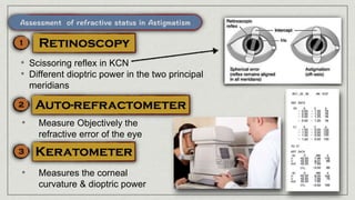
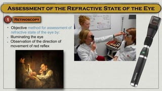
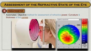
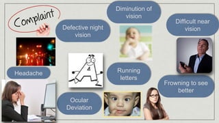
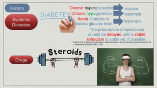
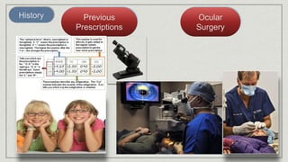
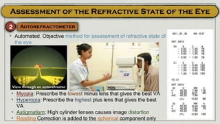
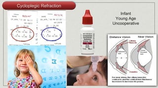
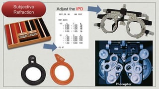
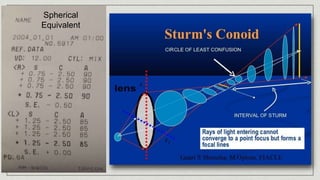
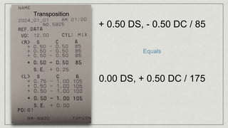
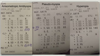
The two Principal Meridians (of highest & lowest power) are NOT at right angle
to each others, and change of power from one meridian to the other is NEITHER
gradual NOR regular
• Corneal scars
• KCN
• Accommodative asthenopia; continuous attempts of the ciliary muscle
to compensate the error
• Blurred vision; near & far
• Running letters
• Distorted objects
• Cornea: scars, stitches, …
• Lens: subluxation
• Retina: Staphyloma
• e.g. Oil droplet in KCN (Charleux)
• Oval optic disc (different refraction along the
two principal meridians)
• Tilted optic disc (Retinal astigmatism)
• Scissoring reflex in KCN
• Different dioptric power in the two principal
meridians
• Measure Objectively the
refractive error of the eye
• Measures the corneal
curvature & dioptric power
• Irregular corneal circles
• Pseudo-colour
mapping of
corneal
curvature,
power &
thickness
• The patient sees the line in one meridian sharper than that on the
perpendicular meridian
Cylindrical Lenses
With its axis perpendicular to the
ocular axis to be corrected
Sphero-Cylindrical Lenses
The spherical lens renders the
error as simple astigmatism
The cylindrical lens corrects the
residual astigmatic error
For low astigmatic errors
In KCN
• A corneal flap is fashioned by an
automated keratome
• Excimer laser is used to ablate the steep
corneal meridian
• The flap is re-positioned
• Partial thickness corneal incisions at the steeper corneal meridian
• Applied in low astigmatic errors
Indications:
A. KCN
A mark on the IOL
denotes the axis of
astigmatism to be
corrected
Using a toric IOL
• Physiological recession of near
point of accommodation
• Increased lens stiffness
• Weak ciliary muscle
• Usually starts around the age of 40 years (earlier in
hyperopes)
• Poor near vision e.g reading, sewing
• Good far vision
• Accommodative asthenopia
• Monfocal: Reading
glasses separate from
distant glasses
Convex Lens
• Bifocal glasses
• Progressive Addition Lenses (PALs)
• 1st Correct vision for FAR
• Measure the Near Point of Accommodation (PP)
• Calculate Amplitude of Accommodation; (1 / PP)
• Keep 1/3 accommodation as reserve to give the presbyope range for reading
• Correct only the remaining 2/3
• Measure the working distance (usually 33 cm)
• Calculate lens power needed to focus at the working distance (1/0.33 = 3D)
• Determine the power of the lens needed for correction of Presbyopia (Power at
working distance - Power at PP)
• Add this power to the power needed for far correction
RAF Rule
• A 45 year old presbyope with + 2D for far
• PP at 40 cm
• Amplitude of Accommodation = 1/0.40 = 2.5D
• 1/3 is kept as reserve i.e 1/3 2.5 = 0.75 D)
• Correct only the remaining 2/3 (2.5 - 0.75 = 1.75 D)
• Measure the working distance (usually 33 cm)
• Calculate lens power needed to focus at the working distance (1/0.33 = 3D)
• Determine the power of the lens needed for correction of Presbyopia (3 -
1.75 = 1.25 D))
• Add this power to the power needed for far correction (1.25 + 2 = 3.25 D)
Convex Lens
• Bifocal lenses
• Excimer laser is used to changes the shape of the cornea to create
different power zones for seeing at varying distances
• Partial thickness scleral pockets are made in each of the 4 eye quadrants
• PMMA (PolyMethyl MethaAcrylate) bands are injected into theses pockets
• The aim is to expand the scleral ring at the zone of ciliary body, thus
stretching the zonules
• Absence of the crystalline lens
• Rare
• Trauma
• Post operative
• Defective far vision; because of hypermetropia
• Defective near vision; due to loss of accommodation
• History of surgery or trauma
• Scar of cataract extraction surgery
• Scar of ruptured globe
• Only two Purkinje Sanson images
absence of the two Purkinje Sanson
images formed by anterior &
posterior surfaces of the lens
• Deep AC
• Tremulous
(Loss of
support by the
lens),
Iridodonesis
• Jet black
colour
(Normally
a greyish
hue
appears in
phakic
clear
lenses)
• High hypermetropia (+ 10
D or more)
• Astigmatism Against the
rule (Because of
contraction of the corneal
scar at 12 o’clock position,
causing flattening of the
vertical meridian)
• Anisometropia (In
unilateral aphakia)
Convex
Lenses
In Bilateral aphakia
Prescribe separate glasses for
distant & near correction or
use bifocals or PALs
Convex
Lenses
In Bilateral or Unilateral aphakia
Prescribe Bifocal Lenses, or Monofocal
lenses with reading glasses
It is the best option (Least image
magnification)
Might be used in Unilateral or Bilateral
aphakia
Implant monofocal IOLs with additional
reading glasses, or implant multifocal
IOL
• Reading ADD in aphakic eyes is + 3D
(Accommodation is lost)
• Disadvantages of Glasses in correction of
Aphakia:
1. Image magnification (30%); Anisometropia in
unilateral cases
2. Peripheral abberation
3. Visual field constriction
4. Thick heavy lenses with poor cosmoses
• A difference of refractive power between the two eyes of 4 D or
more
• Common
• Aphakia
Anis-eiokonia
Different
image size in
both eyes
(>30%)
Binocular Diplopia
Anisometropic Amblyopia
Amblyopic Squint
• Causes less change in image size
(10%)
• LASIK
• IOL Implantation (1% change in image
size)
• Objective method for assessment of
refractive state of the eye by:
Illuminating the eye
Observation of the direction of
movement of red reflex
Retinoscope
Direct
Ophthalmoscop
e
• Put Cycloplegic E.D
• Back of the eye is
illuminated with a
streak light coming
out of the retinoscope
• The Examiner moves
the retinoscope from
side to side & up and
down, observing the
red reflex
• WITH movement
indicates Hyperopia
(Put plus lenses)
• AGAINST movements
indicates Myopia (Put
minus lenses)
• NO movement
indicates emmetropia
(Neutral Point)
• Subtract Working
distance at the end
+ 0.5 DS,
+ 2.00
DC,
Axis 180
+ 1.5 DS,
- 5.00 DC,
Axis 90
- 3.5 DS,
+ 5.00
DC,
Axis 180
• Automated, Objective method for assessment of refractive state of
the eye
• Myopia: Prescribe the lowest minus lens that gives the best VA
• Hyperopia: Prescribe the highest plus lens that gives the best
VA
• Astigmatism: High cylinder lenses causes image distortion
• Reading Correction is added to the spherical component only
• Automated, Objective method for assessment of refractive power, Curvature &
thickness of the cornea
ASSESSME
NT OF
REFRACTIV
E ERRORSMOHAMED ABDELZAHER MD, FRCS
Diminution of
vision
Defective night
vision
Difficult near
vision
Headache Running
letters
Frowning to see
better
Ocular
Deviation
History
Age
History
Systemic
Diseases
Drugs
The prescription of spectacles
should be delayed until a stable
refraction is obtained, if possible.
Chronic hyperglycaemia myopia
Chronic hypoglycemia hyperopia
Acute changes in
plasma glucose level
hyperopia
Fukimi O et al Refractive changes in diabetic patients during intensive glycemic control. Br
J Ophthalmol 2000;84:1097–1102.
History Previous
Prescriptions
Ocular
Surgery
Exam
UAVA
Exam
UAVA
WHY IS THE PATIENT COMPLAINING?!! 😏
To Declare that the patient is seeing a line, he must read
at least ½ of the line correctly.
More evident in Amblyopia
Pin Hole
Vision improves
Vision worsensNo changeRefractive error
Corneal opacity
Cataract
Vitreous hemorrhage
Macular lesion
Thorough Ocular Exam
• Objective method for assessment of
refractive state of the eye by:
Illuminating the eye
Observation of the direction of
movement of red reflex
• Automated, Objective method for assessment of refractive state of
the eye
• Myopia: Prescribe the lowest minus lens that gives the best VA
• Hyperopia: Prescribe the highest plus lens that gives the best
VA
• Astigmatism: High cylinder lenses causes image distortion
• Reading Correction is added to the spherical component only
View through an autorefractor
Cycloplegic Refraction
Infant
Young Age
Uncooperative
Subjective
Refraction
Adjust the IPD
Phoropter
Spherical
Equivalent
Transposition
+ 0.50 DS, - 0.50 DC / 85
0.00 DS, + 0.50 DC / 175
Equals
Hyperopia
Pseudo-myopia
Anisometropic Amblyopia
If you are not willing to learn, No one can help you.
If you are willing to learn, No one can stop you.

Errors 2

  • 1.
    The two PrincipalMeridians (of highest & lowest power) are NOT at right angle to each others, and change of power from one meridian to the other is NEITHER gradual NOR regular • Corneal scars • KCN
  • 2.
    • Accommodative asthenopia;continuous attempts of the ciliary muscle to compensate the error • Blurred vision; near & far • Running letters • Distorted objects
  • 3.
    • Cornea: scars,stitches, … • Lens: subluxation • Retina: Staphyloma • e.g. Oil droplet in KCN (Charleux) • Oval optic disc (different refraction along the two principal meridians) • Tilted optic disc (Retinal astigmatism)
  • 4.
    • Scissoring reflexin KCN • Different dioptric power in the two principal meridians • Measure Objectively the refractive error of the eye • Measures the corneal curvature & dioptric power
  • 5.
    • Irregular cornealcircles • Pseudo-colour mapping of corneal curvature, power & thickness
  • 6.
    • The patientsees the line in one meridian sharper than that on the perpendicular meridian
  • 7.
    Cylindrical Lenses With itsaxis perpendicular to the ocular axis to be corrected
  • 8.
    Sphero-Cylindrical Lenses The sphericallens renders the error as simple astigmatism The cylindrical lens corrects the residual astigmatic error
  • 9.
    For low astigmaticerrors In KCN
  • 10.
    • A cornealflap is fashioned by an automated keratome • Excimer laser is used to ablate the steep corneal meridian • The flap is re-positioned
  • 11.
    • Partial thicknesscorneal incisions at the steeper corneal meridian • Applied in low astigmatic errors
  • 12.
  • 13.
    A mark onthe IOL denotes the axis of astigmatism to be corrected
  • 14.
  • 15.
    • Physiological recessionof near point of accommodation • Increased lens stiffness • Weak ciliary muscle
  • 16.
    • Usually startsaround the age of 40 years (earlier in hyperopes) • Poor near vision e.g reading, sewing • Good far vision • Accommodative asthenopia
  • 17.
    • Monfocal: Reading glassesseparate from distant glasses Convex Lens
  • 18.
    • Bifocal glasses •Progressive Addition Lenses (PALs)
  • 19.
    • 1st Correctvision for FAR • Measure the Near Point of Accommodation (PP) • Calculate Amplitude of Accommodation; (1 / PP) • Keep 1/3 accommodation as reserve to give the presbyope range for reading • Correct only the remaining 2/3 • Measure the working distance (usually 33 cm) • Calculate lens power needed to focus at the working distance (1/0.33 = 3D) • Determine the power of the lens needed for correction of Presbyopia (Power at working distance - Power at PP) • Add this power to the power needed for far correction RAF Rule
  • 20.
    • A 45year old presbyope with + 2D for far • PP at 40 cm • Amplitude of Accommodation = 1/0.40 = 2.5D • 1/3 is kept as reserve i.e 1/3 2.5 = 0.75 D) • Correct only the remaining 2/3 (2.5 - 0.75 = 1.75 D) • Measure the working distance (usually 33 cm) • Calculate lens power needed to focus at the working distance (1/0.33 = 3D) • Determine the power of the lens needed for correction of Presbyopia (3 - 1.75 = 1.25 D)) • Add this power to the power needed for far correction (1.25 + 2 = 3.25 D)
  • 22.
  • 23.
    • Excimer laseris used to changes the shape of the cornea to create different power zones for seeing at varying distances
  • 24.
    • Partial thicknessscleral pockets are made in each of the 4 eye quadrants • PMMA (PolyMethyl MethaAcrylate) bands are injected into theses pockets • The aim is to expand the scleral ring at the zone of ciliary body, thus stretching the zonules
  • 26.
    • Absence ofthe crystalline lens • Rare • Trauma • Post operative
  • 27.
    • Defective farvision; because of hypermetropia • Defective near vision; due to loss of accommodation • History of surgery or trauma
  • 28.
    • Scar ofcataract extraction surgery • Scar of ruptured globe • Only two Purkinje Sanson images absence of the two Purkinje Sanson images formed by anterior & posterior surfaces of the lens
  • 29.
  • 30.
    • Tremulous (Loss of supportby the lens), Iridodonesis
  • 31.
    • Jet black colour (Normally agreyish hue appears in phakic clear lenses)
  • 32.
    • High hypermetropia(+ 10 D or more) • Astigmatism Against the rule (Because of contraction of the corneal scar at 12 o’clock position, causing flattening of the vertical meridian) • Anisometropia (In unilateral aphakia)
  • 33.
    Convex Lenses In Bilateral aphakia Prescribeseparate glasses for distant & near correction or use bifocals or PALs Convex Lenses In Bilateral or Unilateral aphakia Prescribe Bifocal Lenses, or Monofocal lenses with reading glasses
  • 34.
    It is thebest option (Least image magnification) Might be used in Unilateral or Bilateral aphakia Implant monofocal IOLs with additional reading glasses, or implant multifocal IOL
  • 35.
    • Reading ADDin aphakic eyes is + 3D (Accommodation is lost) • Disadvantages of Glasses in correction of Aphakia: 1. Image magnification (30%); Anisometropia in unilateral cases 2. Peripheral abberation 3. Visual field constriction 4. Thick heavy lenses with poor cosmoses
  • 36.
    • A differenceof refractive power between the two eyes of 4 D or more • Common • Aphakia
  • 37.
  • 38.
  • 39.
    • Causes lesschange in image size (10%) • LASIK • IOL Implantation (1% change in image size)
  • 40.
    • Objective methodfor assessment of refractive state of the eye by: Illuminating the eye Observation of the direction of movement of red reflex
  • 41.
  • 42.
    • Put CycloplegicE.D • Back of the eye is illuminated with a streak light coming out of the retinoscope • The Examiner moves the retinoscope from side to side & up and down, observing the red reflex • WITH movement indicates Hyperopia (Put plus lenses) • AGAINST movements indicates Myopia (Put minus lenses) • NO movement indicates emmetropia (Neutral Point) • Subtract Working distance at the end
  • 43.
    + 0.5 DS, +2.00 DC, Axis 180 + 1.5 DS, - 5.00 DC, Axis 90 - 3.5 DS, + 5.00 DC, Axis 180
  • 44.
    • Automated, Objectivemethod for assessment of refractive state of the eye • Myopia: Prescribe the lowest minus lens that gives the best VA • Hyperopia: Prescribe the highest plus lens that gives the best VA • Astigmatism: High cylinder lenses causes image distortion • Reading Correction is added to the spherical component only
  • 45.
    • Automated, Objectivemethod for assessment of refractive power, Curvature & thickness of the cornea
  • 46.
  • 48.
    Diminution of vision Defective night vision Difficultnear vision Headache Running letters Frowning to see better Ocular Deviation
  • 49.
  • 50.
    History Systemic Diseases Drugs The prescription ofspectacles should be delayed until a stable refraction is obtained, if possible. Chronic hyperglycaemia myopia Chronic hypoglycemia hyperopia Acute changes in plasma glucose level hyperopia Fukimi O et al Refractive changes in diabetic patients during intensive glycemic control. Br J Ophthalmol 2000;84:1097–1102.
  • 51.
  • 52.
  • 53.
  • 54.
    WHY IS THEPATIENT COMPLAINING?!! 😏 To Declare that the patient is seeing a line, he must read at least ½ of the line correctly.
  • 55.
    More evident inAmblyopia
  • 56.
    Pin Hole Vision improves VisionworsensNo changeRefractive error Corneal opacity Cataract Vitreous hemorrhage Macular lesion
  • 57.
  • 58.
    • Objective methodfor assessment of refractive state of the eye by: Illuminating the eye Observation of the direction of movement of red reflex
  • 59.
    • Automated, Objectivemethod for assessment of refractive state of the eye • Myopia: Prescribe the lowest minus lens that gives the best VA • Hyperopia: Prescribe the highest plus lens that gives the best VA • Astigmatism: High cylinder lenses causes image distortion • Reading Correction is added to the spherical component only View through an autorefractor
  • 60.
  • 61.
  • 62.
  • 63.
    Transposition + 0.50 DS,- 0.50 DC / 85 0.00 DS, + 0.50 DC / 175 Equals
  • 64.
  • 65.
    If you arenot willing to learn, No one can help you. If you are willing to learn, No one can stop you.