Downloaded 155 times

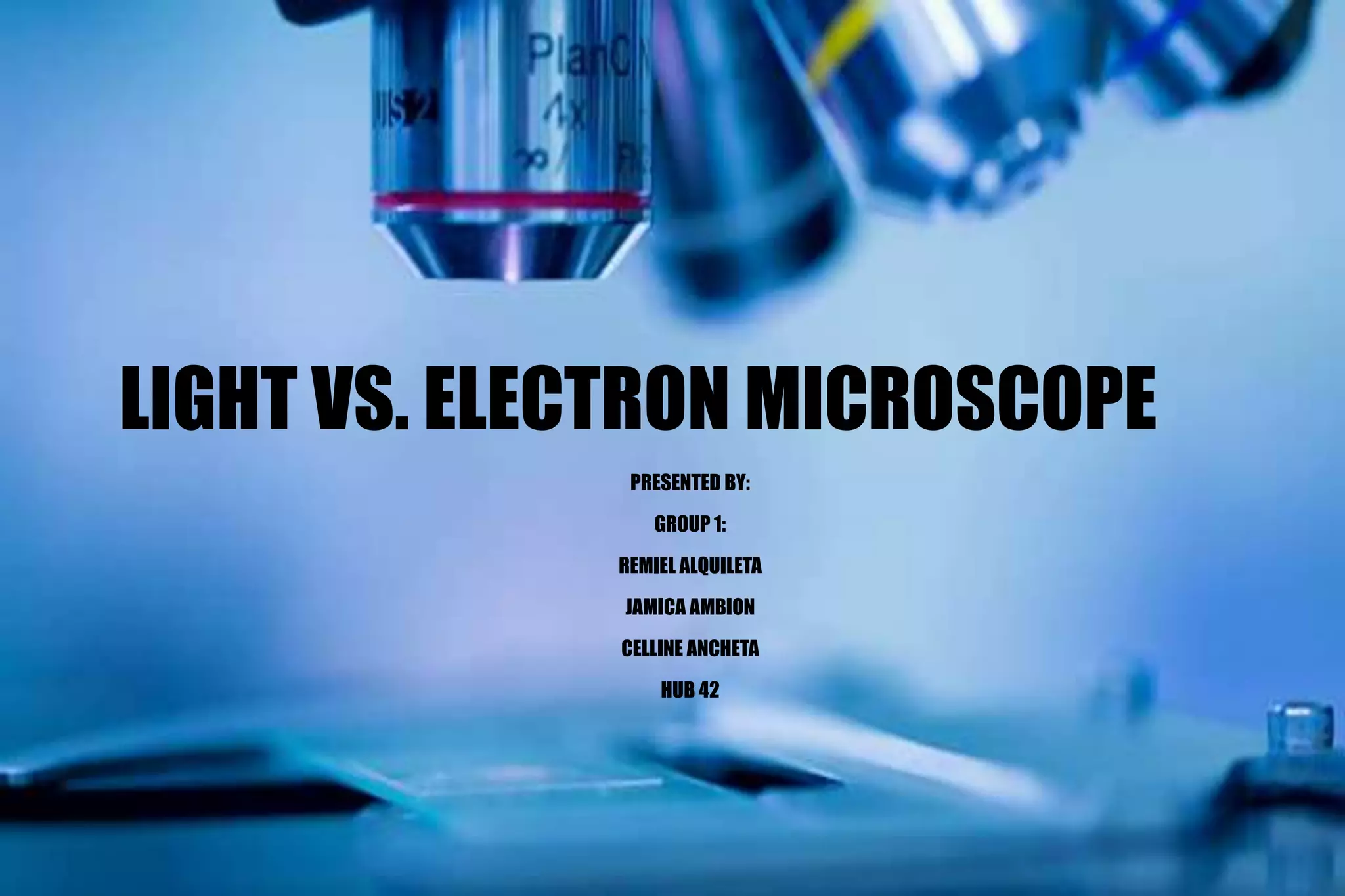


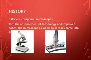
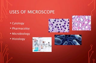
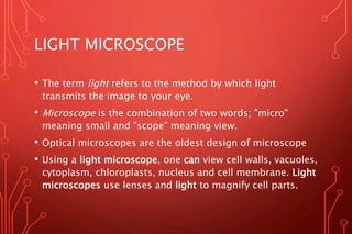
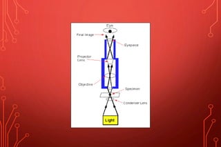
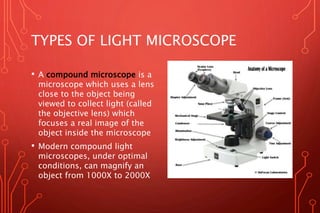
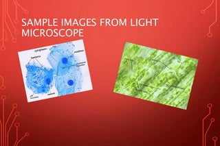
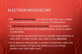
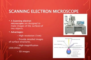
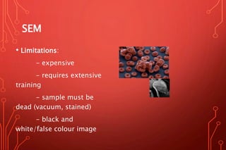
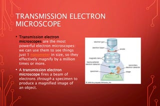
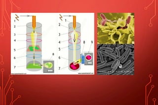
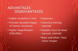
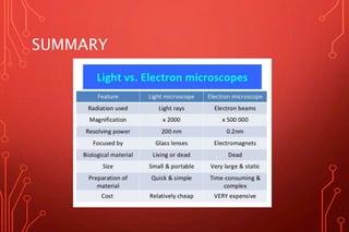

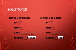

This document provides information on light microscopes and electron microscopes. It discusses the history and development of microscopes from the 1590s to modern technology. Light microscopes use lenses and light to magnify objects up to 2000x, while electron microscopes use beams of electrons to achieve higher magnifications of over 10,000,000x and resolutions of 1 nanometer or less. Scanning electron microscopes provide 3D surface images at magnifications up to 200,000x, while transmission electron microscopes allow viewing of interior structures. Electron microscopes provide higher resolutions and magnifications than light microscopes but are more expensive and require dead samples.


![Polymer [ बहुलक ] Chemistry Notes PDF - Irfanullah Mehar - JJ Sir Chemistry.pdf](https://cdn.slidesharecdn.com/ss_thumbnails/polymerchemistrynotespdf-irfanullahmehar-jjsirchemistry-260210172118-3f9b37f7-thumbnail.jpg?width=640&height=640&fit=bounds)


