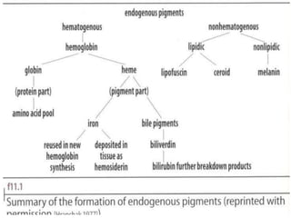
1
Pigments
&
Minerals
By
William Edema
Arua Institute of Health Science
Introduction
• Pigments colored substances deposited in cells
and tissue
• Some pigments are artificial while others are
natural
• Pigments are Classified into
1. Artifact pigments
2. Endogenous pigments
3. Exogenous pigments
Artificial pigments
These are fixation artifacts which include:
• Mercuric chloride
• Chrome deposits
• Pink disease
Exogenous pigments
These originate outside the body, from external
environment and are non physiological
Carbon,
Silica
Asbestos
Dusts e.g coal dust
 Tattoo pigments
Silver
NB
• Carbon is a black pigment commonly seen in
sections of lungs and lymph nodes.
• Carbon is insoluble in conc sulfuric acid and this
distinguishes it from other black pigments.
• Asbestos fibers are bare fringent fibers of
magnesium silicate and may be found in people
exposed to large amounts of asbestos
Exogenous pigments..
• Pneumoconiosis: lung disease due to inhalation
of organic and inorganic particles
• Anthracosis-Coal mining caused by inhalation of
coal dust.
• Tattooing is a form of localized, exogenous
pigmentation of the skin.
• Pigments inoculated are phagocytosed by
dermal macrophages and do not usually evoke
any inflammatory response
Endogenous pigments
• These are produced within tissue and serve a
physiological function or are by products of
normal metabolic processes. They are sub divided
into :
1. Haematogenous (Hemoglobin-derived)
2. Non Haematogenous (Non Hemoglobin-derived)
Haematogenous (Hemoglobin-derived)
These include:-
1. Hemosiderin
2. Bile pigments
3. Porphyrin
4. Hemoglobin
Haematogenous Endogenous pigments
Normal and abnormal Iron
Storage
• Ferritin: protein-iron complex (normal)
• Hemosiderin: ferritin aggregate (abnormal)
Iron(Fe3+) is normally carried by specific transport
proteins, transferrins
•In cells Fe3+ bound to apoferritin to form ferritin micelles
•Ferritin is a constituent of most cell types
•When there is a local or systemic excess of iron, ferritin
forms hemosiderin granules
Normal and abnormal Iron…
• Under normal conditions, hemosiderin can be
seen in the mononuclear phagocytes of the
bone marrow, spleen, and liver
• Actively engaged in red cell break down
• Excesses of iron causes hemosiderin to
accumulate with in cells
• Local excesses of iron and hemosiderin result
from
Hemosiderin
• These pigments are seen as yellow to brown granules
and normally intracelullarly. They contain iron in the
form of ferric hydroxide that is bound to a protein frame
work and is unmasked by various chemicals.
• Ferric iron is stored in bone marrow and spleen as
haemosiderin,
• Causes of Hemosiderin deposits in the body
 Increased absorption of dietary iron
 Impaired iron utilisation
 Excess hemolysis
 Blood transfusion (Exogenous iron load)
Hemosiderin
• Decreased or increased stores of iron are found in
pathologic conditions and are demonstrated by
Prussian blue. (Ferrous iron is demonstrated by
Turnbull’s blue stain)
• Hemosiderosis: This is a condition in which iron
stores in the body become overloaded for
example in recurrent blood transfusions.
• Hemochromatosis is a genetic disease caused by
excessive absorption of dietary iron in amounts
irrelevant to the state of the body x-terized by
excessive hemosiderin.
Demonstration of hemosiderin
• Hemosiderin is distinguished from other yellow pigments
by Purls Prussian blue reaction.
• In un fixed tissue hemosiderin is insoluble in alkaline but
freely soluble in strong acid solutions after fixation in
formalin, it slowly soluble in dilute acids
• Fixatives that contain acids can remove hemosiderin or
alter it in such a way that the reaction are negative
• Some iron found in tissue are not demons tasted using
tradition methods this is because the iron are very tightly
bond e.g in hemoglobin and myoglobin, but if treated
with hydrogen peroxide the iron can be released and
then demonstrated with Perl's Prussian blue.
• NB most Iron in the tissue appears in its ferric state
Other methods used to demonstrate Iron
• Lilies method for Ferric Iron
• Hukill & putts method for ferrous and ferric
• Turnbull’s blue
Demonstration of hemoglobin
• Hemoglobin appears normally in red blood cells
its histological application is not usually normal.
• However there are two types of methods used
to demonstrate it in tissue sections namely:-
1. Leuco patent blue V
2. Amido black technique
Demonstration of bile pigments
• The need to identify bile pigments arises in the
histological examination of the liver where
distinguishing bile pigments from lipofusin may
be important since both appear yellow brown in
H&E stained sections, and the green color of
biliverdin is masked by Eosin.
• Bile pigments are not autofluorescent while
lipofuscin is.
• Bile pigments are demonstrated by modified
Fouchet technique is which the pigment is
coveted to the green color of bilverdin
Demonstration of Porphyrin
• These occur in rare disorders of biosynthesis
of porphyrins and heme in which they are
seen as focal deposits in liver sections.
• The pigment appears as dense dark brown
pigment and in fresh sections shows a bright
red flurosecence that feeds on exposure to UV
Non Haematogenous Endogenous pigments
• These include:
1. Melanin
2. Lipofuscins
3. Chromaffin
4. Pseudomelanosis
5. Dubin Johnson pigment
6. Ceroid type Lipofuscins
7. Hamazaki-Weisenberg bodie.
Melanin
• This pigment varies from light brown to black.
• The pigment is normally found in the skin,
eye, nigra of the brain and hair follicle.
• Under pathological conditions it is found in
benign nevus cell tumors and malignant
melanomas.
• Melanocytes make the pigment called melanin
Production of melanin
• The enzyme Tryosinase acts on Tryonise to
produce DOPA (dihydroxyphenylalanine)
• DOPA is then acted up on by the same enzyme to
produce an intermediate pigment which
polymerises to produce melanin.
Methods used to demonstrate Melanin
1. Reducing methods e.g. Mansoon Fontana, Silver
technique, and Schmorl’s Ferric Ferricyanide
2. Enzyme methods e.g. DOPA
3. Solubility and bleaching characteristics
4. Fluorescent methods
5. Immunocytochemistry
Physical properties of melanin
used to demonstrate it
1. Melanin and melanin precursor are capable of
reducing both silver and acid ferricynaide
solutions
2. Its insoluble in most inorganic solvents sine it is
tightly bound to proteins of the melanocytes
3. Ability to be bleached by strong oxidizing agents
NB: the enzyme tryosinase can be demonstrated by
the DOPA reaction
Lipofuscins.
• Theses are yellow brown to reddish pigments
and are thought to be produced by the oxidation
methods of lipids.
Pigments & minerals in tissue
1.Mercury: mercury fixatives, removed by iodine
and hypo prior to staining.
2.Chrome: chromium fixatives, removed by water
wash before dehydration.
3.Formalin: acidic formalin fixatives; is acid
hematein, which is birefringent, microcrystalline,
gives no iron reaction, is dissolved by alcoholic
picric acid or lithium carbonate in 70% alcohol.
(Note: Hematoidin, pigment found in cells infected
with malaria, resembles formalin pigment.)
…
• Pink disease: Observed in tissue rapidly fixed in
buffered formal saline.
• It is common in lymphoid and epithelia cells.
• It is characterized by the nuclei failing to stain
with haematoxylin and instead taking eosin and
appearing pink.
• Mainly avoided by adding 2% acetic acid to the
fixative or by adding1% HCL to absolute used for
section hydration.
…
Exogenous pigments: formed externally and taken into the
body
1. Carbon (anthracotic): black, found in lungs and
associated nodes; resists bleaching insoluble in concentrate
sulfuric acid.
2. Asbestos fibers: birefringent fibers of magnesium silicate
in lungs, causes reaction that leads to mesothelioma; fibers
become coated with iron-containing protein to become
asbestos bodies; demonstrated by Prussian blue stain.
3.Tattoo pigments: in skin and nodes near tattoo, whether
intentional or accidental tattoo; special stains not
necessary.
…
4.Metals: minerals like Calcium, ferrous or ferric
iron, and copper are metals normally found in the
body; copper and aluminum are found in the body
only in pathologic conditions.
• Minerals normally found in the body:
Calcium: normal in circulating blood and in
bone; demonstrated in tissue by Alizarin red S
and von Kossa’s procedures; pathologically
found in nephrocalcinosis
…
2.Iron: see discussion under hematogenous
endogenous pigments.
3.Copper: normally present in minute amounts;
pathologically present in Wilson’s disease with
copper increased in liver and brain.
Aluminum deposits in bone after long-term dialysis
are demonstrated by aluminon or solochrome
azurine methods.
Endogenous pigments
• Endogenous pigments: produced within tissues and
serves some physiologic function, or the by- products of
metabolic functions:
• Hematogenous: derived from blood.
a. Hemoglobin: normal in RBC’s, abnormal in recent
hemorrhagic tissues; stains with eosin
b. Bile pigment: breakdown product of hemoglobin’s
“heme” part; seen in obstructive jaundice in liver
sections; demonstrated by methods that oxidize
bilirubin (yellow brown) to biliverdin (green). (Note:
hematoidin, another heme breakdown product formed
as a result of hemorrhage is also demonstrated by bile
pigment stains)
PIGMENTS & MINERALS IN TISSUE
.Iron (hemosiderin), ferric iron: stored in bone
marrow and spleen as haemosiderin, ayellow to
brown pigment for use in new blood cell
formation;
Non-hematogenous pigments
Non-hematogenous: not derived from blood
a.Non-lipidic: Melanin is the primary non-lipidic,
non-hematogenous pigment; brown-black
pigment is normal in skin, hair, retina, and
parts of CNS; can be bleached with potassium
permanganate; positive with ferrous iron
procedures; is argentophilic; stains are done
to demonstrate melanomas.
Reference
1. Bancroft’s theory and practice of histological
techniques,7th edition 2013
2. Histotechnology a self instruction text, 4th
edition, 2015, Freida L Carson

PIGMENTS & MINERALS IN TISSUE 2023.ppt

  • 1.
  • 2.
    Introduction • Pigments coloredsubstances deposited in cells and tissue • Some pigments are artificial while others are natural • Pigments are Classified into 1. Artifact pigments 2. Endogenous pigments 3. Exogenous pigments
  • 3.
    Artificial pigments These arefixation artifacts which include: • Mercuric chloride • Chrome deposits • Pink disease
  • 4.
    Exogenous pigments These originateoutside the body, from external environment and are non physiological Carbon, Silica Asbestos Dusts e.g coal dust  Tattoo pigments Silver
  • 5.
    NB • Carbon isa black pigment commonly seen in sections of lungs and lymph nodes. • Carbon is insoluble in conc sulfuric acid and this distinguishes it from other black pigments. • Asbestos fibers are bare fringent fibers of magnesium silicate and may be found in people exposed to large amounts of asbestos
  • 6.
    Exogenous pigments.. • Pneumoconiosis:lung disease due to inhalation of organic and inorganic particles • Anthracosis-Coal mining caused by inhalation of coal dust. • Tattooing is a form of localized, exogenous pigmentation of the skin. • Pigments inoculated are phagocytosed by dermal macrophages and do not usually evoke any inflammatory response
  • 7.
    Endogenous pigments • Theseare produced within tissue and serve a physiological function or are by products of normal metabolic processes. They are sub divided into : 1. Haematogenous (Hemoglobin-derived) 2. Non Haematogenous (Non Hemoglobin-derived)
  • 8.
    Haematogenous (Hemoglobin-derived) These include:- 1.Hemosiderin 2. Bile pigments 3. Porphyrin 4. Hemoglobin
  • 9.
    Haematogenous Endogenous pigments Normaland abnormal Iron Storage • Ferritin: protein-iron complex (normal) • Hemosiderin: ferritin aggregate (abnormal) Iron(Fe3+) is normally carried by specific transport proteins, transferrins •In cells Fe3+ bound to apoferritin to form ferritin micelles •Ferritin is a constituent of most cell types •When there is a local or systemic excess of iron, ferritin forms hemosiderin granules
  • 10.
    Normal and abnormalIron… • Under normal conditions, hemosiderin can be seen in the mononuclear phagocytes of the bone marrow, spleen, and liver • Actively engaged in red cell break down • Excesses of iron causes hemosiderin to accumulate with in cells • Local excesses of iron and hemosiderin result from
  • 12.
    Hemosiderin • These pigmentsare seen as yellow to brown granules and normally intracelullarly. They contain iron in the form of ferric hydroxide that is bound to a protein frame work and is unmasked by various chemicals. • Ferric iron is stored in bone marrow and spleen as haemosiderin, • Causes of Hemosiderin deposits in the body  Increased absorption of dietary iron  Impaired iron utilisation  Excess hemolysis  Blood transfusion (Exogenous iron load)
  • 13.
    Hemosiderin • Decreased orincreased stores of iron are found in pathologic conditions and are demonstrated by Prussian blue. (Ferrous iron is demonstrated by Turnbull’s blue stain) • Hemosiderosis: This is a condition in which iron stores in the body become overloaded for example in recurrent blood transfusions. • Hemochromatosis is a genetic disease caused by excessive absorption of dietary iron in amounts irrelevant to the state of the body x-terized by excessive hemosiderin.
  • 14.
    Demonstration of hemosiderin •Hemosiderin is distinguished from other yellow pigments by Purls Prussian blue reaction. • In un fixed tissue hemosiderin is insoluble in alkaline but freely soluble in strong acid solutions after fixation in formalin, it slowly soluble in dilute acids • Fixatives that contain acids can remove hemosiderin or alter it in such a way that the reaction are negative • Some iron found in tissue are not demons tasted using tradition methods this is because the iron are very tightly bond e.g in hemoglobin and myoglobin, but if treated with hydrogen peroxide the iron can be released and then demonstrated with Perl's Prussian blue. • NB most Iron in the tissue appears in its ferric state
  • 16.
    Other methods usedto demonstrate Iron • Lilies method for Ferric Iron • Hukill & putts method for ferrous and ferric • Turnbull’s blue
  • 17.
    Demonstration of hemoglobin •Hemoglobin appears normally in red blood cells its histological application is not usually normal. • However there are two types of methods used to demonstrate it in tissue sections namely:- 1. Leuco patent blue V 2. Amido black technique
  • 18.
    Demonstration of bilepigments • The need to identify bile pigments arises in the histological examination of the liver where distinguishing bile pigments from lipofusin may be important since both appear yellow brown in H&E stained sections, and the green color of biliverdin is masked by Eosin. • Bile pigments are not autofluorescent while lipofuscin is. • Bile pigments are demonstrated by modified Fouchet technique is which the pigment is coveted to the green color of bilverdin
  • 19.
    Demonstration of Porphyrin •These occur in rare disorders of biosynthesis of porphyrins and heme in which they are seen as focal deposits in liver sections. • The pigment appears as dense dark brown pigment and in fresh sections shows a bright red flurosecence that feeds on exposure to UV
  • 20.
    Non Haematogenous Endogenouspigments • These include: 1. Melanin 2. Lipofuscins 3. Chromaffin 4. Pseudomelanosis 5. Dubin Johnson pigment 6. Ceroid type Lipofuscins 7. Hamazaki-Weisenberg bodie.
  • 21.
    Melanin • This pigmentvaries from light brown to black. • The pigment is normally found in the skin, eye, nigra of the brain and hair follicle. • Under pathological conditions it is found in benign nevus cell tumors and malignant melanomas. • Melanocytes make the pigment called melanin
  • 22.
    Production of melanin •The enzyme Tryosinase acts on Tryonise to produce DOPA (dihydroxyphenylalanine) • DOPA is then acted up on by the same enzyme to produce an intermediate pigment which polymerises to produce melanin.
  • 23.
    Methods used todemonstrate Melanin 1. Reducing methods e.g. Mansoon Fontana, Silver technique, and Schmorl’s Ferric Ferricyanide 2. Enzyme methods e.g. DOPA 3. Solubility and bleaching characteristics 4. Fluorescent methods 5. Immunocytochemistry
  • 24.
    Physical properties ofmelanin used to demonstrate it 1. Melanin and melanin precursor are capable of reducing both silver and acid ferricynaide solutions 2. Its insoluble in most inorganic solvents sine it is tightly bound to proteins of the melanocytes 3. Ability to be bleached by strong oxidizing agents NB: the enzyme tryosinase can be demonstrated by the DOPA reaction
  • 25.
    Lipofuscins. • Theses areyellow brown to reddish pigments and are thought to be produced by the oxidation methods of lipids.
  • 26.
    Pigments & mineralsin tissue 1.Mercury: mercury fixatives, removed by iodine and hypo prior to staining. 2.Chrome: chromium fixatives, removed by water wash before dehydration. 3.Formalin: acidic formalin fixatives; is acid hematein, which is birefringent, microcrystalline, gives no iron reaction, is dissolved by alcoholic picric acid or lithium carbonate in 70% alcohol. (Note: Hematoidin, pigment found in cells infected with malaria, resembles formalin pigment.)
  • 27.
    … • Pink disease:Observed in tissue rapidly fixed in buffered formal saline. • It is common in lymphoid and epithelia cells. • It is characterized by the nuclei failing to stain with haematoxylin and instead taking eosin and appearing pink. • Mainly avoided by adding 2% acetic acid to the fixative or by adding1% HCL to absolute used for section hydration.
  • 28.
    … Exogenous pigments: formedexternally and taken into the body 1. Carbon (anthracotic): black, found in lungs and associated nodes; resists bleaching insoluble in concentrate sulfuric acid. 2. Asbestos fibers: birefringent fibers of magnesium silicate in lungs, causes reaction that leads to mesothelioma; fibers become coated with iron-containing protein to become asbestos bodies; demonstrated by Prussian blue stain. 3.Tattoo pigments: in skin and nodes near tattoo, whether intentional or accidental tattoo; special stains not necessary.
  • 29.
    … 4.Metals: minerals likeCalcium, ferrous or ferric iron, and copper are metals normally found in the body; copper and aluminum are found in the body only in pathologic conditions. • Minerals normally found in the body: Calcium: normal in circulating blood and in bone; demonstrated in tissue by Alizarin red S and von Kossa’s procedures; pathologically found in nephrocalcinosis
  • 30.
    … 2.Iron: see discussionunder hematogenous endogenous pigments. 3.Copper: normally present in minute amounts; pathologically present in Wilson’s disease with copper increased in liver and brain. Aluminum deposits in bone after long-term dialysis are demonstrated by aluminon or solochrome azurine methods.
  • 31.
    Endogenous pigments • Endogenouspigments: produced within tissues and serves some physiologic function, or the by- products of metabolic functions: • Hematogenous: derived from blood. a. Hemoglobin: normal in RBC’s, abnormal in recent hemorrhagic tissues; stains with eosin b. Bile pigment: breakdown product of hemoglobin’s “heme” part; seen in obstructive jaundice in liver sections; demonstrated by methods that oxidize bilirubin (yellow brown) to biliverdin (green). (Note: hematoidin, another heme breakdown product formed as a result of hemorrhage is also demonstrated by bile pigment stains)
  • 32.
    PIGMENTS & MINERALSIN TISSUE .Iron (hemosiderin), ferric iron: stored in bone marrow and spleen as haemosiderin, ayellow to brown pigment for use in new blood cell formation;
  • 33.
    Non-hematogenous pigments Non-hematogenous: notderived from blood a.Non-lipidic: Melanin is the primary non-lipidic, non-hematogenous pigment; brown-black pigment is normal in skin, hair, retina, and parts of CNS; can be bleached with potassium permanganate; positive with ferrous iron procedures; is argentophilic; stains are done to demonstrate melanomas.
  • 34.
    Reference 1. Bancroft’s theoryand practice of histological techniques,7th edition 2013 2. Histotechnology a self instruction text, 4th edition, 2015, Freida L Carson