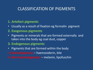
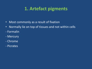
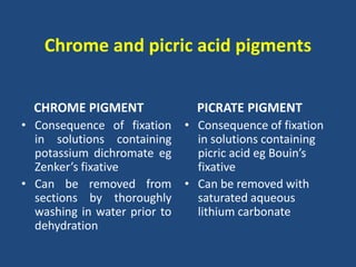
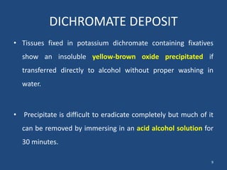
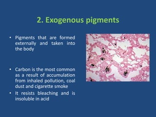
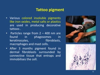
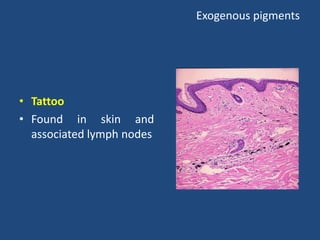
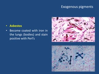
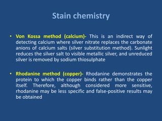
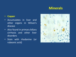
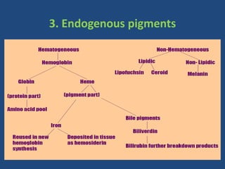
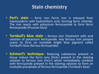
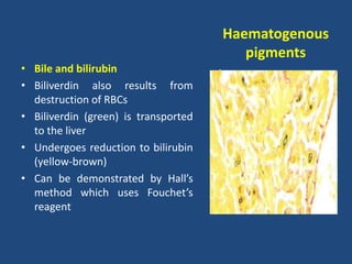
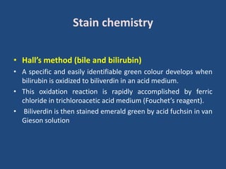
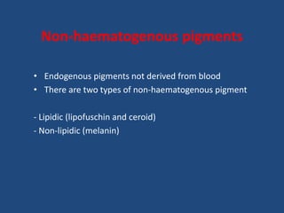
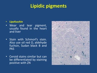
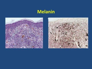
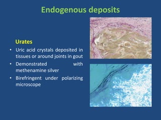
Pigments and minerals are frequently observed in tissues and may be present naturally, may be formed because of a patho- logic condition, may be artifacts caused during fixation, or may be introduced into the body from external sources.
2. CLASSIFICATION OF PIGMENTS
1. Artefact pigments
• Usually as a result of fixation eg formalin pigment
2. Exogenous pigments
• Pigments or minerals that are formed externally and
taken into the body eg coal dust, copper
3. Endogenous pigments
• Pigments that are formed within the body
i. Haematogenous – haemosiderin, bile
ii. Non-haematogenous – melanin, lipofuschin
3. 1. Artefact pigments
• Most commonly as a result of fixation
• Normally lie on top of tissues and not within cells
- Formalin
- Mercury
- Chrome
- Picrates
4. FORMALIN PIGMENT
• Pigment appears as a brown/ brownish black deposit in
tissues fixed in any formalin fixative with an acid pH & is more
likely to occur in blood rich tissues.
• Can be removed by treating with
Saturated alcoholic picric acid for some hours.
Alcoholic solution of Sod. & Pot. Hydroxide.
10% ammonium hydroxide in 70% alcohol for 5-15 min.
4
5. Formalin pigment
• Result of fixation of tissue
in acid formalin
• Also called acid haematin
• Resistant to strong acids
• Usually associated with
blood-containing tissues
• Can be removed from
sections using alcoholic
picric acid
6. MERCURY PIGMENT
• Tissues fixed in fixative mixtures containing mercury have
been found to develop a uniform granular black deposit
known as mercury pigment.
• This can be removed by treatment with Lugol’s iodine and
sodium thiosulphate.
6
7. Mercury pigment
• Occurs with mercury-
containing fixatives such as
Zenker’s and Heidenhain’s
Susa
• Can be removed from
sections using iodine
followed by sodium
thiosulphate (Hypo) solution
• .
8. Chrome and picric acid pigments
CHROME PIGMENT
• Consequence of fixation
in solutions containing
potassium dichromate eg
Zenker’s fixative
• Can be removed from
sections by thoroughly
washing in water prior to
dehydration
PICRATE PIGMENT
• Consequence of fixation
in solutions containing
picric acid eg Bouin’s
fixative
• Can be removed with
saturated aqueous
lithium carbonate
9. DICHROMATE DEPOSIT
• Tissues fixed in potassium dichromate containing fixatives
show an insoluble yellow-brown oxide precipitated if
transferred directly to alcohol without proper washing in
water.
• Precipitate is difficult to eradicate completely but much of it
can be removed by immersing in an acid alcohol solution for
30 minutes.
9
10. 2. Exogenous pigments
• Pigments that are formed
externally and taken into
the body
• Carbon is the most common
as a result of accumulation
from inhaled pollution, coal
dust and cigarette smoke
• It resists bleaching and is
insoluble in acid
• .
11. Tattoo pigment
• Various colored insoluble pigments
like iron oxides, metal salts or plastics
are used in producing decorative
tattoos .
• Particles range from 2 – 400 nm are
found in phagosomes in
keratinocytes, fibroblasts,
macrophages and mast cells.
• After 3 months pigment found in
dermal fibroblasts surrounded by
connective tissue that entraps and
immobilises the cell.
11
14. Minerals
• Metallic and non-metallic ions necessary for growth and other
bodily functions
• Calcium, ferrous and ferric iron, copper, phosphate and
carbonate are the most common
• Some can be deposited pathologically in tissue eg gold, silver,
copper and lead
• Minerals in tissue can be studied using the technique of
microincineration
15. Microincineration
• Study inorganic substances found in tissue
• Cut paraffin sections as usual and mount on high temperature
glass slides
• Slides are incinerated up to 650oC and then allowed to cool
• Slides are removed from oven and coverslipped with glycerol
mountant
• Slides are examined under the microscope
16. Minerals
• Calcium
• Abnormal deposits can be
associated with atherosclerosis,
sarcoid, TB and some tumours
• Also found in joints in
chondrocalcinosis
• Demonstrate with von Kossa
silver method
• .
17. Stain chemistry
• Von Kossa method (calcium)- This is an indirect way of
detecting calcium where silver nitrate replaces the carbonate
anions of calcium salts (silver substitution method). Sunlight
reduces the silver salt to visible metallic silver, and unreduced
silver is removed by sodium thiosulphate
• Rhodanine method (copper)- Rhodanine demonstrates the
protein to which the copper binds rather than the copper
itself. Therefore, although considered more sensitive,
rhodanine may be less specific and false-positive results may
be obtained
18. Minerals
• Copper
• Accumulates in liver and
other organs in Wilson’s
disease
• Also found in primary biliary
cirrhosis and other liver
disorders
• Stain with rhodanine (or
rubeanic acid)
• .
19. Stain chemistry
• Von Kossa method (calcium)
• This is an indirect way of detecting calcium where silver
nitrate replaces the carbonate anions of calcium salts (silver
substitution method). Sunlight reduces the silver salt to visible
metallic silver, and unreduced silver is removed by sodium
thiosulphate
• Rhodanine method (copper)
• Rhodanine demonstrates the protein to which the copper
binds rather than the copper itself. Therefore, although
considered more sensitive, rhodanine may be less specific and
false-positive results may be obtained
21. Haematogenous pigments
• Endogenous pigments derived from blood
• Main ones are haemoglobin, iron, haemosiderin, haematoidin
and bile pigments
• Haemoglobin breaks down into two parts: globin and haem
(iron-containing pigment part)
• The haem portion further splits into iron (haemosiderin) and
bile pigments (biliverdin)
• Haematoidin (similar to bilirubin) is formed in tissue as a
result of haemorrhage
22. Haematogenous
pigments
• Haemosiderin
• Stored as ferric iron in bone
marrow and spleen
• Can be demonstrated using
Perl’s prussian blue stain
which detects ferric iron
• Excessive absorption of
dietary iron causes
haemochromatosis
• Ferrous iron is demonstrated
by Turnbull’s blue
• .
23. Stain chemistry
• Perl’s stain – ferric iron Ferric iron is released from
haemosiderin with hydrochloric acid, forming ferric chloride.
The iron reacts with potassium ferrocyanide to form ferric
ferrocyanide (Prussian blue)
• Turnbull’s blue stain – ferrous iron Treatment with acid
solution of potassium ferricyanide. Any ferrous iron present
reacts to form an insoluble bright blue pigment called
Turnbull’s blue (ferrous ferricyanide)
• Schmorl’s technique- Reducing substances present in
tissue reduce the ferric ions (Fe3+) present in the staining
solution to ferrous ions (Fe2+) which immediately combine
with ferricyanide present in the staining solution to form an
insoluble precipitate of ferrous ferricyanide (Turnbull’s blue)
24. Haematogenous
pigments
• Bile and bilirubin
• Biliverdin also results from
destruction of RBCs
• Biliverdin (green) is transported
to the liver
• Undergoes reduction to bilirubin
(yellow-brown)
• Can be demonstrated by Hall’s
method which uses Fouchet’s
reagent
• .
25. Stain chemistry
• Hall’s method (bile and bilirubin)
• A specific and easily identifiable green colour develops when
bilirubin is oxidized to biliverdin in an acid medium.
• This oxidation reaction is rapidly accomplished by ferric
chloride in trichloroacetic acid medium (Fouchet’s reagent).
• Biliverdin is then stained emerald green by acid fuchsin in van
Gieson solution
26. Non-haematogenous pigments
• Endogenous pigments not derived from blood
• There are two types of non-haematogenous pigment
- Lipidic (lipofuschin and ceroid)
- Non-lipidic (melanin)
27. Lipidic pigments
• Lipofuschin
• Wear and tear pigment,
usually found in the heart
and liver
• Stain with Schmorl’s stain.
Also use oil red O, aldehyde
fuchsin, Sudan black B and
PAS
• Ceroid stains similar but can
be differentiated by staining
positive with ZN
• .
28. Stain chemistry
• Perl’s stain – ferric iron Ferric iron is released from
haemosiderin with hydrochloric acid, forming ferric chloride.
The iron reacts with potassium ferrocyanide to form ferric
ferrocyanide (Prussian blue)
• Turnbull’s blue stain – ferrous iron Treatment with acid
solution of potassium ferricyanide. Any ferrous iron present
reacts to form an insoluble bright blue pigment called
Turnbull’s blue (ferrous ferricyanide)
• Schmorl’s technique- Reducing substances present in tissue
reduce the ferric ions (Fe3+) present in the staining solution
to ferrous ions (Fe2+) which immediately combine with
ferricyanide present in the staining solution to form an
insoluble precipitate of ferrous ferricyanide (Turnbull’s blue)
29. Non-lipidic pigments
• Melanin is the most important and is found in skin, hair,
retina and parts of the CNS
• Melanin can be bleached in the tissue section by treatment
with hydrogen peroxide or potassium permanganate and
oxalic acid
• Pathologically, melanin is found in the cells of malignant
melanomas and various benign naevi
• Melanin reduces silver nitrate to metallic silver in the Masson
Fontana method (argentaffin)
30. Stain chemistry
Masson Fontana and Methenamine silver (argentaffin)
• Certain tissue components such as melanin possess the ability
to bind silver ions from a silver solution and to reduce it to
visible metallic silver without the need for a separate reducing
agent
Grimelius stain (argyrophil)
• Certain tissue components such as reticulin have the ability to
bind silver ions from solution but no inherent ability to reduce
the silver to its visible metallic form.
• An external or chemical reducer is used for this purpose.
These tissue components are referred to as argyrophil
32. Endogenous deposits
Urates
• Uric acid crystals deposited in
tissues or around joints in gout
• Demonstrated with
methenamine silver
• Birefringent under polarizing
microscope
• .
33. REFERENCES
• Bancroft JD & Gamble M. Theory and Practice of Histological
Techniques. 6th ed. Churchill Livingstone.
• Culling CFA, Allison RT, Barr WT. Cellular pathology
Techniques. 2 nd ed.