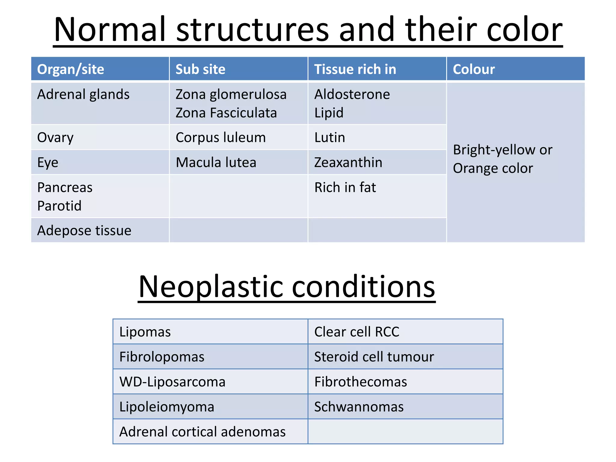
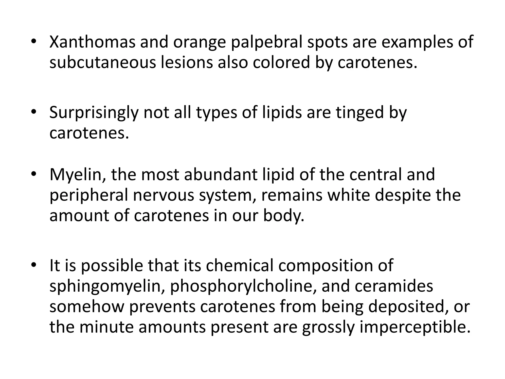
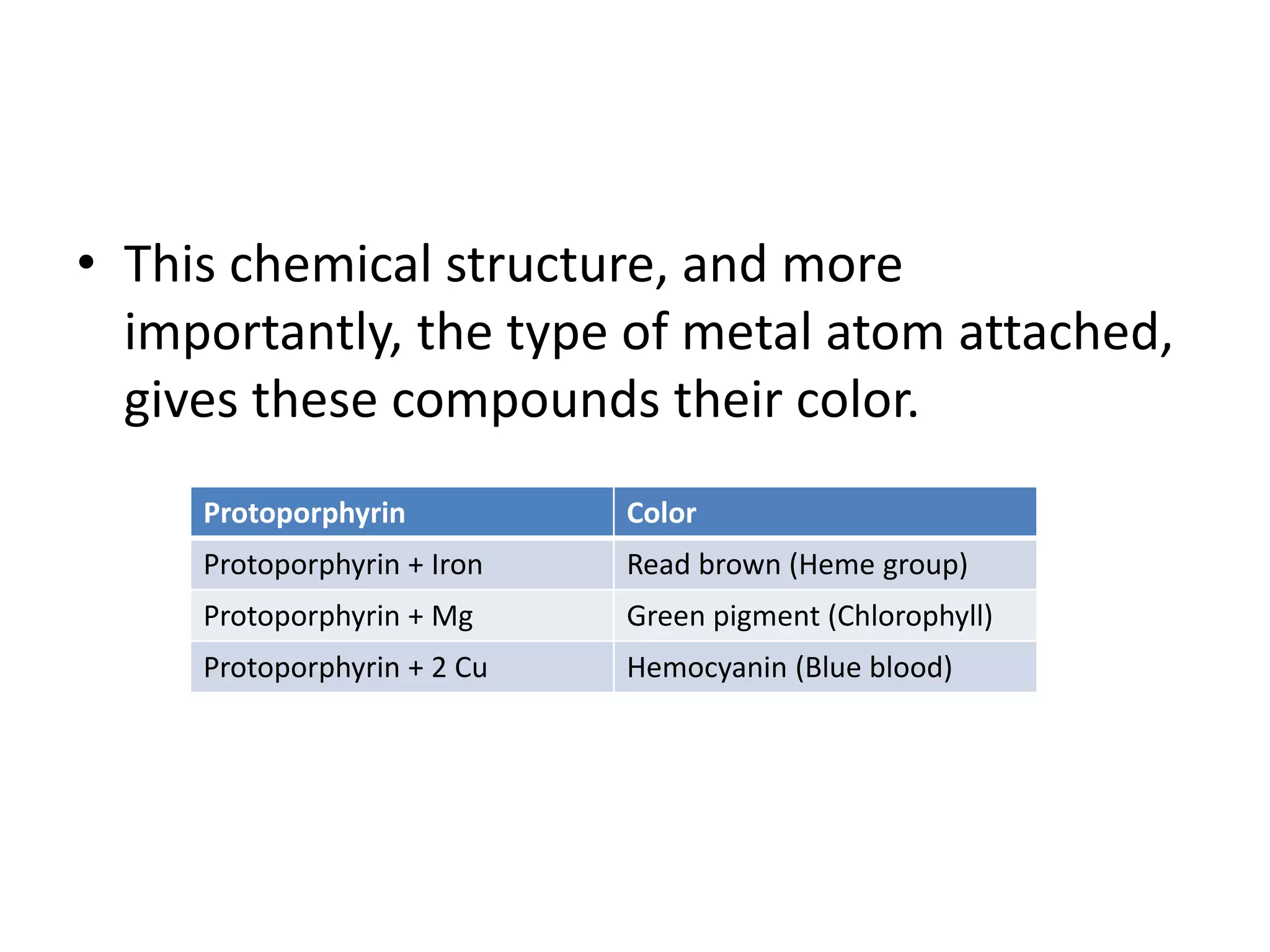
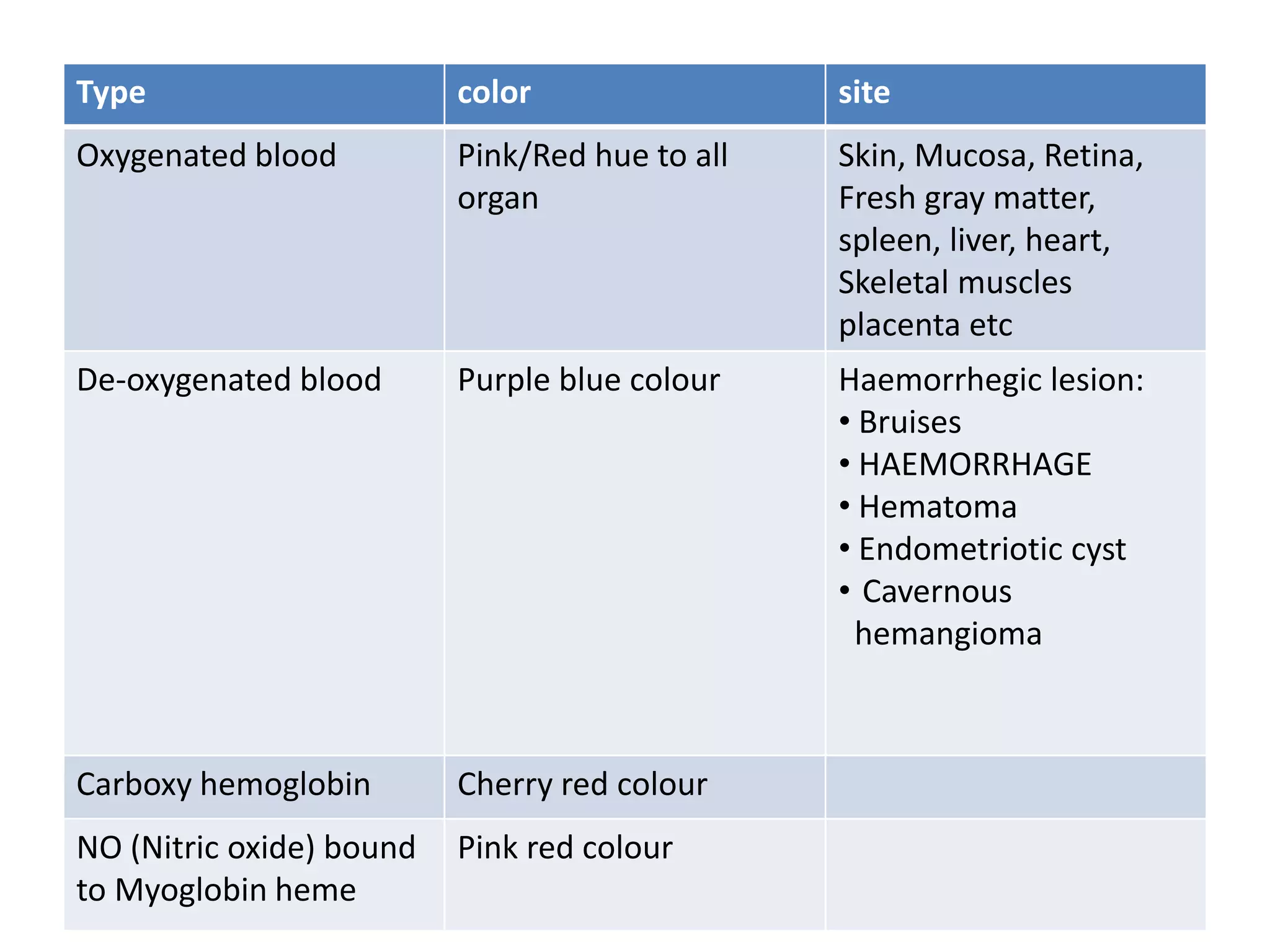
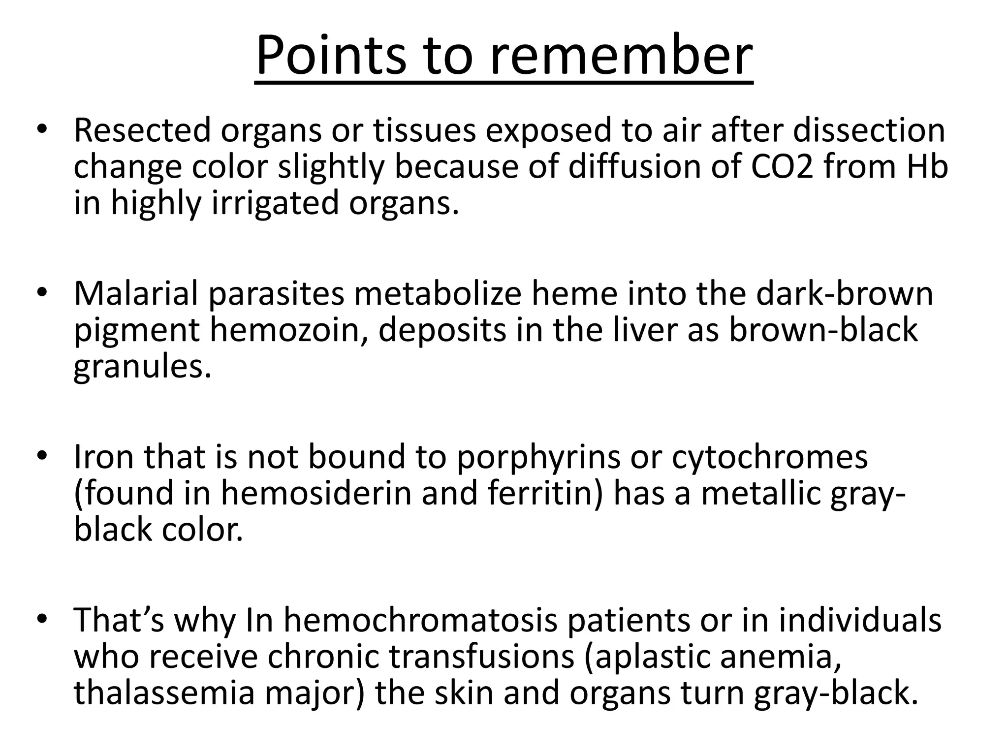
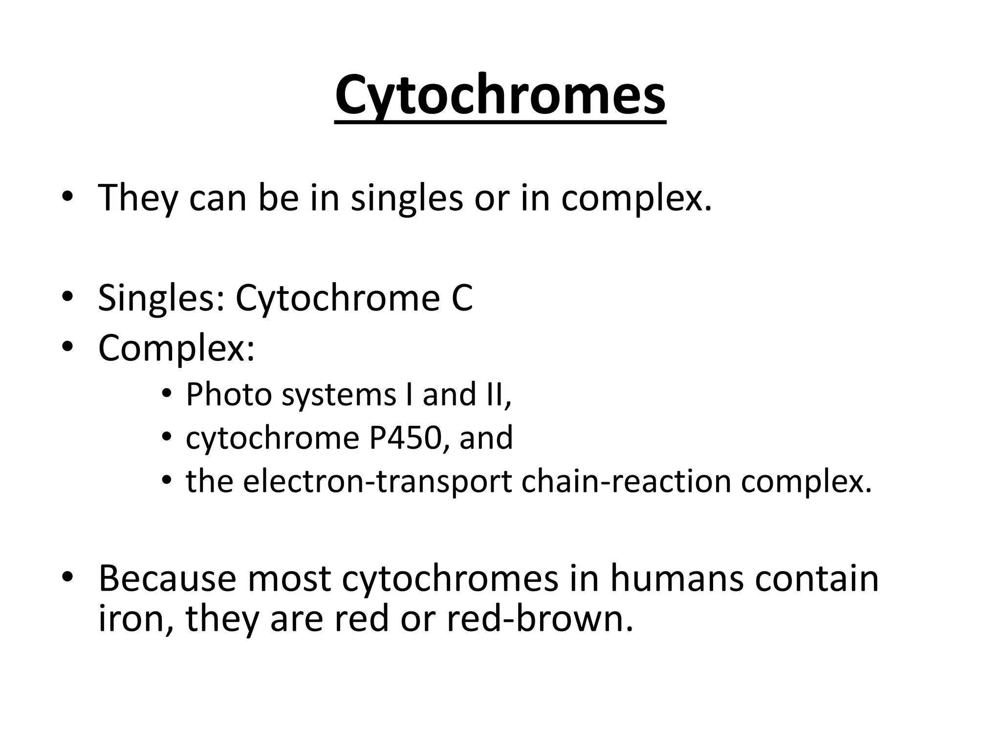
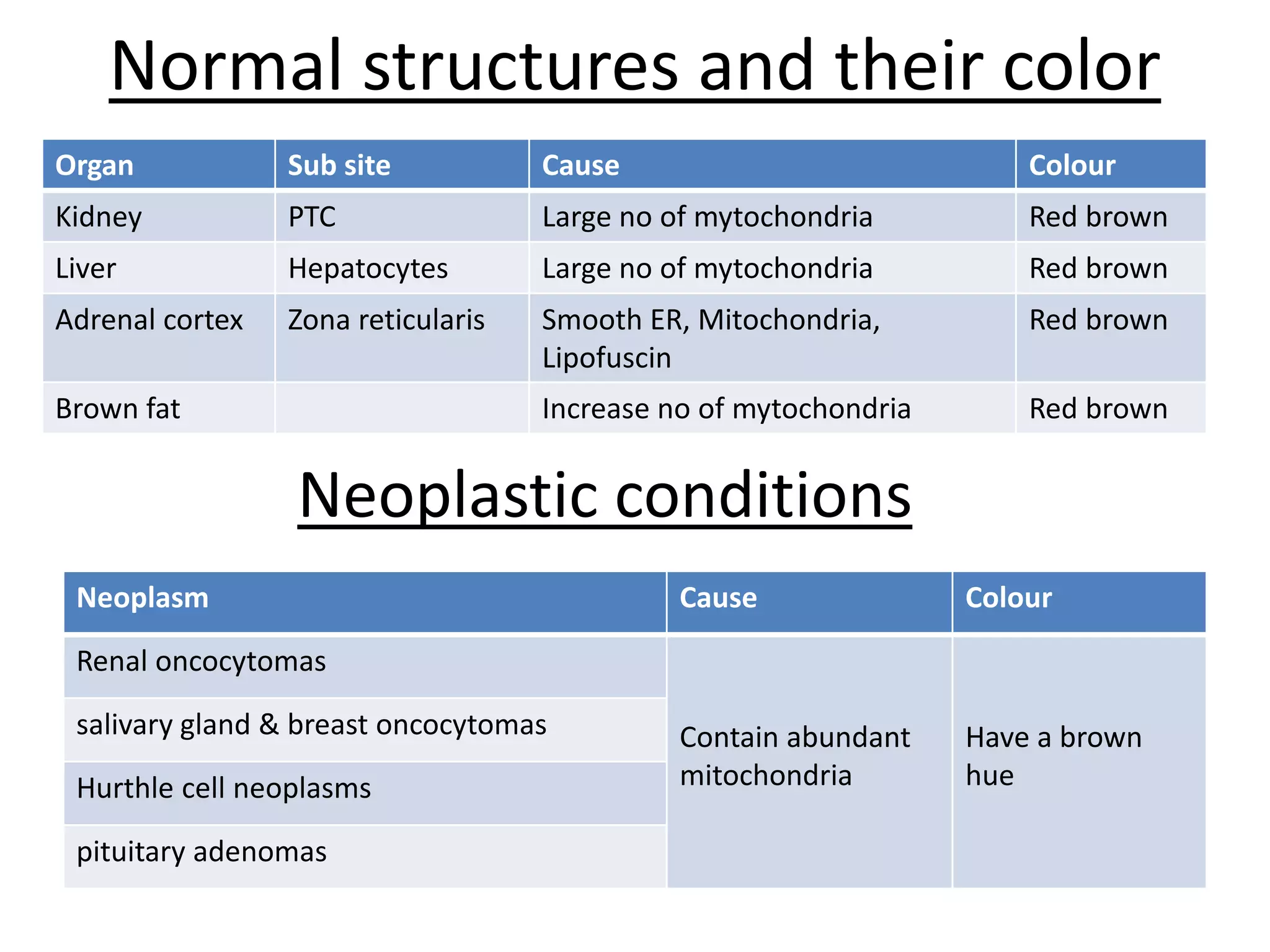
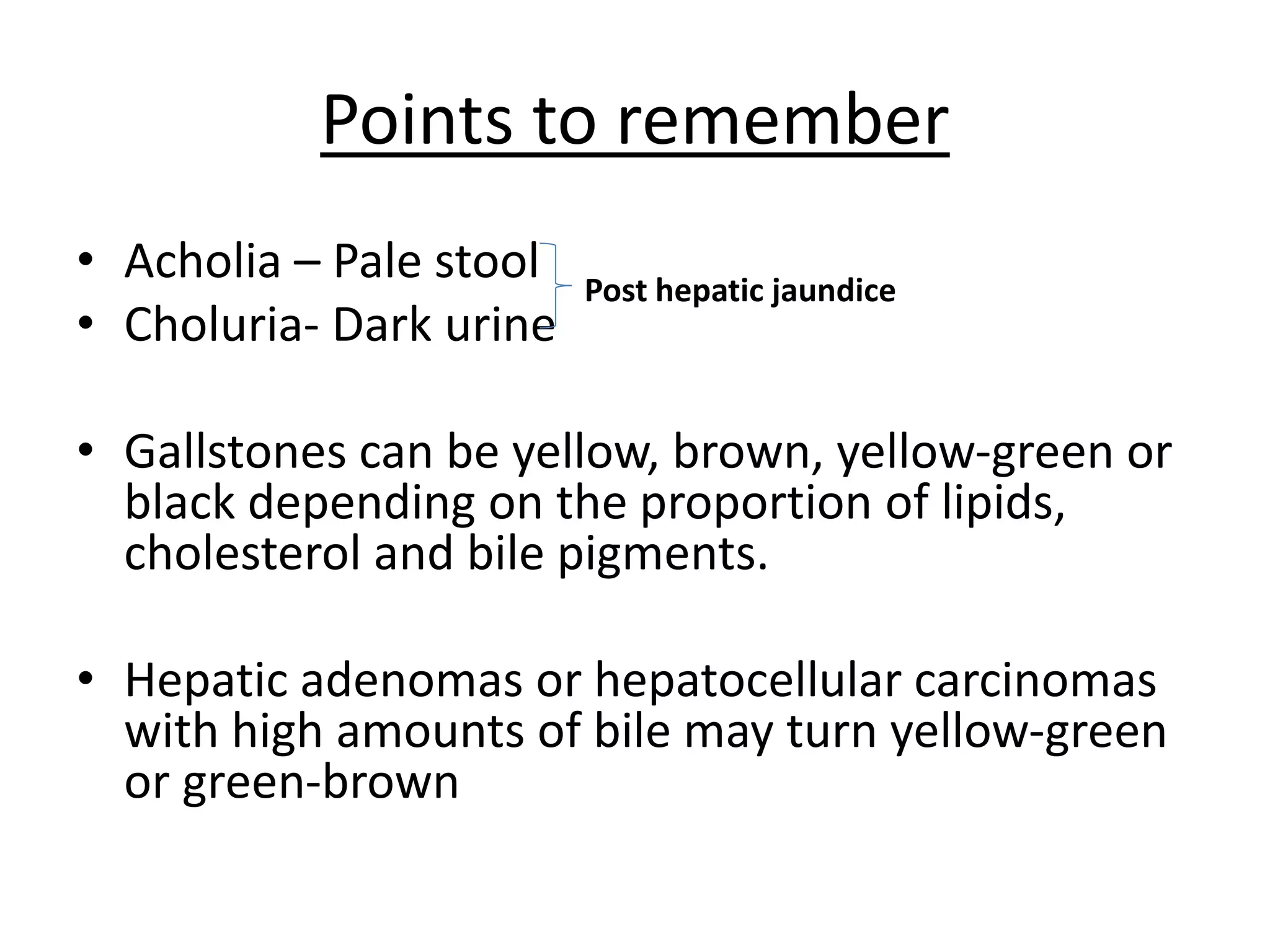
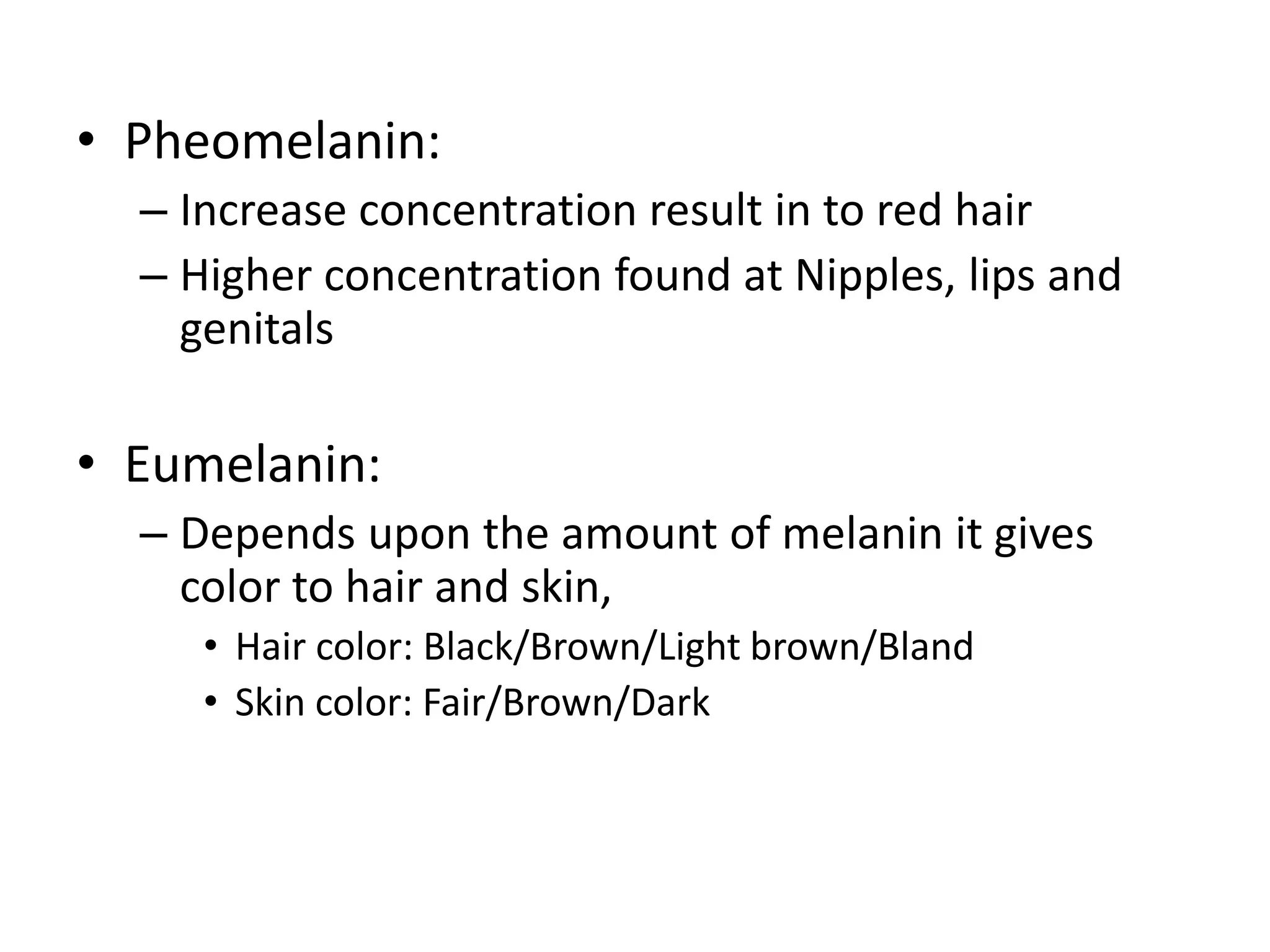
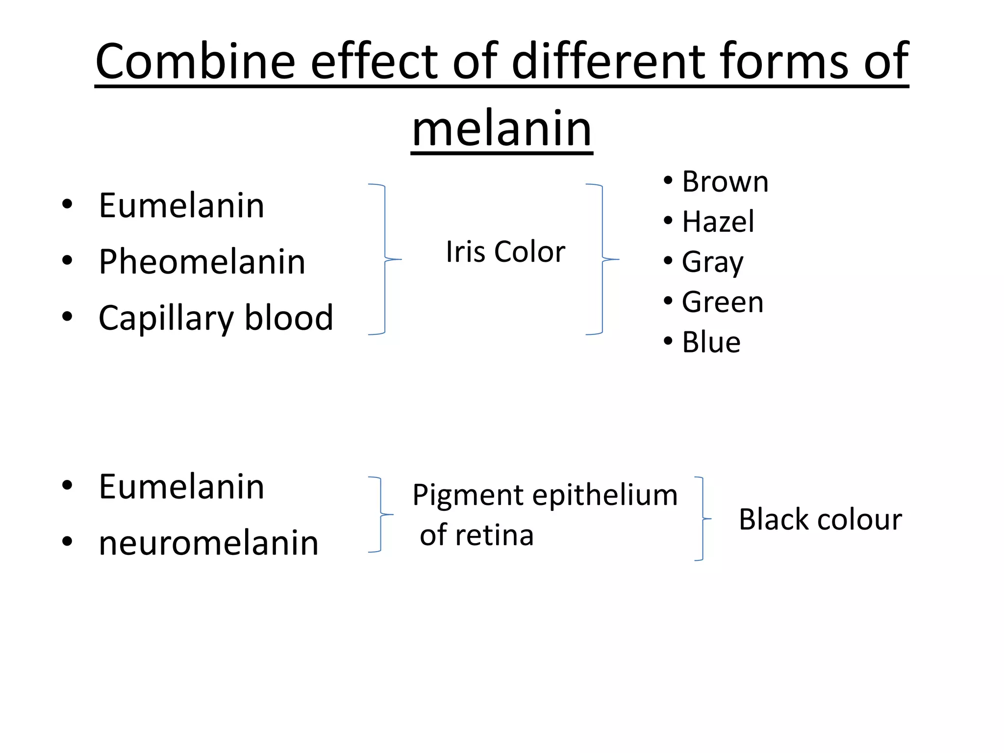
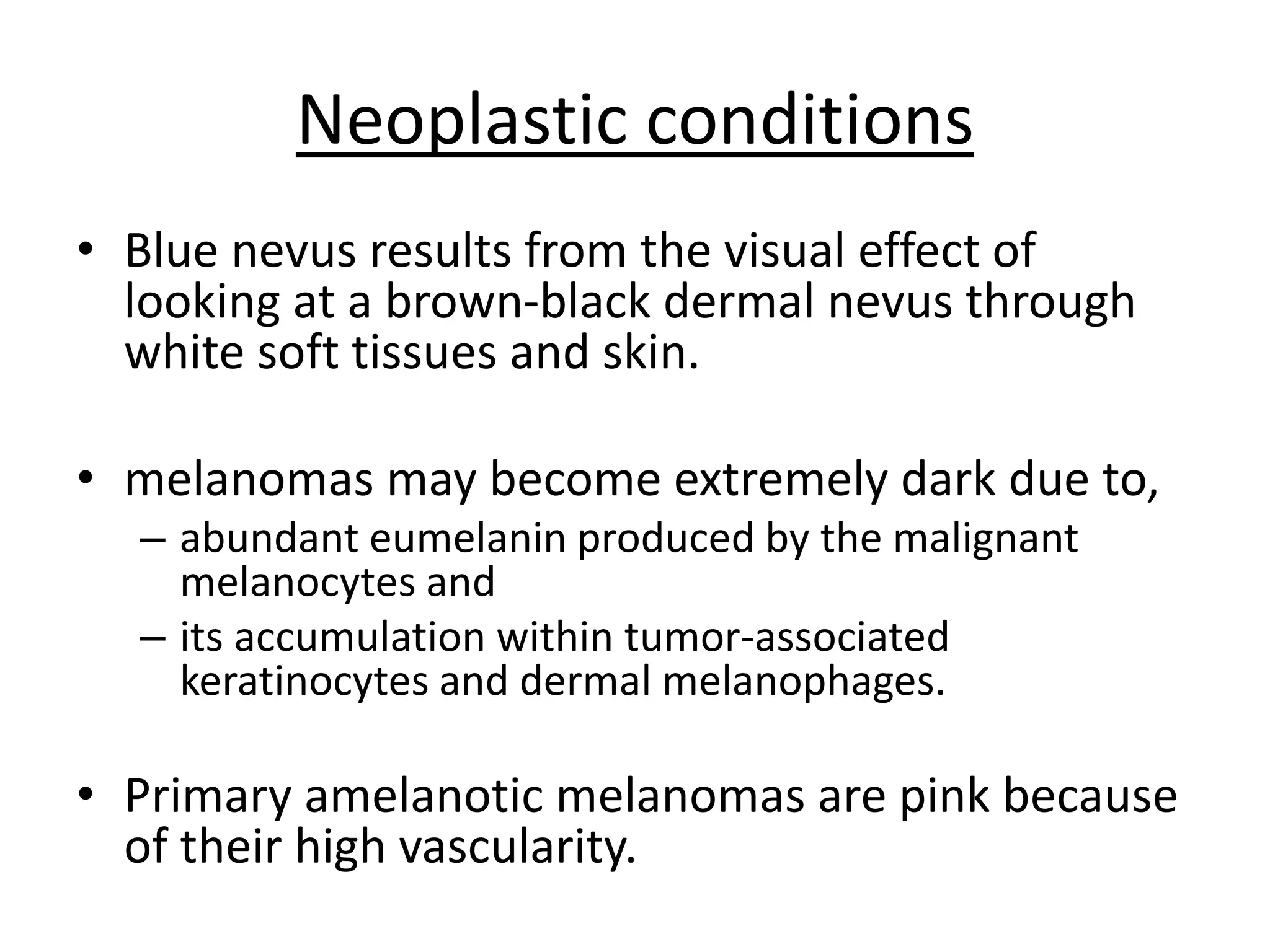
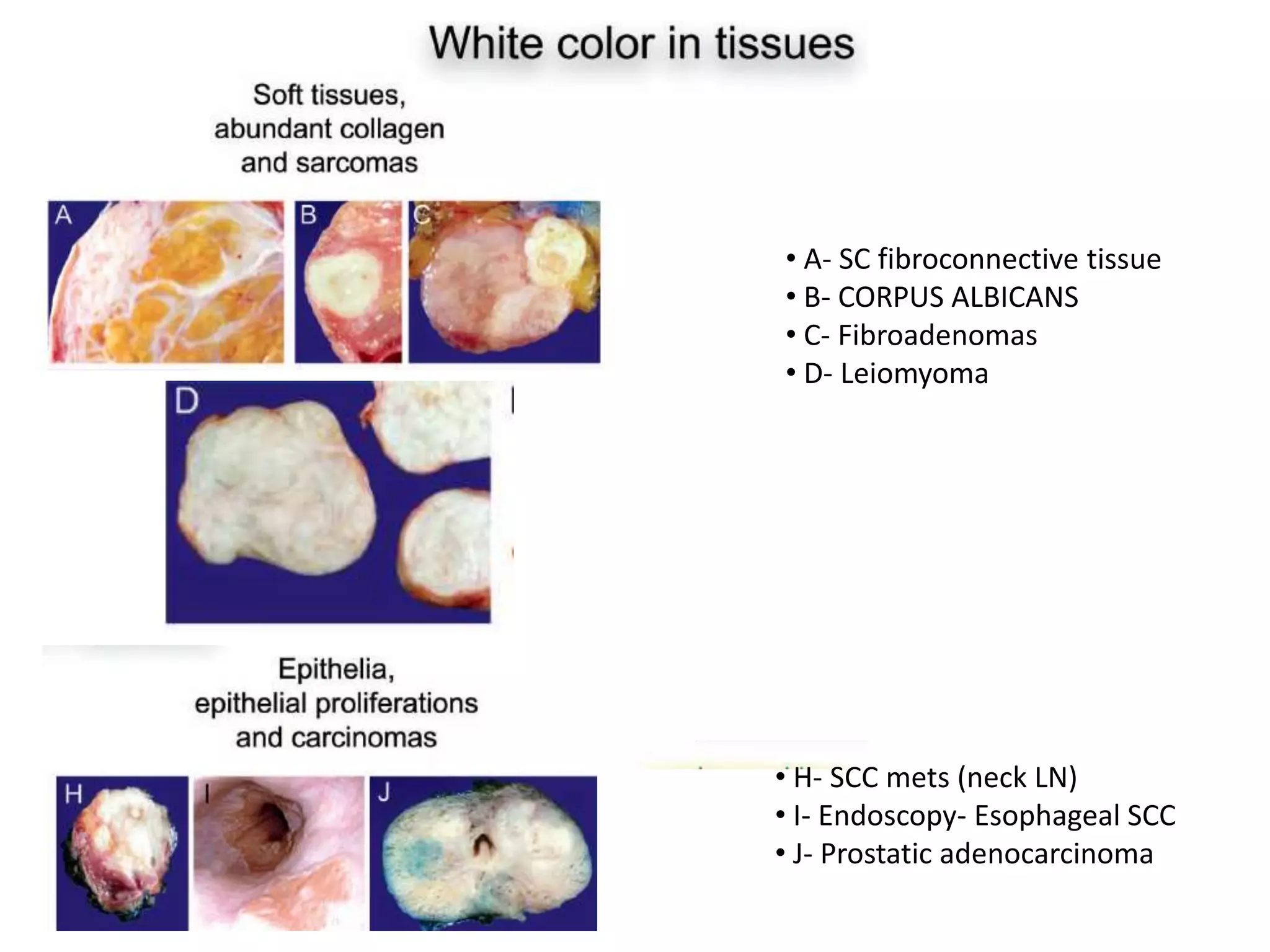
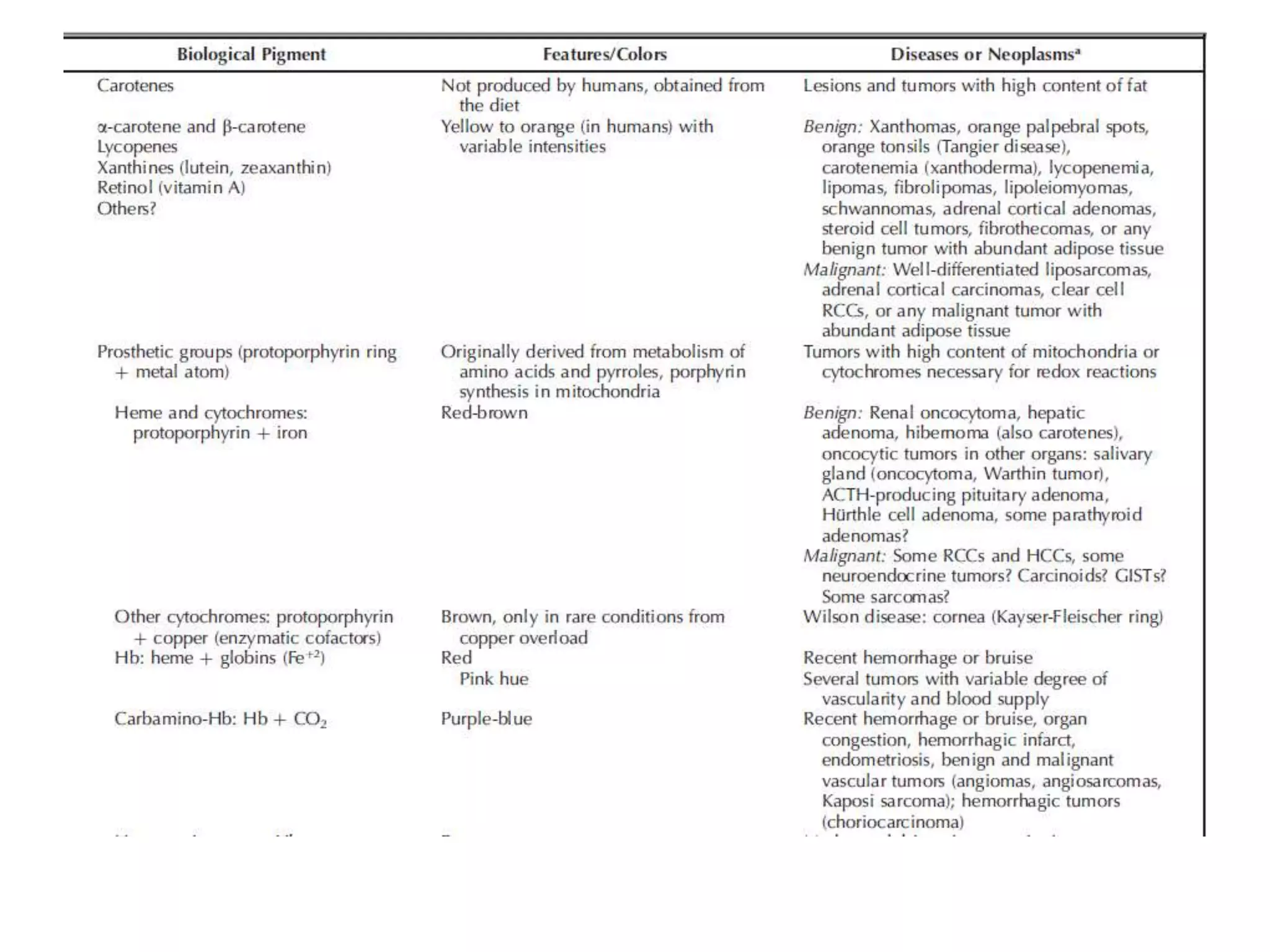
“HUMAN COLORS”
Normal and Pathologic Tissues and Their
Color
DR. Manan Shah
DR. Swaroop Raj
CONTENTS
• Introduction
• Producers of color in healthy and neoplastic
tissue
• Producers of white color in healthy and
neoplastic tissue
• Producers of fluorescence in tissue
• Summary
INTRODUCTION
• Colors are important to all living organisms
• They are crucial for protection, metabolism,
sexual behavior, and communication.
• Human organs obviously have color, that is,
the liver is brown, the heart is red, bones are
white, and so on
INTRODUCTION
• Although this is obvious and established, the
reason why organs have a particular color is
not completely understood.
PRODUCERS OF COLOR IN HEALTHY
AND NEOPLASTIC TISSUE
• Carotenes and carotenoids
• Cytochromes, the Heme Group, Iron, and Bile Pigments
• Lipochromes (Lipofuscin)
• Melanin
• Other color Producers
– Color changes in CNS
– Iodine
– Copper
– Colour of ligaments
– Smooth muscle
Carotenes and carotenoids
• Carotenes are unsaturated hydrocarbons chemically
derived from isopentenyl pyrophosphate and terpenes
and includes carotenes, lycopenes and xanthines.
• Carotenes are fat soluble molecules that can produce
all the colors of the visible spectrum and are
synthesized only by plants.
• Animals including humans obtain them from their diet
and because carotenes are lipophilic, they associate
with lipid-rich tissues.
• Humans metabolize yellow and orange carotenes but
not blue or red ones for unknown reasons.
• The α-carotene and β-carotene, lycopenes, and
xanthines are the most common carotenes in human
tissues.
• They are absorbed and deposited in lipid-rich tissues
even before birth.
• Hypothetically, the adipose tissue of a human never
exposed to carotenes should be white and not bright
yellow, but this scenario does not exist because we
ingest carotenes every day from our diet.
Normal structures and their color
Organ/site Sub site Tissue rich in Colour
Adrenal glands Zona glomerulosa
Zona Fasciculata
Aldosterone
Lipid
Bright-yellow or
Orange color
Ovary Corpus luleum Lutin
Eye Macula lutea Zeaxanthin
Pancreas
Parotid
Rich in fat
Adepose tissue
Neoplastic conditions
Lipomas Clear cell RCC
Fibrolopomas Steroid cell tumour
WD-Liposarcoma Fibrothecomas
Lipoleiomyoma Schwannomas
Adrenal cortical adenomas
breast adipose tissue (D), clear cell renal cell carcinoma (E),
submucosal intestinal lipoma (F)
Adrenal gland cortex.
• Xanthomas and orange palpebral spots are examples of
subcutaneous lesions also colored by carotenes.
• Surprisingly not all types of lipids are tinged by
carotenes.
• Myelin, the most abundant lipid of the central and
peripheral nervous system, remains white despite the
amount of carotenes in our body.
• It is possible that its chemical composition of
sphingomyelin, phosphorylcholine, and ceramides
somehow prevents carotenes from being deposited, or
the minute amounts present are grossly imperceptible.
Cytochromes, Heme Group, Iron And
Bile Pigments
Addition of a metal atom to the central portion of protoporphyrin results
in the formation of an organic prosthetic group.
Forms the tetrapyrrole ring – protoporphyrin.
Assembly of 4 pyrrole rings
composed of a ring of 4 carbon atoms and one nitrogen atom (C4H5N)
Pyrroles are heterocyclic aromatic molecules
• This chemical structure, and more
importantly, the type of metal atom attached,
gives these compounds their color.
Protoporphyrin Color
Protoporphyrin + Iron Read brown (Heme group)
Protoporphyrin + Mg Green pigment (Chlorophyll)
Protoporphyrin + 2 Cu Hemocyanin (Blue blood)
Heme Group
• The degree of oxidation of the iron atom within
these molecules determines the covalency and
color of iron, that is, ferrous iron (Fe+2) is red,
whereas ferric iron (Fe+3) is brown.
• In fair skinned individuals, superficial veins
appear blue-green from the visual effect of
looking at purple-blue blood through a white-
pink vessel wall, yellow fat tissue, and skin.
Type color site
Oxygenated blood Pink/Red hue to all
organ
Skin, Mucosa, Retina,
Fresh gray matter,
spleen, liver, heart,
Skeletal muscles
placenta etc
De-oxygenated blood Purple blue colour Haemorrhegic lesion:
• Bruises
• HAEMORRHAGE
• Hematoma
• Endometriotic cyst
• Cavernous
hemangioma
Carboxy hemoglobin Cherry red colour
NO (Nitric oxide) bound
to Myoglobin heme
Pink red colour
Points to remember
• Resected organs or tissues exposed to air after dissection
change color slightly because of diffusion of CO2 from Hb
in highly irrigated organs.
• Malarial parasites metabolize heme into the dark-brown
pigment hemozoin, deposits in the liver as brown-black
granules.
• Iron that is not bound to porphyrins or cytochromes
(found in hemosiderin and ferritin) has a metallic gray-
black color.
• That’s why In hemochromatosis patients or in individuals
who receive chronic transfusions (aplastic anemia,
thalassemia major) the skin and organs turn gray-black.
Cytochromes
• They can be in singles or in complex.
• Singles: Cytochrome C
• Complex:
• Photo systems I and II,
• cytochrome P450, and
• the electron-transport chain-reaction complex.
• Because most cytochromes in humans contain
iron, they are red or red-brown.
Normal structures and their color
Organ Sub site Cause Colour
Kidney PTC Large no of mytochondria Red brown
Liver Hepatocytes Large no of mytochondria Red brown
Adrenal cortex Zona reticularis Smooth ER, Mitochondria,
Lipofuscin
Red brown
Brown fat Increase no of mytochondria Red brown
Neoplastic conditions
Neoplasm Cause Colour
Renal oncocytomas
Contain abundant
mitochondria
Have a brown
hue
salivary gland & breast oncocytomas
Hurthle cell neoplasms
pituitary adenomas
Bilirubin and Iron
secreted into the small bowel
Direct bilirubin is then secreted to intra hepatic bile ducts, stored in the
gallbladder
converted to direct bilirubin in the liver
indirect bilirubin
biliverdin
Hb
• Biliverdin is green and bilirubin is yellow-red.
• Bilirubin gives a yellow-red color to the liver and tinges
the gallbladder and biliary duct epithelia yellow-red
• Once bilirubin is secreted into the small bowel, it is
metabolized by bacteria into urobilinogen and
stercobilinogen.
• These molecules are later catabolized to urobilin and
stercobilin, which stain stools yellow-brown.
• Urobilinogen is reabsorbed into the bloodstream,
filtered by the kidneys, and its catabolites are then
excreted in urine giving it the characteristic yellow
color.
Points to remember
• Acholia – Pale stool
• Choluria- Dark urine
• Gallstones can be yellow, brown, yellow-green or
black depending on the proportion of lipids,
cholesterol and bile pigments.
• Hepatic adenomas or hepatocellular carcinomas
with high amounts of bile may turn yellow-green
or green-brown
Post hepatic jaundice
Lipochromes (Lipofuscin)
• Lipid Lipofuscin pigment
• Lipofuscin pigment accumulates with age in
organs. eg. Heart, Liver, Retina, Brain.
• It is also known as Wear & tear pigments.
• The amount of lipofuscin found in these organs is
not sufficient to cause dramatic changes in
color—even in older individuals—but may give
them a light-brown.
Oxidation
• For example, Leyding cells from the testis contain
more lipofuscin as they senesce, which may be
responsible for the darker color of the testicular
parenchyma seen sometimes in older men.
• Lipofuscin is also partially responsible for the brown
color of the zona reticularis in the adrenal gland.
• The best condition to appreciate the dark-brown
color of lipofuscin is melanosis coli (‘‘black colon’’),
in which high amounts of lipofuscin— not melanin—
are deposited in the colorectal mucosa after
excessive use of laxatives.
• Another condition is “Black thyroid”, which
occurs in certain individuals after use of
minocycline. Black color is not only because of
the deposition of the drug but also because
lipofuscin accumulates within follicular cells.
• Increased deposition of lipofuscin in the
retinal pigment epithelium is associated with
the pathogenesis of age-related macular
degeneration
Melanin
• Melanin is an intracellular pigment that results from
the metabolism of tyrosine to dopaquinone.
• it is produced by cells derived from neural crest and
neuroectoderm, that is, melanocytes, eye pigment
epithelium, and neurons.
• 3 forms of melanin
– Neuromelanin( Brown-black)
– Eumelanin (Brown-black)
– Pheomelanin (Golden yellow- red)
• Pheomelanin:
– Increase concentration result in to red hair
– Higher concentration found at Nipples, lips and
genitals
• Eumelanin:
– Depends upon the amount of melanin it gives
color to hair and skin,
• Hair color: Black/Brown/Light brown/Bland
• Skin color: Fair/Brown/Dark
Combine effect of different forms of
melanin
• Eumelanin
• Pheomelanin
• Capillary blood
• Eumelanin
• neuromelanin
Iris Color
• Brown
• Hazel
• Gray
• Green
• Blue
Pigment epithelium
of retina Black colour
Neoplastic conditions
• Blue nevus results from the visual effect of
looking at a brown-black dermal nevus through
white soft tissues and skin.
• melanomas may become extremely dark due to,
– abundant eumelanin produced by the malignant
melanocytes and
– its accumulation within tumor-associated
keratinocytes and dermal melanophages.
• Primary amelanotic melanomas are pink because
of their high vascularity.
— Ectopic brain tissue or
Gangliocytomas
ganglioneuromas,
gangliogliomas,
Pheochromocytomas and
mature brain tissue in teratomas
• Central Nervous System
– Increase vascularity of gray matter structures confers
a pink-beige color that turns gray after fixation
have a
color
reminisc
-ent of
gray
matter
Other color Producers
Therefore, it
can be
hypothesize
that neurons
have an
intrinsic beige
or gray color.
• Central Nervous System
– Carcinoids (NET), are usually yellow, pink, or,
sometimes, red because of hemorrhage. why
carcinoids are this color is unknown.
– Do neurosecretory granules, chromogranins, or
synaptophysin have yellow or pink color?
• Iodine
– It can be violet/yellow/red or brown depending on
solvent used.
– when iodine is dissolved in water, it is yellow-brown.
– Increased vascularity, in combination with the amount
of yellow brown iodine, gives the thyroid its color.
• Copper
– Abnormal golden brown color deposition (eg. KF ring)
– Wilsons disease.
• Ligaments
– Light grey to yellow color
– This color is due to the intrinsic yellow color of elastin
– Elastofibroma is a benign tumor composed of elastic fibers
and fibrous tissue that has a typical yellow or sometimes
gray color.
• Smooth muscle:
– Less myoglobin  pale pink in color
– Leiomyosarcoma  retain pink colour
– Leiomyoma  Become white due to high content of
collagen
PRODUCERS OF WHITE COLOR IN
HEALTHY AND NEOPLASTIC TISSUE
• Calcium Phosphate
• Other white molecules and iridescent organs
– Soft tissue, collagen, iridescence
– Epithelial cells
– WBC
– Others
Calcium phosphate
• Elemental calcium is metallic gray, but when combined
with other elements, it turns into white calcium
carbonate (CaCO3) or white calcium phosphate
(CaPO4).
• Bones and enamel are white because they are mostly
composed of calcium phosphate
• Like normal bone and enamel, bone matrix- or enamel
matrix-producing tumors, that is, osteoid osteomas,
osteoblastomas, osteoid-producing osteosarcomas,
odontomas, odontogenic tumors, and ameloblastomas
are white.
Other Molecules and Iridescent Organs
• Several organs, tissues, and cells in the human body are white for
unknown reasons.
• They include soft tissues (collagen), epithelia, myelin and leukocytes.
• Probable reasons are,
1) they cannot absorb carotenes or only absorb minute amounts
that macroscopically do not affect color;
2) they do not have abundant mitochondria or cytochromes;
3) they have very high contents of deoxyribonucleic acid, which is
intrinsically white (lymphomas, leukemias and small round
cell tumors are made of cells with high N:C ratio);
4) they are avascular or require minimal blood supply; and
5) they do not contain melanin, lipofuscin, or any other pigments
(intrinsically white).
Epithelia
• All epithelia are avascular and intrinsically white, without
melanin pigmentation.
• Example,
– Albinism:
• White skin and hair
• Red iris (iris capillaries)
– Psoriasis
– Keratosis
– Leukoplakias
– SCC
• Thymomas are epithelial tumors that can be rich in fat,
epithelial cells, and lymphocytes and have a yellow or pale-
pink color
Not only thickness, but
abnormalities in keratin or
the keratohyaline granules
also have a role.
WBC
• Leukocytes and tumors arising from these cells, such as
leukemia and lymphomas are white.
• Myeloparoxidase is yellow green in color and their high
content in leukemias at extramedullary sites gives
these tumour a yellow-green hue, hence the name
chloromas.
• Reactive lymphocytes  high lymphocytes content 
Fish-flesh appearance.
• Granulomas also white to pale pink if not associated
with other pigments (hemosiderin, anthracotic
pigment).
Collagen
• Type I collagen is the most abundant protein in animal soft
tissues.
• The particular arrangement of collagen fibers in tendons
and fasciae also causes these anatomic structures to be
iridescent
• Benign and malignant proliferations of fibroblasts, smooth
muscle, or stromal cells that produce type I collagen are
white, such as old scars, leiomyomas, and several sarcomas.
• Type II collagen is white and is the main constituent of
cartilage. Because of the high contents of water and
proteoglycans, cartilage may look translucent or bluish.
• H- SCC mets (neck LN)
• I- Endoscopy- Esophageal SCC
• J- Prostatic adenocarcinoma
• A- SC fibroconnective tissue
• B- CORPUS ALBICANS
• C- Fibroadenomas
• D- Leiomyoma
FLUORESCENCE IN TISSUE
• Fluorophores or fluorochromes are molecules that emit
light in a longer wavelength than the one that excited
them.
• Lipofuscin, elastin, and collagen have intrinsic fluorescence
that is not strong enough to be observed at a macroscopic
level.
• In rare congenital erythropoietic porphyria
(uroporphyrinogen -III synthetase deficiency)
– Stools and urine- dark red (porphyrinuria)
– bones and teeth acquire a yellow-orange color.
– The accumulated type I porphyrins in secretions and tissues
emit an impressive pink-red fluorescence under ultraviolet light.
An area of white-silver iridescence
(arrow) is observed in the overlying fascia of muscle
SUMMARY
• Colors in living organisms are result of complex
biochemical reactions with the production of biologic
pigments (cytochromes, porphyrins, melanins,
lipochromes) or because of structural coloration
(iridescence).
• Apart from color pigments may have antioxidant and
cyto-protective effects.
• Although abundant information is available on the
ultraviolet-protective effects of melanin and the
function of cytochromes and heme groups, the role of
other biologic pigments in human organs is still
uncertain.
THANK YOU
Human colors. color of normal and pathologic tissue

Human colors. color of normal and pathologic tissue

  • 1.
    “HUMAN COLORS” Normal andPathologic Tissues and Their Color DR. Manan Shah DR. Swaroop Raj
  • 2.
    CONTENTS • Introduction • Producersof color in healthy and neoplastic tissue • Producers of white color in healthy and neoplastic tissue • Producers of fluorescence in tissue • Summary
  • 3.
    INTRODUCTION • Colors areimportant to all living organisms • They are crucial for protection, metabolism, sexual behavior, and communication. • Human organs obviously have color, that is, the liver is brown, the heart is red, bones are white, and so on
  • 4.
    INTRODUCTION • Although thisis obvious and established, the reason why organs have a particular color is not completely understood.
  • 5.
    PRODUCERS OF COLORIN HEALTHY AND NEOPLASTIC TISSUE • Carotenes and carotenoids • Cytochromes, the Heme Group, Iron, and Bile Pigments • Lipochromes (Lipofuscin) • Melanin • Other color Producers – Color changes in CNS – Iodine – Copper – Colour of ligaments – Smooth muscle
  • 6.
    Carotenes and carotenoids •Carotenes are unsaturated hydrocarbons chemically derived from isopentenyl pyrophosphate and terpenes and includes carotenes, lycopenes and xanthines. • Carotenes are fat soluble molecules that can produce all the colors of the visible spectrum and are synthesized only by plants. • Animals including humans obtain them from their diet and because carotenes are lipophilic, they associate with lipid-rich tissues.
  • 7.
    • Humans metabolizeyellow and orange carotenes but not blue or red ones for unknown reasons. • The α-carotene and β-carotene, lycopenes, and xanthines are the most common carotenes in human tissues. • They are absorbed and deposited in lipid-rich tissues even before birth. • Hypothetically, the adipose tissue of a human never exposed to carotenes should be white and not bright yellow, but this scenario does not exist because we ingest carotenes every day from our diet.
  • 8.
    Normal structures andtheir color Organ/site Sub site Tissue rich in Colour Adrenal glands Zona glomerulosa Zona Fasciculata Aldosterone Lipid Bright-yellow or Orange color Ovary Corpus luleum Lutin Eye Macula lutea Zeaxanthin Pancreas Parotid Rich in fat Adepose tissue Neoplastic conditions Lipomas Clear cell RCC Fibrolopomas Steroid cell tumour WD-Liposarcoma Fibrothecomas Lipoleiomyoma Schwannomas Adrenal cortical adenomas
  • 9.
    breast adipose tissue(D), clear cell renal cell carcinoma (E), submucosal intestinal lipoma (F)
  • 10.
  • 11.
    • Xanthomas andorange palpebral spots are examples of subcutaneous lesions also colored by carotenes. • Surprisingly not all types of lipids are tinged by carotenes. • Myelin, the most abundant lipid of the central and peripheral nervous system, remains white despite the amount of carotenes in our body. • It is possible that its chemical composition of sphingomyelin, phosphorylcholine, and ceramides somehow prevents carotenes from being deposited, or the minute amounts present are grossly imperceptible.
  • 12.
    Cytochromes, Heme Group,Iron And Bile Pigments Addition of a metal atom to the central portion of protoporphyrin results in the formation of an organic prosthetic group. Forms the tetrapyrrole ring – protoporphyrin. Assembly of 4 pyrrole rings composed of a ring of 4 carbon atoms and one nitrogen atom (C4H5N) Pyrroles are heterocyclic aromatic molecules
  • 13.
    • This chemicalstructure, and more importantly, the type of metal atom attached, gives these compounds their color. Protoporphyrin Color Protoporphyrin + Iron Read brown (Heme group) Protoporphyrin + Mg Green pigment (Chlorophyll) Protoporphyrin + 2 Cu Hemocyanin (Blue blood)
  • 14.
    Heme Group • Thedegree of oxidation of the iron atom within these molecules determines the covalency and color of iron, that is, ferrous iron (Fe+2) is red, whereas ferric iron (Fe+3) is brown. • In fair skinned individuals, superficial veins appear blue-green from the visual effect of looking at purple-blue blood through a white- pink vessel wall, yellow fat tissue, and skin.
  • 15.
    Type color site Oxygenatedblood Pink/Red hue to all organ Skin, Mucosa, Retina, Fresh gray matter, spleen, liver, heart, Skeletal muscles placenta etc De-oxygenated blood Purple blue colour Haemorrhegic lesion: • Bruises • HAEMORRHAGE • Hematoma • Endometriotic cyst • Cavernous hemangioma Carboxy hemoglobin Cherry red colour NO (Nitric oxide) bound to Myoglobin heme Pink red colour
  • 16.
    Points to remember •Resected organs or tissues exposed to air after dissection change color slightly because of diffusion of CO2 from Hb in highly irrigated organs. • Malarial parasites metabolize heme into the dark-brown pigment hemozoin, deposits in the liver as brown-black granules. • Iron that is not bound to porphyrins or cytochromes (found in hemosiderin and ferritin) has a metallic gray- black color. • That’s why In hemochromatosis patients or in individuals who receive chronic transfusions (aplastic anemia, thalassemia major) the skin and organs turn gray-black.
  • 19.
    Cytochromes • They canbe in singles or in complex. • Singles: Cytochrome C • Complex: • Photo systems I and II, • cytochrome P450, and • the electron-transport chain-reaction complex. • Because most cytochromes in humans contain iron, they are red or red-brown.
  • 20.
    Normal structures andtheir color Organ Sub site Cause Colour Kidney PTC Large no of mytochondria Red brown Liver Hepatocytes Large no of mytochondria Red brown Adrenal cortex Zona reticularis Smooth ER, Mitochondria, Lipofuscin Red brown Brown fat Increase no of mytochondria Red brown Neoplastic conditions Neoplasm Cause Colour Renal oncocytomas Contain abundant mitochondria Have a brown hue salivary gland & breast oncocytomas Hurthle cell neoplasms pituitary adenomas
  • 22.
    Bilirubin and Iron secretedinto the small bowel Direct bilirubin is then secreted to intra hepatic bile ducts, stored in the gallbladder converted to direct bilirubin in the liver indirect bilirubin biliverdin Hb
  • 23.
    • Biliverdin isgreen and bilirubin is yellow-red. • Bilirubin gives a yellow-red color to the liver and tinges the gallbladder and biliary duct epithelia yellow-red • Once bilirubin is secreted into the small bowel, it is metabolized by bacteria into urobilinogen and stercobilinogen. • These molecules are later catabolized to urobilin and stercobilin, which stain stools yellow-brown. • Urobilinogen is reabsorbed into the bloodstream, filtered by the kidneys, and its catabolites are then excreted in urine giving it the characteristic yellow color.
  • 24.
    Points to remember •Acholia – Pale stool • Choluria- Dark urine • Gallstones can be yellow, brown, yellow-green or black depending on the proportion of lipids, cholesterol and bile pigments. • Hepatic adenomas or hepatocellular carcinomas with high amounts of bile may turn yellow-green or green-brown Post hepatic jaundice
  • 25.
    Lipochromes (Lipofuscin) • LipidLipofuscin pigment • Lipofuscin pigment accumulates with age in organs. eg. Heart, Liver, Retina, Brain. • It is also known as Wear & tear pigments. • The amount of lipofuscin found in these organs is not sufficient to cause dramatic changes in color—even in older individuals—but may give them a light-brown. Oxidation
  • 26.
    • For example,Leyding cells from the testis contain more lipofuscin as they senesce, which may be responsible for the darker color of the testicular parenchyma seen sometimes in older men. • Lipofuscin is also partially responsible for the brown color of the zona reticularis in the adrenal gland. • The best condition to appreciate the dark-brown color of lipofuscin is melanosis coli (‘‘black colon’’), in which high amounts of lipofuscin— not melanin— are deposited in the colorectal mucosa after excessive use of laxatives.
  • 27.
    • Another conditionis “Black thyroid”, which occurs in certain individuals after use of minocycline. Black color is not only because of the deposition of the drug but also because lipofuscin accumulates within follicular cells. • Increased deposition of lipofuscin in the retinal pigment epithelium is associated with the pathogenesis of age-related macular degeneration
  • 29.
    Melanin • Melanin isan intracellular pigment that results from the metabolism of tyrosine to dopaquinone. • it is produced by cells derived from neural crest and neuroectoderm, that is, melanocytes, eye pigment epithelium, and neurons. • 3 forms of melanin – Neuromelanin( Brown-black) – Eumelanin (Brown-black) – Pheomelanin (Golden yellow- red)
  • 30.
    • Pheomelanin: – Increaseconcentration result in to red hair – Higher concentration found at Nipples, lips and genitals • Eumelanin: – Depends upon the amount of melanin it gives color to hair and skin, • Hair color: Black/Brown/Light brown/Bland • Skin color: Fair/Brown/Dark
  • 31.
    Combine effect ofdifferent forms of melanin • Eumelanin • Pheomelanin • Capillary blood • Eumelanin • neuromelanin Iris Color • Brown • Hazel • Gray • Green • Blue Pigment epithelium of retina Black colour
  • 32.
    Neoplastic conditions • Bluenevus results from the visual effect of looking at a brown-black dermal nevus through white soft tissues and skin. • melanomas may become extremely dark due to, – abundant eumelanin produced by the malignant melanocytes and – its accumulation within tumor-associated keratinocytes and dermal melanophages. • Primary amelanotic melanomas are pink because of their high vascularity.
  • 33.
    — Ectopic braintissue or Gangliocytomas ganglioneuromas, gangliogliomas, Pheochromocytomas and mature brain tissue in teratomas • Central Nervous System – Increase vascularity of gray matter structures confers a pink-beige color that turns gray after fixation have a color reminisc -ent of gray matter Other color Producers Therefore, it can be hypothesize that neurons have an intrinsic beige or gray color.
  • 35.
    • Central NervousSystem – Carcinoids (NET), are usually yellow, pink, or, sometimes, red because of hemorrhage. why carcinoids are this color is unknown. – Do neurosecretory granules, chromogranins, or synaptophysin have yellow or pink color? • Iodine – It can be violet/yellow/red or brown depending on solvent used. – when iodine is dissolved in water, it is yellow-brown. – Increased vascularity, in combination with the amount of yellow brown iodine, gives the thyroid its color.
  • 37.
    • Copper – Abnormalgolden brown color deposition (eg. KF ring) – Wilsons disease. • Ligaments – Light grey to yellow color – This color is due to the intrinsic yellow color of elastin – Elastofibroma is a benign tumor composed of elastic fibers and fibrous tissue that has a typical yellow or sometimes gray color. • Smooth muscle: – Less myoglobin  pale pink in color – Leiomyosarcoma  retain pink colour – Leiomyoma  Become white due to high content of collagen
  • 38.
    PRODUCERS OF WHITECOLOR IN HEALTHY AND NEOPLASTIC TISSUE • Calcium Phosphate • Other white molecules and iridescent organs – Soft tissue, collagen, iridescence – Epithelial cells – WBC – Others
  • 39.
    Calcium phosphate • Elementalcalcium is metallic gray, but when combined with other elements, it turns into white calcium carbonate (CaCO3) or white calcium phosphate (CaPO4). • Bones and enamel are white because they are mostly composed of calcium phosphate • Like normal bone and enamel, bone matrix- or enamel matrix-producing tumors, that is, osteoid osteomas, osteoblastomas, osteoid-producing osteosarcomas, odontomas, odontogenic tumors, and ameloblastomas are white.
  • 40.
    Other Molecules andIridescent Organs • Several organs, tissues, and cells in the human body are white for unknown reasons. • They include soft tissues (collagen), epithelia, myelin and leukocytes. • Probable reasons are, 1) they cannot absorb carotenes or only absorb minute amounts that macroscopically do not affect color; 2) they do not have abundant mitochondria or cytochromes; 3) they have very high contents of deoxyribonucleic acid, which is intrinsically white (lymphomas, leukemias and small round cell tumors are made of cells with high N:C ratio); 4) they are avascular or require minimal blood supply; and 5) they do not contain melanin, lipofuscin, or any other pigments (intrinsically white).
  • 41.
    Epithelia • All epitheliaare avascular and intrinsically white, without melanin pigmentation. • Example, – Albinism: • White skin and hair • Red iris (iris capillaries) – Psoriasis – Keratosis – Leukoplakias – SCC • Thymomas are epithelial tumors that can be rich in fat, epithelial cells, and lymphocytes and have a yellow or pale- pink color Not only thickness, but abnormalities in keratin or the keratohyaline granules also have a role.
  • 42.
    WBC • Leukocytes andtumors arising from these cells, such as leukemia and lymphomas are white. • Myeloparoxidase is yellow green in color and their high content in leukemias at extramedullary sites gives these tumour a yellow-green hue, hence the name chloromas. • Reactive lymphocytes  high lymphocytes content  Fish-flesh appearance. • Granulomas also white to pale pink if not associated with other pigments (hemosiderin, anthracotic pigment).
  • 44.
    Collagen • Type Icollagen is the most abundant protein in animal soft tissues. • The particular arrangement of collagen fibers in tendons and fasciae also causes these anatomic structures to be iridescent • Benign and malignant proliferations of fibroblasts, smooth muscle, or stromal cells that produce type I collagen are white, such as old scars, leiomyomas, and several sarcomas. • Type II collagen is white and is the main constituent of cartilage. Because of the high contents of water and proteoglycans, cartilage may look translucent or bluish.
  • 45.
    • H- SCCmets (neck LN) • I- Endoscopy- Esophageal SCC • J- Prostatic adenocarcinoma • A- SC fibroconnective tissue • B- CORPUS ALBICANS • C- Fibroadenomas • D- Leiomyoma
  • 46.
    FLUORESCENCE IN TISSUE •Fluorophores or fluorochromes are molecules that emit light in a longer wavelength than the one that excited them. • Lipofuscin, elastin, and collagen have intrinsic fluorescence that is not strong enough to be observed at a macroscopic level. • In rare congenital erythropoietic porphyria (uroporphyrinogen -III synthetase deficiency) – Stools and urine- dark red (porphyrinuria) – bones and teeth acquire a yellow-orange color. – The accumulated type I porphyrins in secretions and tissues emit an impressive pink-red fluorescence under ultraviolet light.
  • 47.
    An area ofwhite-silver iridescence (arrow) is observed in the overlying fascia of muscle
  • 48.
    SUMMARY • Colors inliving organisms are result of complex biochemical reactions with the production of biologic pigments (cytochromes, porphyrins, melanins, lipochromes) or because of structural coloration (iridescence). • Apart from color pigments may have antioxidant and cyto-protective effects. • Although abundant information is available on the ultraviolet-protective effects of melanin and the function of cytochromes and heme groups, the role of other biologic pigments in human organs is still uncertain.
  • 54.

Editor's Notes

  • #11 breast adipose tissue (D), clear cell renal cell carcinoma (E), submucosal intestinal lipoma (F), atypical lipomatous tumor/well-differentiated liposarcoma (G), schwannoma (H); adrenal cortical adenoma (I); and adrenal gland cortex (J).
  • #12 Function of carotiens- In humans, carotenes not only protect cells from the effects of ultraviolet light but also from the toxic effects of reactive oxygen species.