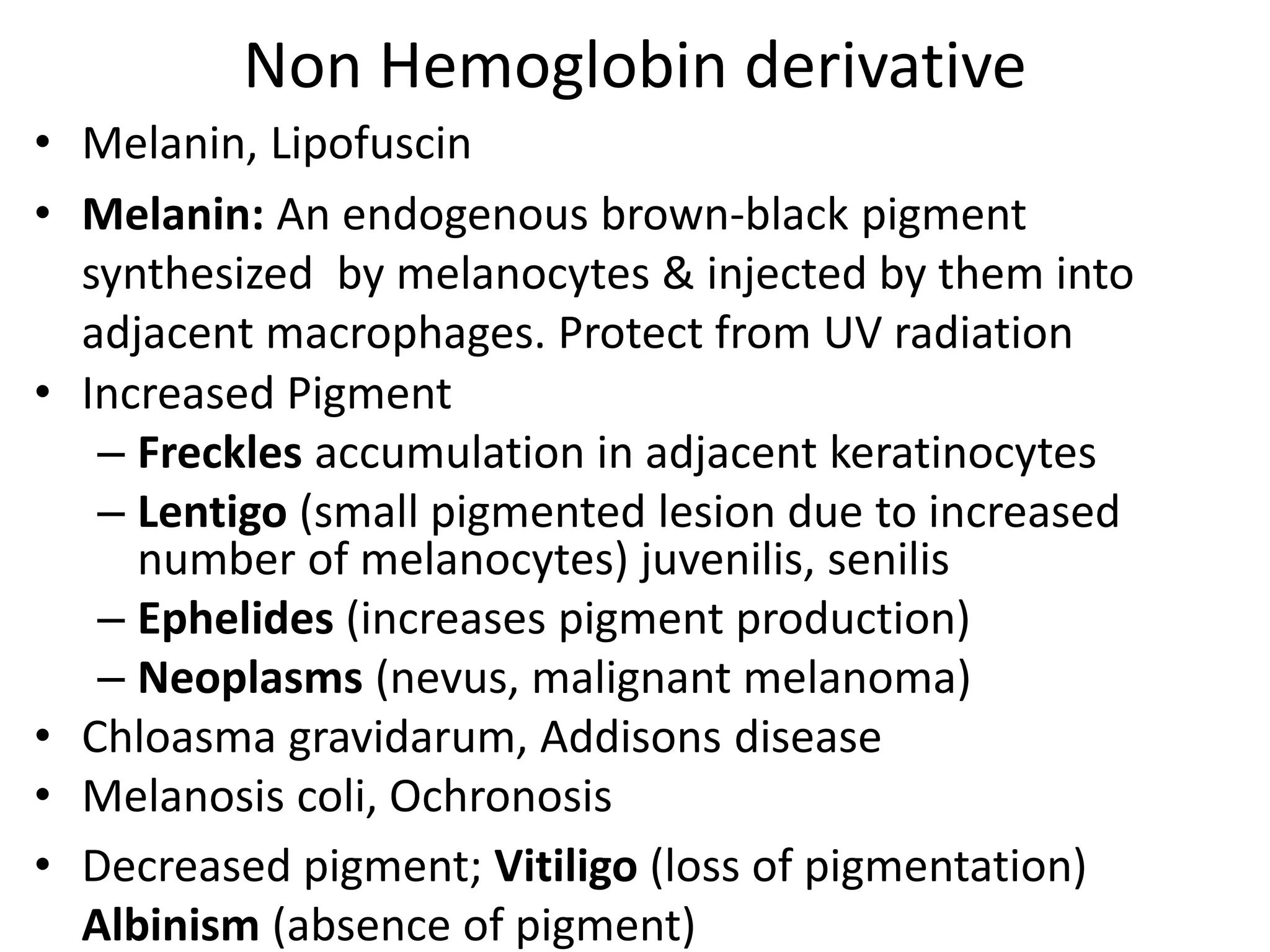
Intracellular Accumulations
L Os
• To identify abnormal accumulations of various
endogenous or exogenous substances in
tissues e.g. pigments
• To explain their causes
• To understand and describe the consequences
of accumulations
Intracellular Accumulations
• Substances within Cytoplasm, organelles (Lysosomes),
Nucleus. May accumulate Transiently or Permanently
• Harmless or toxic.
• Endogenous or exogenous. Produced by cell or elsewhere
• 4 Pathways of abnormal accumulations
1. Inadequate removal of Normal substances due to defects
in packaging and transport. Fatty liver
2. Accumulation of abnormal endogenous substances due
to genetic or acquired defects in folding, packaging
transport and secretion
3. Failure to degrade metabolite due to inherited enzyme
deficiencies. Storage diseases
4. Accumulation of abnormal exogenous substances due to
absence of enzymatic machinery to degrade or transport
e.g. carbon, silicon
Intracellular Accumulations
1. Excessive Normal cellular constituents:
Exogenous or Endogenous substances produced at
increased rate or normal rate but rate of metabolism
is inadequate to remove it. example: fatty change in
liver
water, Lipid, Proteins, carbohydrates
2. Abnormal substances
Exogenous: minerals, products of infectious agents
(viral inclusion bodies)
Endogenous: products of abnormal synthesis or
metabolism
Intracellular Accumulations
3. Pigments
Exogenous:
inhalation - Carbon
injection - Tattooing
ingestion - Beta carotene
Endogenous:
Hemoglobin derivative
Non Hemoglobin derivative
Fatty change (Steatosis)
• Abnormal accumulation of triglycerides within
parenchymal cells.
• May be mild and reversible or Severe producing
irreversible cell injury
• Common sites: liver, Heart, Skeletal muscle, Kidney
• Causes:
• General Hypoxia
• Fat diet – Obesity, Diabetes mellitus, Alcohol abuse
• Starvation, Protein Malnutrition
• Hepatotoxins, Chemicals. drugs, Carbon tetrachloride
• Chronic illnesses like TB
• Late pregnancy
• Reye’s syndrome
Fat Metabolism in liver
Fatty change in liver`
• Decreased processing within the liver: ischemia, toxins
e.g. CCl4
• Decreased removal from liver: hypoproteinemic states
• Significance: Reversible usually, no cell dysfunction
• Morphology
• Gross: Enlarged, yellow and greasy liver
• Microscopic:
– early change: fat accumulation as clear vacuoles in
cytoplasm (microvesicular) in centrilobular
hepatocytes, extending outward
– late stage: vacuoles coalesce to form a single large
vacuole (macrovesicular) or signet ring appearance;
rupture of vesicles (fatty cysts)
Microscopic features
Morphology
• Gross:
Accumulation of Cholesterol and
Cholesteryl esters
• As membrane bound Cholestrol vacuoles in
macrophages – Foam cells
• Fibrofatty plaques of atherosclerosis
• Xanthomas. Cholestrol rich yellow patch /nodule on
skin
• Inflammation / Necrosis
Protein accumulation
• Excessive presentation to cell or excessive formation
by cells
• Improper formation /folding cell injuries
• Proteinuria in nephrotic syndrome, as Pink hyaline
cytoplasmic droplets/vesicles in renal tubular cells.
• Amyloid
• Excess Immunoglobulins formation in RER of Plasma
cells as eosinophilic Russell bodies
• α1-antitrypsin deficiency
• Mallory-Denk body (keratin) – Alcoholic hyaline in
liver
• Neurofibrillary tangles in neurons - Alzheimer
disease - brain
Carbohydrate accumulation
• Glycogen deposits due to abnormal glucose or
glycogen metabolism in
– Diabetes mellitus (renal tubule, cardiac myocytes
and islets cell
– Glycogen storage disorders
Pigments
• Pigments are normal/abnormal colored substances that
accumulate within the cells either due to increased
production/decreased removal/lack of metabolic
enzymes
• Classification of pigments: Endogenous and Exogenous
• Endogenous
– Hemoglobin derivative
• Hemosiderin: Localised or Generalised:
• Acid heamatin
• Bilirubin and Porphyrins
– Non hemoglobin derivative: Melanin and Lipofuscin
• Exogenous: carbon
Hemoglobin derived pigments
• Hemolysis or red cell destruction in macrophages.
Hb released, Hemoglobinuria
• globin – to protein pool,
• Porphyrin ring yields bilirubin & iron gives
hemosiderin.
• Hemochromatosis (hemosiderin + heme free
pigment in liver cirhosis, heart failure, diabetes –
bronze skin
• Cyanosis increased reduced Hb,
Hemosiderin
• granular, golden-yellow to
brown pigment, accumulates
in tissues in case of local or
systemic iron excess.
• It represents large aggregates of ferritin micelles
• Causes: excessive dietary iron absorption, transfusion,
hemolytic anemias, local deposits at sites of hemorrhage
e.g. bruise
• Hemosiderosis: accumulation in organs & tissues.
• Macrophages of liver, bone marrow, spleen & lymph
nodes Siderophage. Later parenchymal cells involved
• Perl’s stain: special stain used for detection
• Prussian blue reaction: used to stain iron deposits in
tissues
Bruise
Hemosiderin brown
coarsely granular
material in macrophages
in alveolus.
Accumulated as a result
of RBC's breakdown.
Macrophages clear up
debris to be recycled.
Hemosiderin in
alveoli
A Prussian blue iron
staining of liver
demonstrate large
amounts of hemosiderin
in hepatocytes and
Kupffer cells
Hemosiderin in renal
tubules
demonstrated by
iron stain.
Bilirubin
• Accumulation of Bilirubin in the interstitial tissues gives
yellow green discoloration
• Accumulation in parenchymal cells leads to dysfunction
Increased amounts of circulating
Bilirubin in the blood lead to physical
examination finding of "icterus" or
jaundice as seen here as yellowish hue
of the skin..
The normally white sclerae of eyes
is an easiest place on physical
examination, is yellow
Bilirubin pigment yellow-green globular material seen
in small bile ductules in liver
Non Hemoglobin derivative
• Melanin, Lipofuscin
• Melanin: An endogenous brown-black pigment
synthesized by melanocytes & injected by them into
adjacent macrophages. Protect from UV radiation
• Increased Pigment
– Freckles accumulation in adjacent keratinocytes
– Lentigo (small pigmented lesion due to increased
number of melanocytes) juvenilis, senilis
– Ephelides (increases pigment production)
– Neoplasms (nevus, malignant melanoma)
• Chloasma gravidarum, Addisons disease
• Melanosis coli, Ochronosis
• Decreased pigment; Vitiligo (loss of pigmentation)
Albinism (absence of pigment)
Melanin
Non Hb derived
• Lipofuscin: ‘wear & tear pigment’. An insoluble,
brownish-yellow, granular intracellular pigment.
• Protein-lipid complexes derived from free radical
catalyzed peroxidation of polyunsaturated lipids of
subcellular membranes. Aging & atrophy in heart, liver,
brain etc. Brown Atrophy. Not injurious to cell. indicates
free radical induced injury. Appearance by E/M, seen as
perinuclear granules
Lipofuscin
Exogenous pigments
• Tattooing
• Coal workers Pneumoconiosis
• Anthracosis: Aggregates of of carbon or coal dust as black
pigment in lymph nodes and lung parenchyma due either
to atmospheric pollution or smoking or occupational
exposure. Inhaled, Injected, Ingested. Most common
• Morphology in lung :
• Gross: blackening of parenchyma
• Microscopic: macrophages full of black pigment
• Outcome:
– Mild to moderate depositions - without any effects
– Heavy depositions - occupational exposure lead to
serious lung disease
Carbon
Anthracotic pigment in
macrophages in a hilar
lymph node. most
pronounced in Smokers . It
looks bad, causes no major
organ dysfunction.
Anthracotic pigment seen asblack streaks between lobules
of lung beneath the pleural surface. Anthracosis not
harmful, occurs due to inhalation of carbonaceous
material in dirty air typical of industrialized regions
Tattooing
• Intentional
introduction of
pigment into skin for
cosmetic reasons.
• The pigment is taken
up by dermal
macrophages and
remains there
Pathologic Calcification
• Abnormal deposition of calcium salts together with small
amount of Iron, magnesium and other minerals in
soft tissues of body, other than osteoid or enamel.
Types: (i) Dystrophic (ii) Metastatic
Metastatic calcification: occurs in normal tissues secondary to
derangement in calcium metabolism (hypercalcemia)
• Causes:
• Hyperparathyroidism: Parathyroid hyperplasia, adenoma,
carcinoma
• Bone destruction due to increase turn over. Pagets disease,
immobilization, bone tumors and metastasis Multiple
myeloma, Leukemia
• Vitamin D related disorders intoxication, Sarcoidosis
• Milk alkali syndrome and Renal failure→ PO4 retention →
secondary hyperparathyroidism
Metastatic Calcification
• Sites: normal tissues, acid secreting areas, having
background alkalinity. Gastric mucosa, walls of major blood
vessels, kidney, lungs
• Morphology: Gross: white, gritty to feel, fine granules or
clumps
• Microscopic: intensely basophilic structureless deposits
• Stainnig: red with Alizarin,
blue with Von Kossa
• Extensive, shadows on X rays
• Outcome: generally no
cell/organ dysfunction.
May cause respiratory
deficit or Renal damage.
lung
Dystrophic Calcification
• occurs at dead or degenerate tissues
• Pathogenesis
• Two Steps: Initiation & Propagation – extra or intra-cellular
• Initiation: at extra-cellular occurs as matrix vesicles of
200nm size derived from cartilage, bone and degenerated
cells. Calcium affinity to phospholipids, phosphate
accumulate due to action of phosphatases. intra-cellular
calcification occurs in mitochondria of dead or dying cell.
• Propagation: progressively Calcium Phosphate crystals
formation, which depends on
– Calcium and Phosphate conc. in extracellular space
– Degree of collagenization
– Osteopontin (ca binding phosphoprotein)
– mineral inhibitors.
• outcome: cell/organ dysfunction
Dystrophic Calcification
• Morphology:
• Gross: fine white granules or
clumps, gritty to feel. s/t as stone
• Microscopic: intracellular or
extracellular intensely basophilic
structure less deposits. s/t as bone
in necrotic areas.
• Conditions
• fat and caseous necrosis,
dead parasites , infarcts
• CVS: Atheromas, thrombi,
damaged valves
• Tissue damage :
medium sized arteries, muscles,
tendons, hematomas
• Neoplasms, Scars
• Senile: costal cartilages, aorta
dystrophic calcification in wall of
stomach. At far left - artery with
calcification. Irregular bluish-purple
calcium deposits in submucosa.
Cellular aging
• It is the result of progressive decline in the life span and
functional capacity of cells and accumulation of cellular
damage.
• One of the strongest risk factor for many chronic diseases
• Mechanisms:
• DNA damage: accumulation of metabolic insults lead to
nuclear and mitochondrial DNA damage. Defects in DNA
repair.
• Decreased cellular replication – limited replication capacity
of cells. Fixed number of divisions- arrest – replicative
senescence - telomere shortening –cell cycle arrest. Role of
telomerase in maintaining length by adding nucleotides.
(activity present in germ, stem and cancer cells. Absent in
somatic cells)
Cellular aging
• Defective protein homeostasis. Due to decreased
synthesis and increased turnover. Accumulation of
misfolded proteins triggering apoptosis
• Factors effecting aging
• Environmental stresses like calorie restriction
• Reduced signaling by insulin like growth factor
receptors, reduced activation of kinases and altered
transcriptional activity. Counteract aging by
improving DNA repair, protein homeostasis and
enhanced immunity.
• Environmental stresses activate proteins of Sirtuin
family Sir2 - protein deacytalase – activate DNA
repair and stabilization
5. intracellular accumulation.pptx

5. intracellular accumulation.pptx

  • 1.
  • 3.
    Intracellular Accumulations L Os •To identify abnormal accumulations of various endogenous or exogenous substances in tissues e.g. pigments • To explain their causes • To understand and describe the consequences of accumulations
  • 4.
    Intracellular Accumulations • Substanceswithin Cytoplasm, organelles (Lysosomes), Nucleus. May accumulate Transiently or Permanently • Harmless or toxic. • Endogenous or exogenous. Produced by cell or elsewhere • 4 Pathways of abnormal accumulations 1. Inadequate removal of Normal substances due to defects in packaging and transport. Fatty liver 2. Accumulation of abnormal endogenous substances due to genetic or acquired defects in folding, packaging transport and secretion 3. Failure to degrade metabolite due to inherited enzyme deficiencies. Storage diseases 4. Accumulation of abnormal exogenous substances due to absence of enzymatic machinery to degrade or transport e.g. carbon, silicon
  • 5.
    Intracellular Accumulations 1. ExcessiveNormal cellular constituents: Exogenous or Endogenous substances produced at increased rate or normal rate but rate of metabolism is inadequate to remove it. example: fatty change in liver water, Lipid, Proteins, carbohydrates 2. Abnormal substances Exogenous: minerals, products of infectious agents (viral inclusion bodies) Endogenous: products of abnormal synthesis or metabolism
  • 6.
    Intracellular Accumulations 3. Pigments Exogenous: inhalation- Carbon injection - Tattooing ingestion - Beta carotene Endogenous: Hemoglobin derivative Non Hemoglobin derivative
  • 7.
    Fatty change (Steatosis) •Abnormal accumulation of triglycerides within parenchymal cells. • May be mild and reversible or Severe producing irreversible cell injury • Common sites: liver, Heart, Skeletal muscle, Kidney • Causes: • General Hypoxia • Fat diet – Obesity, Diabetes mellitus, Alcohol abuse • Starvation, Protein Malnutrition • Hepatotoxins, Chemicals. drugs, Carbon tetrachloride • Chronic illnesses like TB • Late pregnancy • Reye’s syndrome
  • 8.
  • 9.
    Fatty change inliver` • Decreased processing within the liver: ischemia, toxins e.g. CCl4 • Decreased removal from liver: hypoproteinemic states • Significance: Reversible usually, no cell dysfunction • Morphology • Gross: Enlarged, yellow and greasy liver • Microscopic: – early change: fat accumulation as clear vacuoles in cytoplasm (microvesicular) in centrilobular hepatocytes, extending outward – late stage: vacuoles coalesce to form a single large vacuole (macrovesicular) or signet ring appearance; rupture of vesicles (fatty cysts)
  • 10.
  • 11.
  • 12.
    Accumulation of Cholesteroland Cholesteryl esters • As membrane bound Cholestrol vacuoles in macrophages – Foam cells • Fibrofatty plaques of atherosclerosis • Xanthomas. Cholestrol rich yellow patch /nodule on skin • Inflammation / Necrosis
  • 13.
    Protein accumulation • Excessivepresentation to cell or excessive formation by cells • Improper formation /folding cell injuries • Proteinuria in nephrotic syndrome, as Pink hyaline cytoplasmic droplets/vesicles in renal tubular cells. • Amyloid • Excess Immunoglobulins formation in RER of Plasma cells as eosinophilic Russell bodies • α1-antitrypsin deficiency • Mallory-Denk body (keratin) – Alcoholic hyaline in liver • Neurofibrillary tangles in neurons - Alzheimer disease - brain
  • 15.
    Carbohydrate accumulation • Glycogendeposits due to abnormal glucose or glycogen metabolism in – Diabetes mellitus (renal tubule, cardiac myocytes and islets cell – Glycogen storage disorders
  • 16.
    Pigments • Pigments arenormal/abnormal colored substances that accumulate within the cells either due to increased production/decreased removal/lack of metabolic enzymes • Classification of pigments: Endogenous and Exogenous • Endogenous – Hemoglobin derivative • Hemosiderin: Localised or Generalised: • Acid heamatin • Bilirubin and Porphyrins – Non hemoglobin derivative: Melanin and Lipofuscin • Exogenous: carbon
  • 17.
    Hemoglobin derived pigments •Hemolysis or red cell destruction in macrophages. Hb released, Hemoglobinuria • globin – to protein pool, • Porphyrin ring yields bilirubin & iron gives hemosiderin. • Hemochromatosis (hemosiderin + heme free pigment in liver cirhosis, heart failure, diabetes – bronze skin • Cyanosis increased reduced Hb,
  • 18.
    Hemosiderin • granular, golden-yellowto brown pigment, accumulates in tissues in case of local or systemic iron excess. • It represents large aggregates of ferritin micelles • Causes: excessive dietary iron absorption, transfusion, hemolytic anemias, local deposits at sites of hemorrhage e.g. bruise • Hemosiderosis: accumulation in organs & tissues. • Macrophages of liver, bone marrow, spleen & lymph nodes Siderophage. Later parenchymal cells involved • Perl’s stain: special stain used for detection • Prussian blue reaction: used to stain iron deposits in tissues Bruise
  • 19.
    Hemosiderin brown coarsely granular materialin macrophages in alveolus. Accumulated as a result of RBC's breakdown. Macrophages clear up debris to be recycled. Hemosiderin in alveoli
  • 20.
    A Prussian blueiron staining of liver demonstrate large amounts of hemosiderin in hepatocytes and Kupffer cells Hemosiderin in renal tubules demonstrated by iron stain.
  • 21.
    Bilirubin • Accumulation ofBilirubin in the interstitial tissues gives yellow green discoloration • Accumulation in parenchymal cells leads to dysfunction Increased amounts of circulating Bilirubin in the blood lead to physical examination finding of "icterus" or jaundice as seen here as yellowish hue of the skin.. The normally white sclerae of eyes is an easiest place on physical examination, is yellow
  • 22.
    Bilirubin pigment yellow-greenglobular material seen in small bile ductules in liver
  • 23.
    Non Hemoglobin derivative •Melanin, Lipofuscin • Melanin: An endogenous brown-black pigment synthesized by melanocytes & injected by them into adjacent macrophages. Protect from UV radiation • Increased Pigment – Freckles accumulation in adjacent keratinocytes – Lentigo (small pigmented lesion due to increased number of melanocytes) juvenilis, senilis – Ephelides (increases pigment production) – Neoplasms (nevus, malignant melanoma) • Chloasma gravidarum, Addisons disease • Melanosis coli, Ochronosis • Decreased pigment; Vitiligo (loss of pigmentation) Albinism (absence of pigment)
  • 25.
  • 26.
    Non Hb derived •Lipofuscin: ‘wear & tear pigment’. An insoluble, brownish-yellow, granular intracellular pigment. • Protein-lipid complexes derived from free radical catalyzed peroxidation of polyunsaturated lipids of subcellular membranes. Aging & atrophy in heart, liver, brain etc. Brown Atrophy. Not injurious to cell. indicates free radical induced injury. Appearance by E/M, seen as perinuclear granules
  • 27.
  • 28.
    Exogenous pigments • Tattooing •Coal workers Pneumoconiosis • Anthracosis: Aggregates of of carbon or coal dust as black pigment in lymph nodes and lung parenchyma due either to atmospheric pollution or smoking or occupational exposure. Inhaled, Injected, Ingested. Most common • Morphology in lung : • Gross: blackening of parenchyma • Microscopic: macrophages full of black pigment • Outcome: – Mild to moderate depositions - without any effects – Heavy depositions - occupational exposure lead to serious lung disease
  • 29.
    Carbon Anthracotic pigment in macrophagesin a hilar lymph node. most pronounced in Smokers . It looks bad, causes no major organ dysfunction.
  • 30.
    Anthracotic pigment seenasblack streaks between lobules of lung beneath the pleural surface. Anthracosis not harmful, occurs due to inhalation of carbonaceous material in dirty air typical of industrialized regions
  • 31.
    Tattooing • Intentional introduction of pigmentinto skin for cosmetic reasons. • The pigment is taken up by dermal macrophages and remains there
  • 32.
    Pathologic Calcification • Abnormaldeposition of calcium salts together with small amount of Iron, magnesium and other minerals in soft tissues of body, other than osteoid or enamel. Types: (i) Dystrophic (ii) Metastatic Metastatic calcification: occurs in normal tissues secondary to derangement in calcium metabolism (hypercalcemia) • Causes: • Hyperparathyroidism: Parathyroid hyperplasia, adenoma, carcinoma • Bone destruction due to increase turn over. Pagets disease, immobilization, bone tumors and metastasis Multiple myeloma, Leukemia • Vitamin D related disorders intoxication, Sarcoidosis • Milk alkali syndrome and Renal failure→ PO4 retention → secondary hyperparathyroidism
  • 33.
    Metastatic Calcification • Sites:normal tissues, acid secreting areas, having background alkalinity. Gastric mucosa, walls of major blood vessels, kidney, lungs • Morphology: Gross: white, gritty to feel, fine granules or clumps • Microscopic: intensely basophilic structureless deposits • Stainnig: red with Alizarin, blue with Von Kossa • Extensive, shadows on X rays • Outcome: generally no cell/organ dysfunction. May cause respiratory deficit or Renal damage. lung
  • 34.
    Dystrophic Calcification • occursat dead or degenerate tissues • Pathogenesis • Two Steps: Initiation & Propagation – extra or intra-cellular • Initiation: at extra-cellular occurs as matrix vesicles of 200nm size derived from cartilage, bone and degenerated cells. Calcium affinity to phospholipids, phosphate accumulate due to action of phosphatases. intra-cellular calcification occurs in mitochondria of dead or dying cell. • Propagation: progressively Calcium Phosphate crystals formation, which depends on – Calcium and Phosphate conc. in extracellular space – Degree of collagenization – Osteopontin (ca binding phosphoprotein) – mineral inhibitors. • outcome: cell/organ dysfunction
  • 35.
    Dystrophic Calcification • Morphology: •Gross: fine white granules or clumps, gritty to feel. s/t as stone • Microscopic: intracellular or extracellular intensely basophilic structure less deposits. s/t as bone in necrotic areas. • Conditions • fat and caseous necrosis, dead parasites , infarcts • CVS: Atheromas, thrombi, damaged valves • Tissue damage : medium sized arteries, muscles, tendons, hematomas • Neoplasms, Scars • Senile: costal cartilages, aorta dystrophic calcification in wall of stomach. At far left - artery with calcification. Irregular bluish-purple calcium deposits in submucosa.
  • 36.
    Cellular aging • Itis the result of progressive decline in the life span and functional capacity of cells and accumulation of cellular damage. • One of the strongest risk factor for many chronic diseases • Mechanisms: • DNA damage: accumulation of metabolic insults lead to nuclear and mitochondrial DNA damage. Defects in DNA repair. • Decreased cellular replication – limited replication capacity of cells. Fixed number of divisions- arrest – replicative senescence - telomere shortening –cell cycle arrest. Role of telomerase in maintaining length by adding nucleotides. (activity present in germ, stem and cancer cells. Absent in somatic cells)
  • 37.
    Cellular aging • Defectiveprotein homeostasis. Due to decreased synthesis and increased turnover. Accumulation of misfolded proteins triggering apoptosis • Factors effecting aging • Environmental stresses like calorie restriction • Reduced signaling by insulin like growth factor receptors, reduced activation of kinases and altered transcriptional activity. Counteract aging by improving DNA repair, protein homeostasis and enhanced immunity. • Environmental stresses activate proteins of Sirtuin family Sir2 - protein deacytalase – activate DNA repair and stabilization