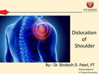
Dear all,
This ppt contains the cause, types, clinical and radiological features, treatment and complication of dislocation of the shoulder. I hope this is useful to you.
Thank you
2. Mechanism
• As it is very incongruent joint, it is very
common to dislocate
• FOOSH injury
• Occassionally thorugh direct hitting
• Epileptic convulsion and electric shock
5. Bankart’s lesion
• Stripping of glenoid
labrum along with
periosteum from
ant-inf surface.
• Head comes in
front of the
scapular neck
Hill-sachs lesion
• It is a depression
on the humeral
head in its postero
lateral quadrant
• Caused by anterior
edge of the glenoid
of the glenoid
Rounding off
• Rounding off of the
anterior glenoid
rim occurs
6.
7.
8.
9. Diagnosis
• History
– Patient’s shoulder is supported in adduction and
elbow supported with the opposite hand.
– History of fall
– Pain and inability
to move the shoulder
10. • On examination in anterior
dislocation
– Normal round contour is lost
– Fullness below clavicle can be felt due
to displaced hand. Can be confirmed by
rotating hand
– Dugas test :- inability to touch opposite
shoulder
– Hamilton ruler test :- Ruler can be
placed on lateral side of shoulder. This
touches acromion process and lateral
condyle of humerus simultaneously.
11.
12. • On examination in posterior dislocation
– Loss of external rotation. Injury is often missed in
x-ray.
13. Treatment
• Treatment of acute dislocation is reduction
under sedation or general anesthesia,
followed by immobilization of the shoulder in
chest arm bandage for 3 weeks.
14. • Techniques of reduction of shoulder
dislocation
• Kocher’s maneuver :-
Traction is applied
along with long axis
of humerus
Arm is rotated
externally
Arm is adducted by
carrying the elbow
across the body
towards the
midline
Arm is rotated
internally so that
hand falls across
the opposite
shoulder
15.
16. • Hippocrates maneuver :-
Surgeon
applies firm
and steady pull
on semi-
abducted arm
He keeps foot
in axilla against
the chest wall
Head of
humerus is
levered back
into position
using the foot
as fulcrum
17.
18. Complication
• Early complication
– Injury to axillary nerve
• Late complication
– Recurrent dislocation may be due to
1. Anatomically unstable joint e.g. Marfan’s syndrome
2. Inadequate healing after first dislocation
3. An epileptic patient.