
Recommended
More Related Content
What's hot
What's hot (20)
Similar to Heart sounds
Similar to Heart sounds (20)
More from PRAVEEN GUPTA
More from PRAVEEN GUPTA (20)
Recently uploaded
Recently uploaded (20)
Heart sounds
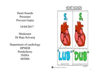
- 1. Heart Sounds Presenter Praveen Gupta 18/04/2017 Moderator Dr Raja Selvaraj Department of cardiology JIPMER Pondicherry INDIA 605006 1
- 2. Heart sound Two types High-frequency, abrupt terminal checking of valves,closing or opening Mitral and tricuspid closing sounds (M1, T1), nonejection sounds, opening snaps, aortic and pulmonic closure sounds (A2, P2) and early valvular ejection sounds Low-frequency, S3 and S3 gallop ,S4 gallop 2 Hurst J, Fuster V, Walsh RA. Hurst's the Heart. McGraw-Hill Medical; 2011.
- 3. Heart Sound S1 Two components Audible at left lower sternal border Louder M1 followed by T1 Deceleration of blood setting cardiohemic into vibration 3 Hurst J, Fuster V, Walsh RA. Hurst's the Heart. McGraw-Hill Medical; 2011.
- 4. Spltting of S1 Normal wide splitting,normal (M1, T1) Right bundle-branch block LV pacing Ectopic beats Idioventricular rhythms from LV Reversed splitting (T1, M1) Pacing from the RV Ectopic beats and idioventricular rhythms from RV 4 Hurst J, Fuster V, Walsh RA. Hurst's the Heart. McGraw-Hill Medical; 2011.
- 5. Factors determining intensity of S1 Integrity of valve closure Mobility of the valve Velocity of valve closure Status of ventricular contraction Transmission characteristics of the thoracic cavity and chest wall Physical characteristics of the vibrating structures 5 Hurst J, Fuster V, Walsh RA. Hurst's the Heart. McGraw-Hill Medical; 2011.
- 6. Integrity of Valve Closure In severe MR, inadequate coaptation of the mitral leaflets to a degree that valve closure is not effective, S1 markedly attenuated 6 Hurst J, Fuster V, Walsh RA. Hurst's the Heart. McGraw-Hill Medical; 2011.
- 7. Mobility of the Valve Severe calcific fixation of the mitral valve with severe MS complete immobilization, attenuated M1 7Hurst J, Fuster V, Walsh RA. Hurst's the Heart. McGraw-Hill Medical; 2011.
- 8. Velocity of Valve Closure Relation of S1 with PR interval PR decreases from 130 to 30 ms increase in the intensity of M1 Mitral leaflets are maximally separated At longer PR intervals, there is less separation of the mitral valve leaflets Variable S1 Complete AV block with AV dissociation Mobitz type I AV block Ventricular tachycardia with AV dissociation Atrial fibrillation 8 Hurst J, Fuster V, Walsh RA. Hurst's the Heart. McGraw-Hill Medical; 2011.
- 9. Status of Ventricular Contraction Exercise and catecholamine infusion increase the amplitude of S1 β-blocking agents decreases S1 is increased in anemia, arteriovenous fistulas, pregnancy, anxiety, and fever. Loud T1 in an ASD decrease in the intensity of S1 myxedema, cardiomyopathy, and acute MI 9 Hurst J, Fuster V, Walsh RA. Hurst's the Heart. McGraw-Hill Medical; 2011.
- 10. Transmission Characteristics of the Thoracic Cavity and Chest Wall Obesity Emphysema Large pleural Pericardial effusions Decrease the intensity of all auscultatory events, Thin body habitus increase the intensity Physical Characteristics of the Vibrating Structures MI and ischemia induced by pacing decrease the intensity of S1 10 Hurst J, Fuster V, Walsh RA. Hurst's the Heart. McGraw-Hill Medical; 2011.
- 11. S1 in Mitral Stenosis A loud M1 Loud OS, Calcific fixation of the stenotic MV occurs, M1 is soft, and the OS is absent 11 Hurst J, Fuster V, Walsh RA. Hurst's the Heart. McGraw-Hill Medical; 2011.
- 12. S1 in Mitral Valve Prolapse Loud M1 heard over apex with nonrheumatic MR; indicate holosystolic MVP Increased amplitude of leaflet excursion with prolapse beyond the line of closure explains the loud M1 associated with holosystolic prolapse Middle to late systolic prolapse have a normal S1 Soft or absent S1 indicate a flail mitral leaflet 12 Hurst J, Fuster V, Walsh RA. Hurst's the Heart. McGraw-Hill Medical; 2011.
- 13. S1 in LBBB M1 decreased in intensity and delayed, reversal S1 sequence LBBB (Delay in onset of LV contraction,LV dysfunction) Acute AR attenuation or absence of M1 (Increase in the LVEDP, premature closure of the mitral valve) 13 Hurst J, Fuster V, Walsh RA. Hurst's the Heart. McGraw-Hill Medical; 2011.
- 14. Systolic Ejection Sounds Originate from left or right of the heart Valvular from deformed aortic or pulmonic valves Vascular or root, rapid, forceful ejection of blood into the great vessels Root ejection sounds indicate abnormalities of great vessels with or without systemic or PHT Definine level of outflow tract obstruction 14 Hurst J, Fuster V, Walsh RA. Hurst's the Heart. McGraw-Hill Medical; 2011.
- 15. Aortic Valvular Ejection Sounds Nonstenotic congenital bicuspid valves (Mild to severe stenosis ) With ejection murmur of AS Widely transmitted Heard best at the apex 20 to 40 ms after pressure rise onset in aorta Ejection click associated with aortic stenosis due to a congenitally bicuspid valve. Note the high-frequency, high-amplitude sound that follows S1 and is coincident with the onset of ejection into the aorta. The aortic ejection sound is formed by sudden cessation of the opening motion of the abnormal valve leaflets (doming). Note also the delayed carotid upstroke and long systolic murmur. (From Abrams J: Synopsis of Cardiac Physical Diagnosis. 2nd ed. Boston, Butterworth Heinemann, 2001, p 135.) 15Hurst J, Fuster V, Walsh RA. Hurst's the Heart. McGraw-Hill Medical; 2011.
- 16. Aortic Valvular Ejection Sounds With sharp anacrotic notch on the upstroke of the aortic pressure curve With maximal excursion of the domed valve when elastic limits are met Intensity of sound correlates directly with mobility of valve No correlation between intensity and severity of the obstruction Ejection click associated with aortic stenosis due to a congenitally bicuspid valve. Note the high-frequency, high-amplitude sound that follows S1 and is coincident with the onset of ejection into the aorta. The aortic ejection sound is formed by sudden cessation of the opening motion of the abnormal valve leaflets (doming). Note also the delayed carotid upstroke and long systolic murmur. (From Abrams J: Synopsis of Cardiac Physical Diagnosis. 2nd ed. Boston, Butterworth Heinemann, 2001, p 135.) 16Hurst J, Fuster V, Walsh RA. Hurst's the Heart. McGraw-Hill Medical; 2011.
- 17. Pulmonic Valvular Ejection Sounds Occurs at maximal excursion of the stenotic pulmonic valve Pulmonic ejection click decreases with inspiration in mild to moderate PS 17 Hurst J, Fuster V, Walsh RA. Hurst's the Heart. McGraw-Hill Medical; 2011.
- 18. Vascular Ejection Sounds Originating from aortic root Common in HTN with tortuous sclerotic aortic root Coincident with the upstroke of central aortic pressure Sound occurs at the moment of complete opening of the aortic valve Tend to be poorly transmitted from the aortic area and are not heard well at the apex Interpreted as an exaggeration of the ejection component of the normal S1 18 Hurst J, Fuster V, Walsh RA. Hurst's the Heart. McGraw-Hill Medical; 2011.
- 19. Pulmonary Vascular Ejection Sounds From pulmonary artery & due to dilatation of the pulmonary artery Dilatation can be idiopathic or secondary to severe PH Louder during expiration Louder in 2ND and 3RD left intercostal spaces Occurring during upstroke of pulmonary artery pressure recording 19 Hurst J, Fuster V, Walsh RA. Hurst's the Heart. McGraw-Hill Medical; 2011.
- 20. Nonejection Sounds Midsystolic click Prolapse of the mitral or tricuspid valve With a systolic regurgitant murmur 20 Hurst J, Fuster V, Walsh RA. Hurst's the Heart. McGraw-Hill Medical; 2011.
- 21. Nonejection Sounds Sharp High-frequency Clicking quality Confined to the apex Transmitted widely on the precordium Can isolated finding In middle to late systole Can be multiple clicks 21 Hurst J, Fuster V, Walsh RA. Hurst's the Heart. McGraw-Hill Medical; 2011.
- 22. Nonejection Sounds Occurs at time of maximal prolapse Upright posture, click moves earlier Squatting, click toward S2 Differentiating nonejection click from early ejection sounds, a split S2, or an S3 22Hurst J, Fuster V, Walsh RA. Hurst's the Heart. McGraw-Hill Medical; 2011.
- 23. Heart sound (S2) High-frequency Two component, A2 and P2 Produced by the sudden deceleration of retrograde flow of the blood column in the aorta and pulmonary artery Increased intensity of A2 and P2 in systemic and PH 23 Hurst J, Fuster V, Walsh RA. Hurst's the Heart. McGraw-Hill Medical; 2011.
- 24. Normal Physiologic Splitting In expiration, A2 & P2 separated by <30 ms Heard by the clinician as a single sound During inspiration, both components audible ,caused by a delayed P2 P2 softer than A2 and rarely audible at apex When P2 is heard at the apex significant PH is present Single S2 during both phases of respiration normal in subjects older than 40 years of age 24 Hurst J, Fuster V, Walsh RA. Hurst's the Heart. McGraw-Hill Medical; 2011.
- 25. Abnormal Splitting of S2 Exists by presence of audible expiratory splitting (>30 ms) Must be present in both the supine and upright There are three causes of audible expiratory splitting (1) wide physiologic splitting primarily caused by delayed P2, (2) Reversed splitting primarily caused by delayed A2 (3) narrow physiologic splitting as seen in PH, where A2 and P2 are heard as two distinct sounds during expiration at a narrow splitting interval. 25 Hurst J, Fuster V, Walsh RA. Hurst's the Heart. McGraw-Hill Medical; 2011.
- 26. Wide physiologic splitting of S2 Right bundle-branch block Severe PH and PS ASD Acute MR Idiopathic dilatation of the pulmonary artery Mild PS with aneurysmal dilatation of the pulmonary artery 26 Hurst J, Fuster V, Walsh RA. Hurst's the Heart. McGraw-Hill Medical; 2011.
- 27. Reversed splitting of S2 Caused by a delay in A2 P2 preceding A2. Paradoxical movement of A2 and P2 with respiration During inspiration, P2 moves toward A2, and the splitting interval narrows During expiration, the two components separate, and audible expiratory splitting is present Indicates cardiovascular disease 27 Hurst J, Fuster V, Walsh RA. Hurst's the Heart. McGraw-Hill Medical; 2011.
- 28. Reversed splitting of S2 RV ectopic and paced beats Complete LBBB Hypertrophic cardiomyopathy Valvular AS, Hypertensive cardiovascular diseas ( rare) Ischemic heart disease Episodes of angina pectoris Poststenotic dilatation of the aorta Chronic AR Patent ductus arteriosus Type B Wolff-Parkinson-White syndrome 28 Hurst J, Fuster V, Walsh RA. Hurst's the Heart. McGraw-Hill Medical; 2011.
- 29. Narrow Physiologic Splitting Common finding in severe PH In contrast to the normal situation, where only a single sound is heard during expiration, both A2 and P2 are easily heard, even though the splitting interval is less than 30 ms because of the increased intensity and high-frequency composition of P2 Wide, persistent splitting sign of abnormal RV performance in patients with primary PH Fixed splitting of S2 occasionally has been documented in severe RV failure secondary to PH. 29 Hurst J, Fuster V, Walsh RA. Hurst's the Heart. McGraw-Hill Medical; 2011.
- 30. Single S2 Delay A2 produce when splitting interval <30 ms One component of S2 is either absent or inaudible Eisenmenger VSD Inability to hear the fainter of the two components of the sound (usually P2) because of emphysema, obesity, or respiratory noise Seen in older than 50 years of age 30 Hurst J, Fuster V, Walsh RA. Hurst's the Heart. McGraw-Hill Medical; 2011.
- 31. Opening Snaps Opening of AV valve silent event With thickening and deformity of the leaflets sound is generated in early diastole High-frequency Early diastolic sound Absent in thickened and immobile valves 31 Hurst J, Fuster V, Walsh RA. Hurst's the Heart. McGraw-Hill Medical; 2011.
- 32. Opening snap Crisp Sharp sound Heard in the midprecordial location Best in the area from the left sternal border to just inside the apex Often heard well at the base of the heart Diastolic rumble follows opening snap No variation in the intensity or timing of the mitral opening snap with respiration 32 Hurst J, Fuster V, Walsh RA. Hurst's the Heart. McGraw-Hill Medical; 2011.
- 33. Opening snap Intensity correlates with valve mobility Loud in mobile stenotic valves Absent with severe calcific valve Intensity of M1 parallels the intensity of the opening snap The opening snap occurs at the maximal mitral valve opening shortly after LV–left atrial pressure crossovers. 33 Hurst J, Fuster V, Walsh RA. Hurst's the Heart. McGraw-Hill Medical; 2011.
- 34. Factors that influence the timing of the opening snap relative to A2 Rate of LV pressure decline Level of the LV pressure at the time of A2 Level of the left atrial pressure Increasing severity of MS,shortening of the A2–opening snap interval Imperfect correlation between A2– opening snap interval and mitral area A2–opening snap interval in atrial fibrillation vary with cycle length 34 Hurst J, Fuster V, Walsh RA. Hurst's the Heart. McGraw-Hill Medical; 2011.
- 35. Differential diagnosis of opening snap Differentiated from other early diastolic sounds S3, the pulmonary component of a widely split S2 35
- 36. Third and Fourth Heart Sounds Low-frequency Related to early and late diastolic filling of the ventricles Disease states called gallop sounds Gives information of ventricular function and compliance 36 Hurst J, Fuster V, Walsh RA. Hurst's the Heart. McGraw-Hill Medical; 2011.
- 37. Third heart sound (S3) Physiologic S3 benign finding Commonly in children, adolescents, and young adults Rarely after 40 years of age, when present associated with a thin, asthenic body Low-frequency sound that Follows A2 by 120 to 200 ms 37 Hurst J, Fuster V, Walsh RA. Hurst's the Heart. McGraw-Hill Medical; 2011.
- 38. Third heart sound (S3) Occurs during rapid filling of the ventricle Best heard at the apex In left lateral position Stethoscope's bell pressed lightly against skin Differentiated from the pathologic S3 primarily by the "company it keeps." 38 RV S3 heard at the lower left sternal edge and increase in intensity with inspiration LV systolic dysfunction Diuresis, S3 decreases S3 with cardiomyopathy/Myocardial infacrtion ominous sign Hurst J, Fuster V, Walsh RA. Hurst's the Heart. McGraw-Hill Medical; 2011.
- 39. Third heart sound(S3) Chronic AR Acute AR AV valve regurgitation Large left-to-right shunts VSD Patent ductus arteriosus ASD Restrictive cardiomyopathy Hypertrophic cardiomyopathy 39 Hurst J, Fuster V, Walsh RA. Hurst's the Heart. McGraw-Hill Medical; 2011.
- 40. Fourth Heart Sound (S4) Best heard at the apex In left lateral position Varies with respiration Heard best during expiration S4 just prior to S1 Also termed atrial diastolic gallop or the presysolic gallop Atrial contraction must be present for S4 Absent in atrial fibrillation 40 Hurst J, Fuster V, Walsh RA. Hurst's the Heart. McGraw-Hill Medical; 2011.
- 41. Fourth Heart Sound (S4) Audibility depends on its intensity and frequency, separation from S1 Degree of separation is determined primarily by PR interval A loud S1 also can mask the audibility of a preceding softer S4 Left-sided S4 and S3 augmented post-tussively and sustained handgrip exercise Maneuvers that increase venous return increase the audibility by increasing the intensity of the sound and by causing it to occur earlier, thereby separating it further from S1. Decreased venous return does the opposite Accompanied by a palpable presystolic apical impulse S4 generated by right atrial contraction heard best at the lower left sternal border Accentuated with inspiration 41 Hurst J, Fuster V, Walsh RA. Hurst's the Heart. McGraw-Hill Medical; 2011.
- 42. Fourth Heart Sound(S4) Systemic hypertension Severe valvular AS Hypertrophic cardiomyopathy 42 Hurst J, Fuster V, Walsh RA. Hurst's the Heart. McGraw-Hill Medical; 2011.
- 43. Prosthetic valves sound Type of valve, its position, whether it is functioning normally Mechanical valves produce opening and closing clicks Are easily audible Can be heard even without a stethoscope. Ball-in-cage valves produce the loudest and most distinctive opening and closing clicks The metallic ball of the Starr-Edwards valve also produces multiple early systolic clicks Absence or decrease in intensity of clicks occur with valve obstruction or LV dysfunction. A decrease in the intensity of the opening and closing clicks, and the absence of the opening click are also indications of valve malfunction. 43Hurst J, Fuster V, Walsh RA. Hurst's the Heart. McGraw-Hill Medical; 2011.
- 45. Extracardiac Sounds Pacemaker Sounds High-frequency sounds of brief duration with transvenous pacemakers located in the RV apex Extracardiac in origin within 6-10 ms with the pacemaker spike caused by stimulation of intercostal nerves adjacent to endocardial electrodes Should suggest myocardial perforation by the endocardial lead 45 Hurst J, Fuster V, Walsh RA. Hurst's the Heart. McGraw-Hill Medical; 2011.
- 46. Pericardial Friction Rub Inflammation of the pericardial sac with or without fluid Very high pitched Leathery Scratchy in nature Seem close to the ear Auscultated best with the patient leaning forward or knee–chest position Holding his or her breath after forced expiration 46 Hurst J, Fuster V, Walsh RA. Hurst's the Heart. McGraw-Hill Medical; 2011.
- 47. Pericardial Friction Rub Three components Atrial systole, Ventricular contraction Rapid early diastolic filling Uremic pericarditis Acute phase of transmural MI 47 Hurst J, Fuster V, Walsh RA. Hurst's the Heart. McGraw-Hill Medical; 2011.
- 48. Mediastinal Crunch: Hamman Sign When air is present in the mediastinum Scratchy sounds (Hamman sign) occur Related indirectly to both heartbeat and respiratory excursion Occur most frequently during ventricular systole Caused by air in the mediastinum Common after cardiac surgery 48 Hurst J, Fuster V, Walsh RA. Hurst's the Heart. McGraw-Hill Medical; 2011.
- 49. Approach with cardiac sound 49
- 50. Reference Hurst J, Fuster V, Walsh RA. Hurst's the Heart. McGraw-Hill Medical; 2011. Google Images 50
- 51. 51 THANK YOU