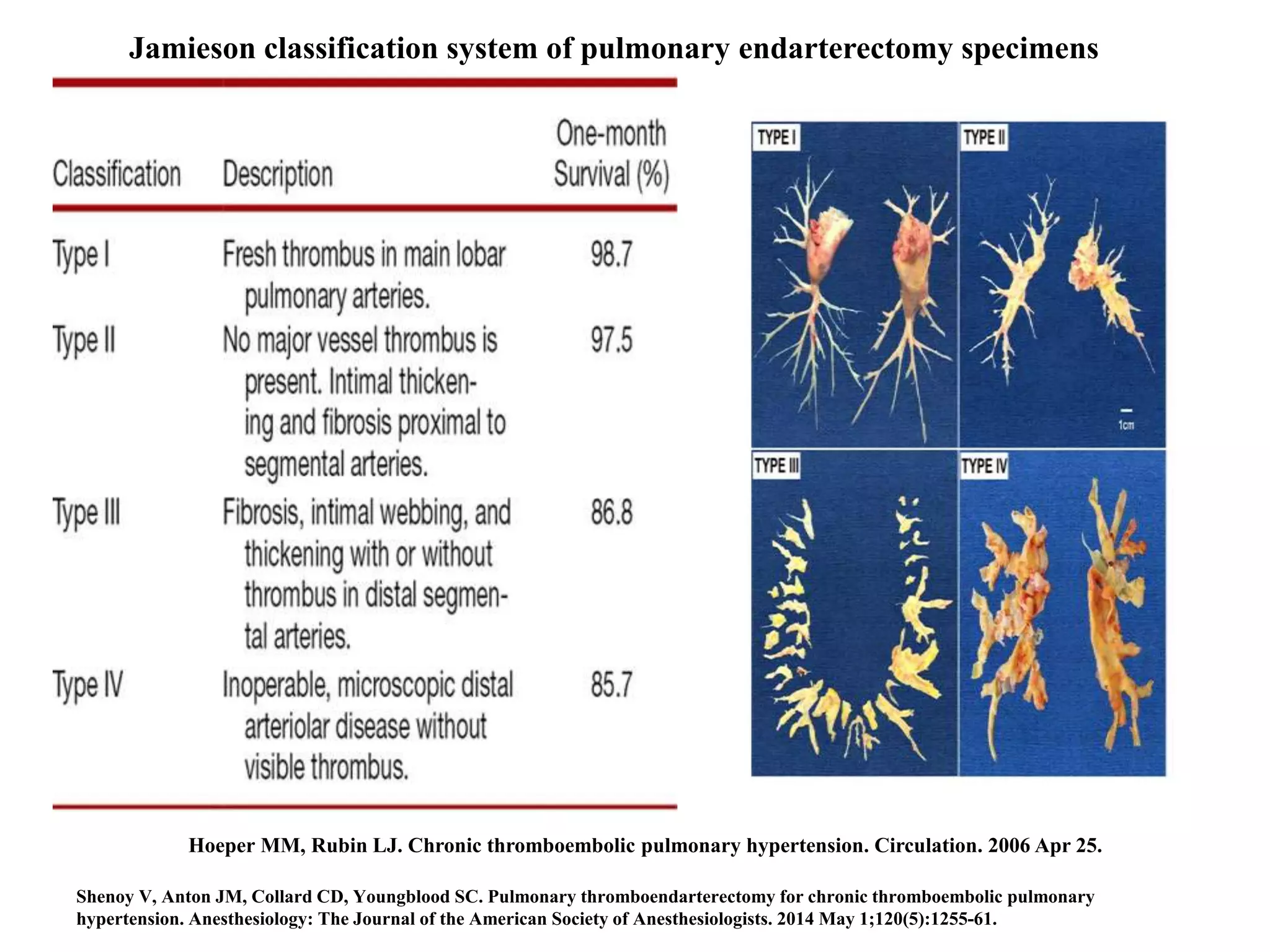
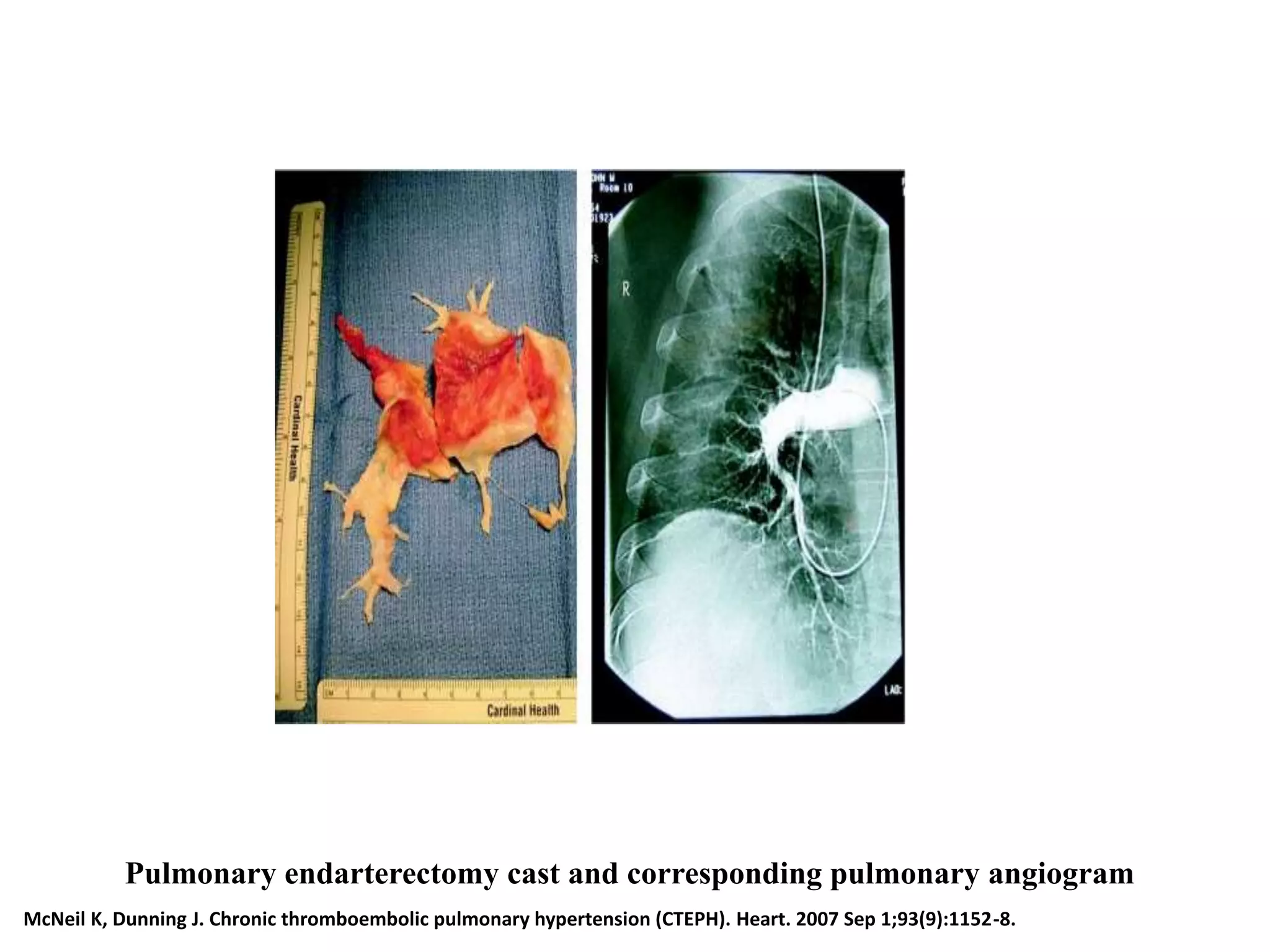
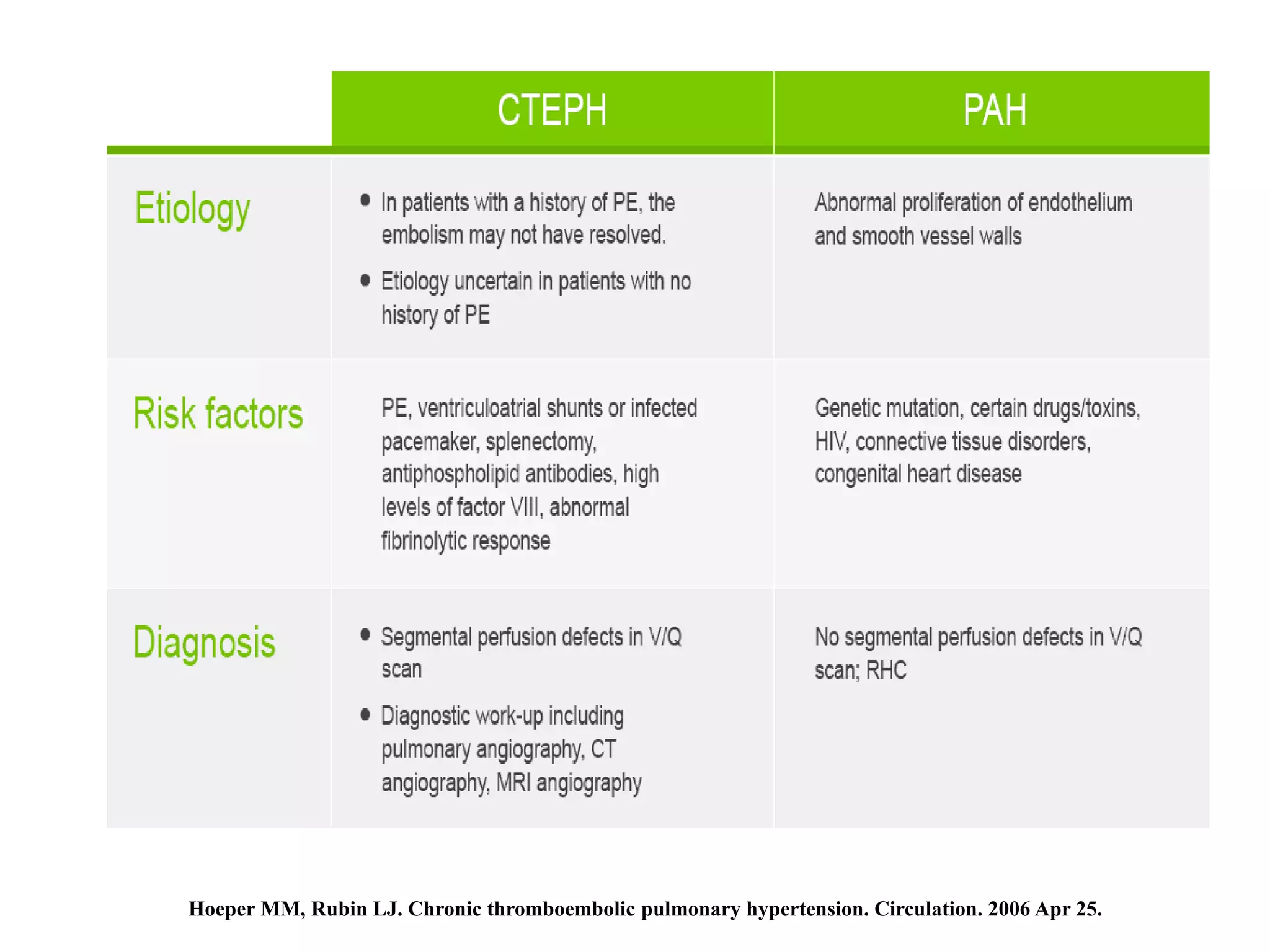
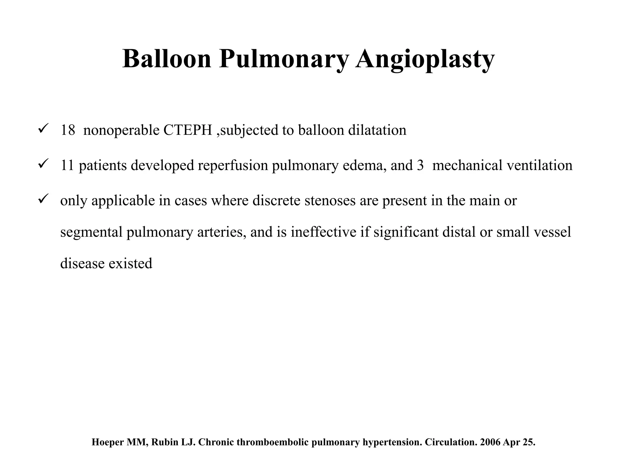
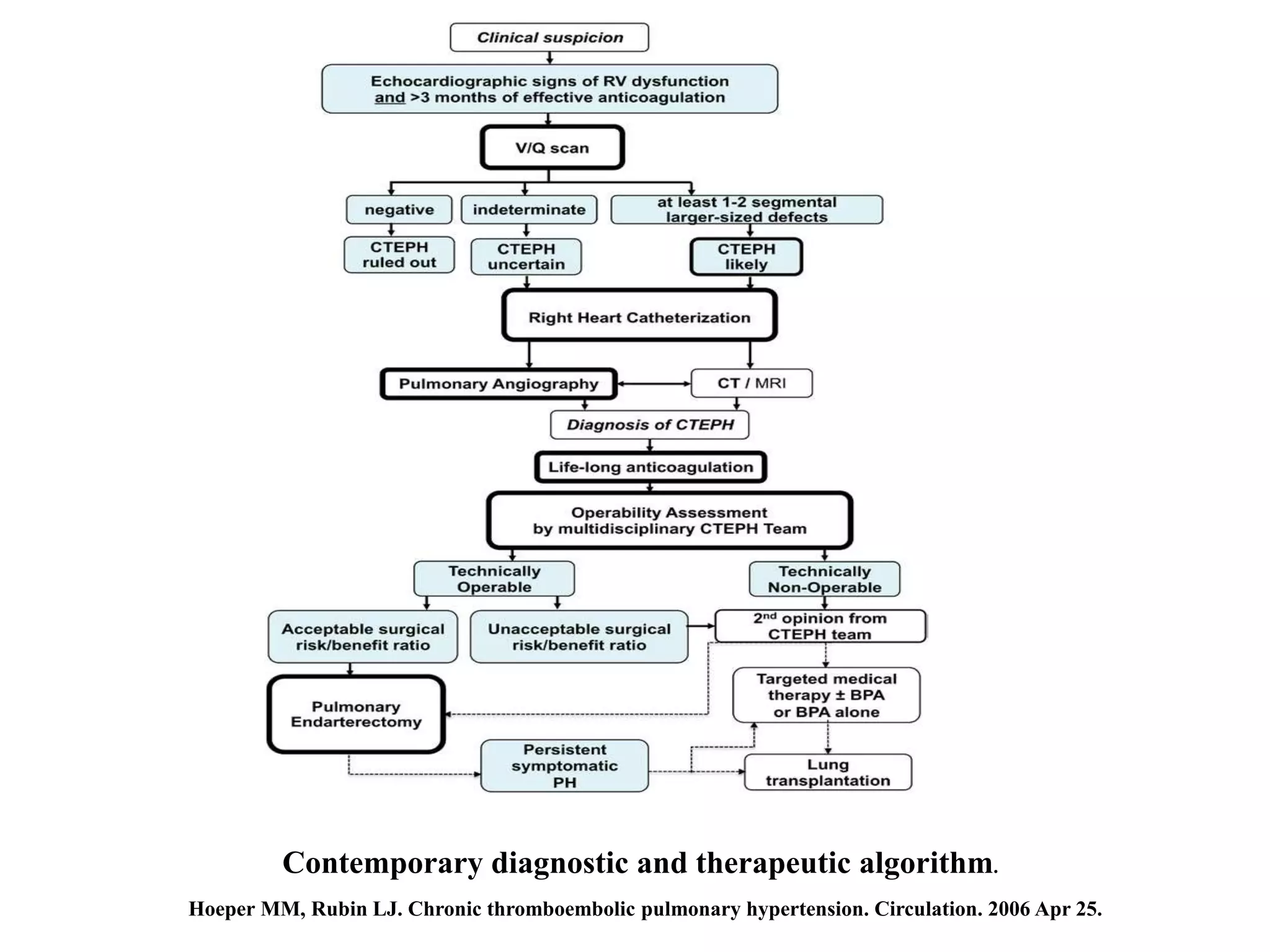
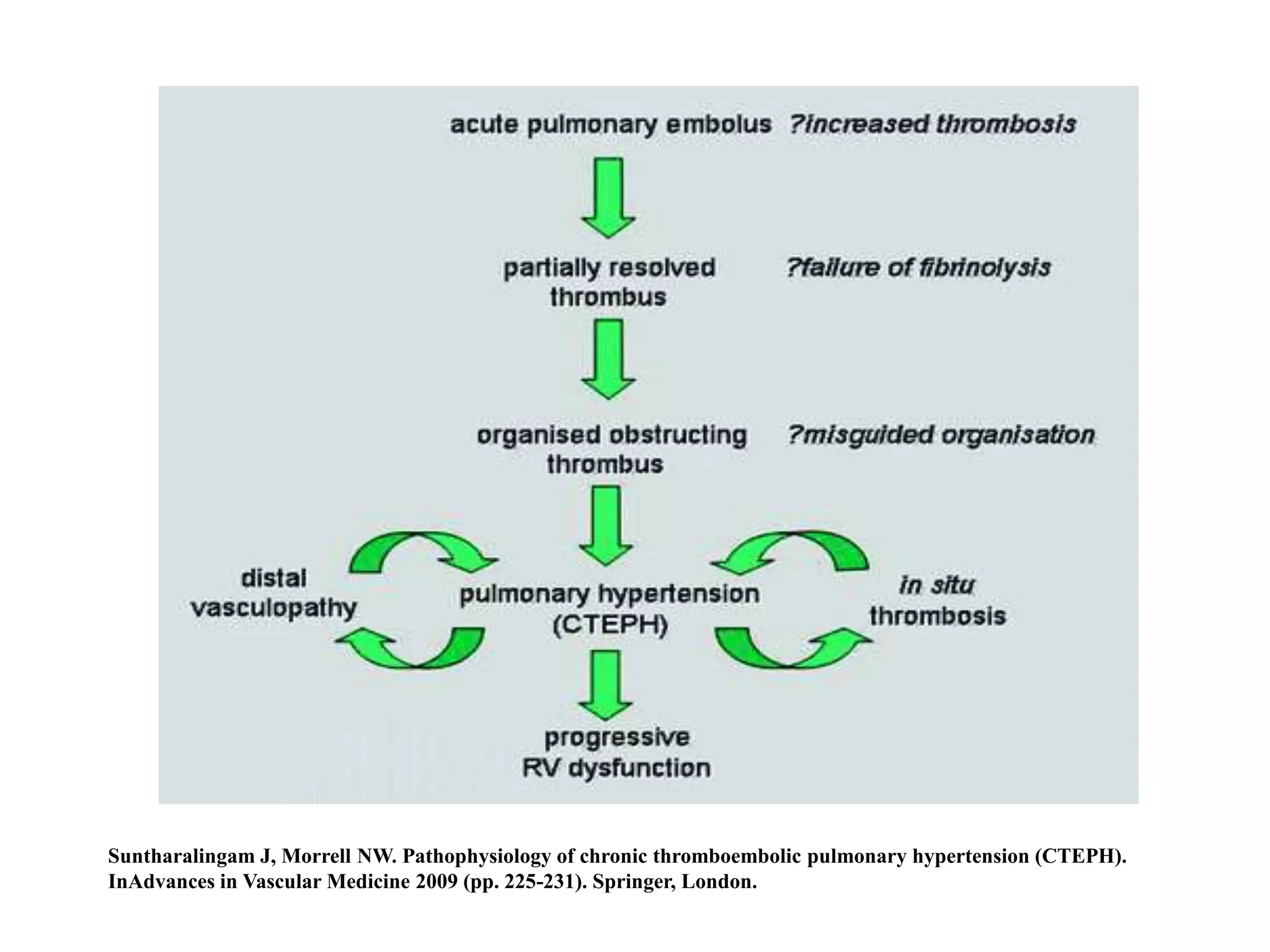
Chronic thromboembolic pulmonary hypertension (CTEPH) is caused by chronic obstruction of the major pulmonary arteries by thrombi. It is amenable to cure by pulmonary endarterectomy (PEA). Diagnosis involves ventilation/perfusion scanning, CT pulmonary angiography, pulmonary angiography, and right heart catheterization to assess pulmonary hypertension. PEA aims to surgically remove the obstructive thrombi and can cure CTEPH if performed at an expert center, where the condition is often underdiagnosed or referred to late.
Pulmonary arterial hypertension in congenital heart disease Ramachandra Barik
Pulmonary hypertension (PH) is an increase of blood pressure in the pulmonary artery, pulmonary vein, or pulmonary capillaries, together known as the lung vasculature, leading to shortness of breath, dizziness, fainting, leg swelling and other symptoms. Pulmonary hypertension can be a severe disease with a markedly decreased exercise tolerance and heart failure. It was first identified by Ernst von Romberg in 1891. According to the most recent classification, it can be one of five different types: arterial, venous, hypoxic, thromboembolic or miscellaneous.
In this ppt i am going to discuss various spotters, including ECG, X-ray, fluroscopy images and there answers. These spotter now days asked in various DM cardiology exam conducted all over India, so it will help you in your DM Cardiology exam preperationn.
Point of critical care Ultrasound play a pivotal role in management of critically ill patients admitted in ICU . Its usage in this regard is ever growing . Here we discus about pearls and pitfalls of POCUS in Intensive care medicine.
DIAGNOSIS & MANAGEMENT OF PULMONARY HYPERTENSIONKamal Bharathi
Pulmonary hypertension (PH) is defined by a mean pulmonary artery pressure ≥25 mm Hg at rest, measured during right heart catheterization. There is still insufficient evidence to add an exercise criterion to this definition. The term pulmonary arterial hypertension (PAH) describes a subpopulation of patients with PH characterized hemodynamically by the presence of pre-capillary PH including an end-expiratory pulmonary artery wedge pressure (PAWP) ≤15 mm Hg and a pulmonary vascular resistance >3 Wood units. Right heart catheterization remains essential for a diagnosis of PH or PAH. This procedure requires further standardization, including uniformity of the pressure transducer zero level at the midthoracic line, which is at the level of the left atrium. One of the most common problems in the diagnostic workup of patients with PH is the distinction between PAH and PH due to left heart failure with preserved ejection fraction (HFpEF). A normal PAWP does not rule out the presence of HFpEF. Volume or exercise challenge during right heart catheterization may be useful to unmask the presence of left heart disease, but both tools require further evaluation before their use in general practice can be recommended. Early diagnosis of PAH remains difficult, and screening programs in asymptomatic patients are feasible only in high-risk populations, particularly in patients with systemic sclerosis, for whom recent data suggest that a combination of clinical assessment and pulmonary function testing including diffusion capacity for carbon monoxide, biomarkers, and echocardiography has a higher predictive value than echocardiography alone.
PowerPoint presentation describing various aspects of Pulmonary Hypertension. Please mail me your feedback on this presentation to following Email ID: tinkujoseph2010@gmail.com.
Deep Venous Thrombosis and Pulmonary Embolism : Diagnostic Approach and Curre...Bassel Ericsoussi, MD
Acute pulmonary embolism: Overview, Diagnosis, Treatment
DVT/PE in pregnancy
Prevalence of PE in COPD exacerbations
Diagnostic vascular ultrasonography
Pulmonary embolism - Diagnosis and managementDr Vivek Baliga
Pulmonary embolism is a common problem seen in medical practice. This presentation by Dr Vivek Baliga discusses the basic aspects and evidence behind current management.
In this ppt, I am going to discuss the role of ICD in the patient with Non-ischemic cardiomyopathy. I am going to discuss all the major trials done in the patient with non-ischemic cardiomyopathy.
2015 Jones criteria for the diagnosis of rheumatic feverPRAVEEN GUPTA
In this ppt, I am going to discuss 2015 Jones criteria for the diagnosis of rheumatic fever and various modification made into it and basis for those modifications
Palestine last event orientationfvgnh .pptxRaedMohamed3
An EFL lesson about the current events in Palestine. It is intended to be for intermediate students who wish to increase their listening skills through a short lesson in power point.
Francesca Gottschalk - How can education support child empowerment.pptxEduSkills OECD
Francesca Gottschalk from the OECD’s Centre for Educational Research and Innovation presents at the Ask an Expert Webinar: How can education support child empowerment?
Biological screening of herbal drugs: Introduction and Need for
Phyto-Pharmacological Screening, New Strategies for evaluating
Natural Products, In vitro evaluation techniques for Antioxidants, Antimicrobial and Anticancer drugs. In vivo evaluation techniques
for Anti-inflammatory, Antiulcer, Anticancer, Wound healing, Antidiabetic, Hepatoprotective, Cardio protective, Diuretics and
Antifertility, Toxicity studies as per OECD guidelines
Synthetic Fiber Construction in lab .pptxPavel ( NSTU)
Synthetic fiber production is a fascinating and complex field that blends chemistry, engineering, and environmental science. By understanding these aspects, students can gain a comprehensive view of synthetic fiber production, its impact on society and the environment, and the potential for future innovations. Synthetic fibers play a crucial role in modern society, impacting various aspects of daily life, industry, and the environment. ynthetic fibers are integral to modern life, offering a range of benefits from cost-effectiveness and versatility to innovative applications and performance characteristics. While they pose environmental challenges, ongoing research and development aim to create more sustainable and eco-friendly alternatives. Understanding the importance of synthetic fibers helps in appreciating their role in the economy, industry, and daily life, while also emphasizing the need for sustainable practices and innovation.
The Roman Empire A Historical Colossus.pdfkaushalkr1407
The Roman Empire, a vast and enduring power, stands as one of history's most remarkable civilizations, leaving an indelible imprint on the world. It emerged from the Roman Republic, transitioning into an imperial powerhouse under the leadership of Augustus Caesar in 27 BCE. This transformation marked the beginning of an era defined by unprecedented territorial expansion, architectural marvels, and profound cultural influence.
The empire's roots lie in the city of Rome, founded, according to legend, by Romulus in 753 BCE. Over centuries, Rome evolved from a small settlement to a formidable republic, characterized by a complex political system with elected officials and checks on power. However, internal strife, class conflicts, and military ambitions paved the way for the end of the Republic. Julius Caesar’s dictatorship and subsequent assassination in 44 BCE created a power vacuum, leading to a civil war. Octavian, later Augustus, emerged victorious, heralding the Roman Empire’s birth.
Under Augustus, the empire experienced the Pax Romana, a 200-year period of relative peace and stability. Augustus reformed the military, established efficient administrative systems, and initiated grand construction projects. The empire's borders expanded, encompassing territories from Britain to Egypt and from Spain to the Euphrates. Roman legions, renowned for their discipline and engineering prowess, secured and maintained these vast territories, building roads, fortifications, and cities that facilitated control and integration.
The Roman Empire’s society was hierarchical, with a rigid class system. At the top were the patricians, wealthy elites who held significant political power. Below them were the plebeians, free citizens with limited political influence, and the vast numbers of slaves who formed the backbone of the economy. The family unit was central, governed by the paterfamilias, the male head who held absolute authority.
Culturally, the Romans were eclectic, absorbing and adapting elements from the civilizations they encountered, particularly the Greeks. Roman art, literature, and philosophy reflected this synthesis, creating a rich cultural tapestry. Latin, the Roman language, became the lingua franca of the Western world, influencing numerous modern languages.
Roman architecture and engineering achievements were monumental. They perfected the arch, vault, and dome, constructing enduring structures like the Colosseum, Pantheon, and aqueducts. These engineering marvels not only showcased Roman ingenuity but also served practical purposes, from public entertainment to water supply.
Introduction to AI for Nonprofits with Tapp NetworkTechSoup
Dive into the world of AI! Experts Jon Hill and Tareq Monaur will guide you through AI's role in enhancing nonprofit websites and basic marketing strategies, making it easy to understand and apply.
2. Introduction
CTEPH caused by chronic obstruction of major pulmonary arteries
Amenable to cure by pulmonary endarterectomy (PEA)
≈5 individuals per million per year (Orphan disease)
Cumulative incidence, 0.1% and 9.1% after symptomatic PE
Routine screening after acute PE not recommended
Most common subsets of precapillary PH
Underdiagnosis
common delay or lack of referral to expert centers for surgery
Hoeper MM, Rubin LJ. Chronic thromboembolic pulmonary hypertension. Circulation. 2006 Apr 25.
3.
4. Definition
Precapillary PH by invasive right heart catheterization (mean pulmonary artery pressure
≥25 mm Hg, mean pulmonary arterial wedge pressure ≤15 mm Hg)
Presence of chronic/organized flow-limiting thrombi in the elastic pulmonary arteries
(main, lobar, segmental, subsegmental)
After at least 3 months of effective anticoagulation.
Hoeper MM, Rubin LJ. Chronic thromboembolic pulmonary hypertension. Circulation. 2006 Apr 25.
5. Thrombophilia and CTEPH
Antithrombin deficiency
Protein C deficiency
Protein S deficiency
Factor V Leiden
Plasminogen deficiency
Anticardiolipin antibodies
Blood group types A, B, and AB
Non-O blood group carriers also have higher levels of von Willebrand factor and factor VIII
Elevated plasma levels of lipoprotein(a)
Hoeper MM, Rubin LJ. Chronic thromboembolic pulmonary hypertension. Circulation. 2006 Apr 25.
6. Dartevelle P, Fadel E, Mussot S, Chapelier A, Herve P, De Perrot M, Cerrina J, Ladurie FL, Lehouerou D, Humbert M, Sitbon O. Chronic
thromboembolic pulmonary hypertension. European Respiratory Journal. 2004 Apr 1;23(4):637-48.
7. Suntharalingam J, Morrell NW. Pathophysiology of chronic thromboembolic pulmonary hypertension (CTEPH).
InAdvances in Vascular Medicine 2009 (pp. 225-231). Springer, London.
8. Vascular obstructive lesions in acute pulmonary embolism (A) and chronic thromboembolic pulmonary hypertension (CTEPH; B). The CTEPH
patient underwent bilateral pulmonary endarterectomy; however, only the right-side specimen is shown. Images courtesy of Dr. Joyce Johnson (A)
and Tarek Absi (B)
Matthews DT, Hemnes AR. Current concepts in the pathogenesis of chronic thromboembolic pulmonary hypertension.
Pulmonary circulation. 2016 Jun;6(2):145-54.
9. Microvasculopathy in chronic thromboembolic pulmonary hypertension involving pulmonary arterioles, venules and capillaries. Schematic representation of
anastomosis between systemic and pulmonary circulation through hypertrophic bronchial arteries and vasa vasorum. PA: pulmonary artery; PVOD: pulmonary
veno-occlusive disease; PAH: pulmonary arterial hypertension. Reproduced and modified from [67] with permission.
Simonneau G, Torbicki A, Dorfmüller P, Kim N. The pathophysiology of chronic thromboembolic pulmonary hypertension. European
Respiratory Review. 2017 Mar 31;26(143):160112.
10. Pulmonary endarterectomy specimens stained with hematoxylin and eosin. A, Neointimal thickening, 18×; B, region of fibrotic chronic
thromboembolic pulmonary hypertension clot containing extracellular matrix and spindle-shaped cells, 290×; C, fresh red clot within an area of
organized clot, 100×; D, obstructing lesion with areas of recanalization, 170×.
Matthews DT, Hemnes AR. Current concepts in the pathogenesis of chronic thromboembolic pulmonary hypertension.
Pulmonary circulation. 2016 Jun;6(2):145-54.
11. Jamieson classification system of pulmonary endarterectomy specimens
Shenoy V, Anton JM, Collard CD, Youngblood SC. Pulmonary thromboendarterectomy for chronic thromboembolic pulmonary
hypertension. Anesthesiology: The Journal of the American Society of Anesthesiologists. 2014 May 1;120(5):1255-61.
Hoeper MM, Rubin LJ. Chronic thromboembolic pulmonary hypertension. Circulation. 2006 Apr 25.
12. Clinical Characteristics
Median age is 63 years, and surgical cases are younger (57 years)
Rare in children
In Europe, both sexes are equally affected, whereas women in Japan
Nonspecific or absent in early stage
Right heart failure in advanced disease stages
Early diagnosis is difficult
Median time of 14 months between symptom onset and diagnosis
Hoeper MM, Rubin LJ. Chronic thromboembolic pulmonary hypertension. Circulation. 2006 Apr 25.
13. Clinical Characteristics
Episodic disease course in CTEPH
Symptomatic thromboembolic event, followed by honeymoon period
Hemoptysis more frequently with idiopathic PAH
Differentiate CTEPH from PAH
Comparative survival in CTEPH has been reported to be twice that of PAH
Hoeper MM, Rubin LJ. Chronic thromboembolic pulmonary hypertension. Circulation. 2006 Apr 25.
15. Diagnostic Procedures
Unexplained PH should be subjected to a CTEPH evaluation
Suspicion should be high with history of previous VTE
PE survivors with persistent dyspnea
Hoeper MM, Rubin LJ. Chronic thromboembolic pulmonary hypertension. Circulation. 2006 Apr 25.
18. Echocardiography
Recommend first diagnostic step
Estimating peak velocity of tricuspid valve regurgitation
Atrioventricular pressure gradients
Right atrial dilatation
Right ventricular dilatation
Reduced right ventricular contractility
Doppler flow abnormalities in the right ventricular outflow tract
Can miss pulmonary hypertension in as many as 10–31% of cases
Hoeper MM, Rubin LJ. Chronic thromboembolic pulmonary hypertension. Circulation. 2006 Apr 25.
20. Ventilation/perfusion scintigraphy
Imaging modality of choice for exclusion of CTEPH
Ventilation/perfusion scan planar images on at least 6 views combined with single-photon
emission CT remain the preferred initial diagnostic test for CTEPH
Normal V/Q scan excludes CTEPH with sensitivity of 90–100% and specificity of 94–100%
Hoeper MM, Rubin LJ. Chronic thromboembolic pulmonary hypertension. Circulation. 2006 Apr 25.
21. Tanabe N, Sugiura T, Tatsumi K. Recent progress in the diagnosis and management of chronic thromboembolic pulmonary
hypertension. Respiratory investigation. 2013 Sep 1;51(3):134-46.
22. Diagnosis of CTEPH
Subsequent investigations aim to define the nature and extent of thromboembolic disease
To assess surgical suitability.
Pulmonary angiography, MRI angiography and CTPA
Hoeper MM, Rubin LJ. Chronic thromboembolic pulmonary hypertension. Circulation. 2006 Apr 25.
23. CT pulmonary angiography
Sensitivity of 51% compared with >96% sensitivity of V/Q scan
Absence or sudden loss of contrast filled vessels,
Best appreciated by following individual vessels while scrolling sequential slices.
Filling defects are not commonly seen.
If filling defects are present, they occur eccentrically within the vessel lumen
In acute pulmonary embolism CTPA typically shows central filling defects
Hoeper MM, Rubin LJ. Chronic thromboembolic pulmonary hypertension. Circulation. 2006 Apr 25.
24. CT pulmonary angiography
Diagnosis missed unless the radiologist is specifically asked to look
Pulmonary artery webs and bands, wall irregularities, stenoses, and, as well as
bronchial collaterals
Identify LMCA compression
Identifies emphysema, bronchial disease, and mediastinal or interstitial lung
disease, as well as infarcts, vascular and pericardial malformations, and thoracic
wall deformities
Perfusion inequalities manifest as a mosaic parenchymal pattern with dark areas
corresponding to relatively decreased perfusion.
Hoeper MM, Rubin LJ. Chronic thromboembolic pulmonary hypertension. Circulation. 2006 Apr 25.
25. Diagnostic Procedures
MRI is inferior to CT
Intravascular imaging, with ultrasound and optical coherence tomography providing
new insights into the vascular obstructions of CTEPH
Hoeper MM, Rubin LJ. Chronic thromboembolic pulmonary hypertension. Circulation. 2006 Apr 25.
26. Pulmonary angiography
Definitive for the diagnosis and assessment of surgically correctable CTEPH
Safe, even with severe pulmonary hypertension.
Findings include vascular webs ,abrupt vascular cut‐offs
Filling defects per se are less common
Anteroposterior and lateral views taken to define the extent of disease
Hoeper MM, Rubin LJ. Chronic thromboembolic pulmonary hypertension. Circulation. 2006 Apr 25.
28. Patient with inoperable chronic thromboembolic pulmonary hypertension showing a typical aspect of subpleural hypoperfusion at the capillary phase of pulmonary
angiography. Image kindly provided by P. Dartevelle, French National Reference Centre of Pulmonary Hypertension, Hôpital Bicêtre, Le Kremlin-Bicêtre, France
Simonneau G, Torbicki A, Dorfmüller P, Kim N. The pathophysiology of chronic thromboembolic pulmonary hypertension. European Respiratory Review. 2017 Mar 31;26(143):160112.
29. Right heart catheterisation
Mandatory
Measurement of PAOP to exclude post-capillary PH
LVEDP by left ventricular catheterisation
Measure cardiac output , because PVR assess prognosis and the risks associated with PEA
Early stages patients to have a near‐normal resting mPAP, with significant pulmonary
hypertension manifesting only with exercise
Hoeper MM, Rubin LJ. Chronic thromboembolic pulmonary hypertension. Circulation. 2006 Apr 25.
30. Diagnostic Procedures
DSA remains the gold standard for assessment of pulmonary revascularization
Combination of imaging with the indispensable hemodynamic assessment
Ring-like stenosis, pouches, webs, occlusions, and distal lesions
Distribution of disease (ie, subpleural perfusion) have been associated with
postoperative outcome
Hoeper MM, Rubin LJ. Chronic thromboembolic pulmonary hypertension. Circulation. 2006 Apr 25.
32. Completed diagnostic pathways of chronic thromboembolic pulmonary hypertension imaging and their therapeutic consequences, according to the algorithm shown in Figure
3, illustrating an operable and a nonoperable patient case. Examples of technetium-99 m–labeled aerosol ventilation (A) and perfusion (B) images are shown, labeled as high
probability under Prospective Investigation of Pulmonary Embolism Diagnosis criteria. High-resolution computed tomographies (C) and spiral computed tomographic
angiographies (D), as well as side-selective pulmonary angiographies in the anteroposterior right (E), left (G), and lateral projections (F and H), are shown. Patient 1
in A1 through I1 is a 69-year-old man with past medical history of deep vein thrombosis and acute pulmonary embolism. He underwent pulmonary endarterectomy (specimen
in I1) in addition to coronary artery bypass grafting in the rewarming period. At the time of surgery, type II disease was encountered, with a larger amount of thromboembolic
material than was expected on the basis of the imaging studies, which is commonly the case. He was discharged home 1 week postoperatively with normal right ventricular
function and pulmonary vascular resistance of 70 dyne·s/cm5 compared with 450 dyne·s/cm5 preoperatively. Patient 2 in A2through I2 is 49-year-old woman with history of
progressive shortness of breath on exertion for many years. After initiation of pulmonary arterial hypertension therapy, right heart catheterization revealed only moderate
pulmonary hypertension and a few distal segmental and subsegmental obstructions. Surgery was not believed to be beneficial for this patient. In contrast to A, there is no
pulmonary endarterectomy specimen in B.
Hoeper MM, Rubin LJ. Chronic thromboembolic pulmonary hypertension. Circulation. 2006 Apr 25.
33. Treatment Algorithm
PEA is the only curative treatment
Periprocedural mortality <2% to 5%
A center adequate expertise :20 PEAs per year with a mortality rate <10%
Decision should be made by a CTEPH team
Should not be considered nonoperable if not reviewed by at least 2 independent
experienced PEA surgeons
Hoeper MM, Rubin LJ. Chronic thromboembolic pulmonary hypertension. Circulation. 2006 Apr 25.
34. Principle of PEA
Endarterectomy must be bilateral
Approach median sternotomy.
Identification of the correct dissection
plane
Perfect visualization is essential by use
of circulatory arrest that is usually
limited to 20 minutes at a time and
supported by cooling to 18°C.
A complete endarterectomy is essential
Hoeper MM, Rubin LJ. Chronic thromboembolic pulmonary hypertension. Circulation. 2006 Apr 25.
35. PEA
Tricuspid repair is not performed
Long term outcomes excellent
significant improvements in haemodynamics and functional capacity
Reverse pulmonary vascular remodelling can occur for up to a year post‐PEA
. Recurrent CTEPH after successful PEA is extremely rare
Redo PEA is commonly not as successful
Hoeper MM, Rubin LJ. Chronic thromboembolic pulmonary hypertension. Circulation. 2006 Apr 25.
36. Factors determining the outcome of PEA
Increasing severity of right ventricular dysfunction preoperatively, poorer outcome
Immediate postoperative PVR <590 dyne·s/cm5 had better long-term outcomes
than PVR ≥590 dyne·s/cm5
Higher downstream and lower upstream resistance increased risk for persistent PH
and poor outcome after PEA.
upstream resistance <60% are at highest risk for adverse outcomes after PEA
Hoeper MM, Rubin LJ. Chronic thromboembolic pulmonary hypertension. Circulation. 2006 Apr 25.
37. PEA
Occurrence of reperfusion lung injury due to loss of endothelial integrity
Mild (non‐cardiogenic) pulmonary odema to a severe diffuse alveolar damage
Up to a third of patients will require prolonged (>2 days) ventilatory support
Responsible for approximately half of the mortality associated with the procedure
Minimal use of catecholamines
utilizing low tidal volumes and low peak inspiratory pressures
Hoeper MM, Rubin LJ. Chronic thromboembolic pulmonary hypertension. Circulation. 2006 Apr 25.
38. PEA
Cerebral injury associated with cardiopulmonary bypass and circulatory arrest.
Minimal circulatory arrest times
Reduced morbidity are achieved with increased surgical experience
Hoeper MM, Rubin LJ. Chronic thromboembolic pulmonary hypertension. Circulation. 2006 Apr 25.
39. Lung Transplantation
Rare intervention
Transplantation may be considered for young patients who are not candidates for
other treatments
Hoeper MM, Rubin LJ. Chronic thromboembolic pulmonary hypertension. Circulation. 2006 Apr 25.
40. Balloon Pulmonary Angioplasty
18 nonoperable CTEPH ,subjected to balloon dilatation
11 patients developed reperfusion pulmonary edema, and 3 mechanical ventilation
only applicable in cases where discrete stenoses are present in the main or
segmental pulmonary arteries, and is ineffective if significant distal or small vessel
disease existed
Hoeper MM, Rubin LJ. Chronic thromboembolic pulmonary hypertension. Circulation. 2006 Apr 25.
41. Balloon Pulmonary Angioplasty
Japanese investigators refined BPA by using smaller balloons, balloon inflations per session
to 1 or 2 pulmonary vascular segments, and by use of intravascular imaging
Pulmonary Edema Predictive Scoring Index reduced the incidence of reperfusion pulmonary
edema to 2%
BPA gaining attention in elderly and frail, for low-risk palliation
Hoeper MM, Rubin LJ. Chronic thromboembolic pulmonary hypertension. Circulation. 2006 Apr 25.
42. Medical Treatments
Life-long anticoagulation (INR 2 to 3 )
IVC filter placement is not mandatory because origin of clot may be other sites
Riociguat, met the primary end point for nonoperable CTEPH or
persistent/recurrent PH after PEA
In the CTREPH trial , subcutaneous treprostinil has confirmed improvement in
exercise capacity, hemodynamics, and quality of life at 6 months
Hoeper MM, Rubin LJ. Chronic thromboembolic pulmonary hypertension. Circulation. 2006 Apr 25.
43. Ghofrani HA, D'armini AM, Grimminger F, Hoeper MM, Jansa P, Kim NH, Mayer E, Simonneau G, Wilkins MR, Fritsch A,
Neuser D. Riociguat for the treatment of chronic thromboembolic pulmonary hypertension. New England Journal of Medicine.
2013 Jul 25;369(4):319-29.
44. Contemporary diagnostic and therapeutic algorithm.
Hoeper MM, Rubin LJ. Chronic thromboembolic pulmonary hypertension. Circulation. 2006 Apr 25.
46. Take home message
More research to understand the mechanisms of fibrotic vascular remodeling
Methods to prevent and predict microvascular disease
Riociguat for nonoperable CTEPH and persistent/recurrent PH after PEA
BPA has been refined in Japan ,
A complete bilateral PEA remains the best treatment option