SlideShare a Scribd company logo
1 of 26
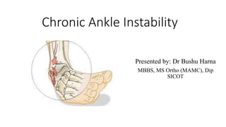
Chronic Ankle Instability
Presented by: Dr Bushu Harna
MBBS, MS Ortho (MAMC), Dip
SICOT
• Chronic lateral ankle instability (CLAI) is defined as recurrent sprains or repeated
“giving way” resulting from trauma for at least 1 year.
• Giving way is considered an uncontrolled or unpredictable excessive inversion of
the ankle joints that occurs at heel strike or toe-off during walking or running.
• It is estimated that 80 to 85% of ankle sprains occur to the lateral ligaments an
inversion ankle
• Only risk factor is previous ankle sprain
• Lateral ankle ligaments and bone congruency are main static stabilizers.
• Peroneal tendons provide dynamic stability.
• ATFL is most common injured ligament
LATERAL LIGAMENTS INJURY
• Inversion type of sprains
• Happens due to unstable landing after a jump or running/walking on an uneven
surface. It results from the inversion and plantarflexion of the foot injuring the
ATFL.
• A lot of force applied to the ATFL during the inversion sprain may break it and
affect the CFL. The effect occurs because the CFL is the next ligament supposed to
take the stress.
• The CFL can be injured if the inversion sprain is extreme enough.
• Anterior Talofibular Ligament
• ATFL primarily resists translational laxity of the talus in the sagittal plane.
• ATFL has the lowest threshold for failure and thus is the most commonly injured
ligament.
• Calcaneofibular Ligament
• CFL resists excessive supination at both the mortise and the subtalar joint.
• Posterior Talofibular Ligament
• PTFL is a robust ligament with a broad surface conferring a high tensile strength.
• Provides resistance to inversion and internal rotation.
• Injury to the PTFL is the least likely to occur.
Pathogenesis
• Usually the sequelae of an acute sprain that fails to heal properly.
• Ligaments may remain torn.
• Most commonly, heal but in an elongated fashion.
• Can result from loss of proprioception, muscle weakness, nerve injury, or tendon
damage.
Two main types of instability can be distinguished:
• Mechanical instability related to anatomic abnormalities of the ankle, usually related
to ligament laxity.
• Functional instability related to posture defects or tendon and muscle adjustment,
usually related to a proprioceptive deficit.
Mechanical instability
• Bone instability: a congruence defect as well as a more anterior position of the
talus in relation to the tibia on the loading.
• The lateral malleolus seems to be in a posterior position because of distension or
rupture of the anterior talofibular ligament.
Functional Instability
• Can result from alteration in proprioception, neuromuscular control, postural
control and the resultant strength deficits after sustaining an ankle sprain.
• The end result is a propensity for recurrent ankle sprains, persistent pain, and
muscular weakness.
Presentation
• history of injuries
• occurrence of repeated episodes of lateral ankle instability
• pain
• swelling
• ‘‘giving way”
• loss of motion
Physical Exam
• Lateral sided ankle tenderness
• Range of motion
• Provocative tests including anterior drawer (sulcus sign) and
• Talar tilt test
• Neuromuscular exam
• Assessment for hindfoot alignment, equinus contracture
• Weakness of peronei
• generalized signs of hypermobility
• Romberg test for proprioception
Other Lateral Injuries
• 5th metatarsal avulsion fracture
• Jones fracture
• Peroneus brevis tendon tear
• Peroneus longus tendinopathy
• Cuboid fracture
Anterior drawer test:
• The patient needs to be relaxed, seated with the knee flexed and
the ankle plantar flexed around 10-20 degrees.
• One of the examiner’s hands stabilises the tibia and, with the
other hand, the foot is pulled forwards.
• Stabilise the heel on the bed with one hand and push tibia with
the other hand.
• The test should be compared with the contralateral ankle.
• A sulcus sign is present when a dimple is seen in the lateral
gutter on testing.
Talar tilt test: Can evaluates both ATFL and CFL.
• With the ankle plantigrade the hindfoot is tilted one way.
• Pain or gap at palpation.
Imaging Studies
• Plain standing films--Alignment and bone injury
• Stress radiographs comparing the affected side to the
unaffected sign are the gold standard for mechanical
stability.
Stress Views
• A difference of >4mm in anterior drawer and >6 degrees on
talar tilt is significant for instability.
• >15 degrees of talar tilt or >1 cm anterior translation also
usually considered abnormal.
• Ultrasound
• MRI: Allows not only the assessment of the ATFL and CFL but also of
local tendons and bones.
Treatment
• Non-operative treatment is the first line of therapy.
• Strength training and proprioception instruction are used, focusing on taking the ankle
through a full range of normal motion.
• Bracing may help protect from reinjury but does not correct underlying pathology.
• Taping/ lateral heal wedges
• Peroneal strengthening
Surgical Treatment
• Indicated if instability symptoms persist after 3-5 months of rehab attempts.
• FI with no MI = no surgery
• MI with no FI = no surgery
• MI with FI = proprioception, strength training. If no improvement then surgery
Two main types: anatomic repair or non anatomic/ tenodesis stabilization
• Anatomic repair restores normal anatomy and biomechanics while
maintaining ankle motion
• Tenodesis stabilization restricts laxity and pathologic motion but ignores
the underlying ligamentous pathology. Stabilization is achieved at the
expense of altering normal ankle and hindfoot biomechanics.
Anatomic Ligament repair
• Anatomical repair gives the best results and is indicated in most patients. Anatomical
repair of the existing ATFL and CFL to bone was described by Brostrom but the
modification by Gould is currently most popular. Gould recommended reinforcement
of the repair using inferior extensor retinaculum.
• Anatomical repair is not only simple, it also restores ankle kinematics to near normal
compared to ligament reconstruction techniques.
• Arthroscopic Brostrom-Gould repair.
Ligament reconstruction: Can be performed using a number of options: native
autografts, allografts or synthetic implants.
• May be a role for early ligament reconstruction in patients with a high BMI, heavy
manual jobs, sportsmen or those with generalised ligament laxity who may
overstress the repaired ligament and are therefore at high risk of failure of primary
repair.
• Reconstruction is also indicated when there is insufficient native ligament left for
adequate repair.
• Arthroscopic assessment reveals the ATFL to be highly irregular in appearance.
Non-anatomic lateral ligament reconstruction: A number of procedures have been
described.
• Watson Jones described re-routing of the peroneus brevis (PB) tendon between the
talus and the calcaneus.
• This was subsequently modified by Evans, who preferred re-routing via the distal
fibula.
• Chrisman and Snook detached only a portion of PB, keeping its distal attachment
intact.
• The main criticism: Restrict the movement of the subtalar joint and also sacrifice the
PB tendon, which is an important lateral stabiliser of the ankle.
Recommendations
1.Surgery is suggested when patients continue to have symptoms of CLAI after 3 to 6
months of nonsurgical treatment and have indications of CLAI on physical and
imaging examinations (stress radiography or MRI).
2.Simultaneous surgery for CLAI and OCL lesions of the talus or tibia is suggested
when both are present.
3.Open and arthroscopic procedures are both recommended in patients with CLAI
who undergo anatomic repair or reconstruction.
4.Autograft and allograft are both recommended for anatomic reconstruction in
patients with CLAI.
5.When patients with CLAI have subtalar joint instability and both the ATFL and the
CFL are ruptured, ATFL and CFL reconstruction is suggested.
6. For patients with CLAI who undergo anatomic repair or reconstruction, ROM
(mostly dorsiflexion) and partial weightbearing with a brace from the second day after
surgery are recommended; for those with OCL who undergo anatomic repair or
reconstruction and additional microfracture, early ROM and relatively delayed
weightbearing are recommended.
7. Joint stability, muscle strength, and full ROM are recommended prior to returning to
sport after surgery.
Thank You for Patient Listening

More Related Content

What's hot

Rotator cuff Tear and its management
Rotator cuff Tear and its managementRotator cuff Tear and its management
Rotator cuff Tear and its managementRohan Vakta
 
SPINAL STABILIZATION PPT
SPINAL STABILIZATION PPTSPINAL STABILIZATION PPT
SPINAL STABILIZATION PPTssuser2f50ef
 
Arthroscopic Rotator Cuff Repair
Arthroscopic Rotator Cuff RepairArthroscopic Rotator Cuff Repair
Arthroscopic Rotator Cuff RepairBijayendra Singh
 
Seminar recent advances reverse shoulder arthroplasty
Seminar recent  advances reverse shoulder arthroplastySeminar recent  advances reverse shoulder arthroplasty
Seminar recent advances reverse shoulder arthroplastyBipulBorthakur
 
Frieberg’s metatarsalgia
Frieberg’s metatarsalgiaFrieberg’s metatarsalgia
Frieberg’s metatarsalgiaorthoprince
 
Hand rehabilitation following flexor tendon injuries
Hand rehabilitation following flexor tendon injuriesHand rehabilitation following flexor tendon injuries
Hand rehabilitation following flexor tendon injuriesAbey P Rajan
 
High tibial osteotomy
High tibial osteotomyHigh tibial osteotomy
High tibial osteotomyorthoprince
 
Techniques in primary total knee arthroplasty
Techniques in primary total knee arthroplastyTechniques in primary total knee arthroplasty
Techniques in primary total knee arthroplastyHBGMedical
 
Total elbow arthroplasty
Total elbow arthroplastyTotal elbow arthroplasty
Total elbow arthroplastySudheer Kumar
 
Knee Arthrodesis
Knee ArthrodesisKnee Arthrodesis
Knee Arthrodesisdrsp46
 
Limb length discrepancy
Limb length discrepancyLimb length discrepancy
Limb length discrepancyMadhukar Reddy
 
Arthroscopic bankart’s repair
Arthroscopic bankart’s repairArthroscopic bankart’s repair
Arthroscopic bankart’s repairorthoprince
 
Principles of deformity correction
Principles of deformity correctionPrinciples of deformity correction
Principles of deformity correctionAbdulla Kamal
 
Graft choices for Anterior Cruciate Ligament - ACL Reconstruction - Dr Chinta...
Graft choices for Anterior Cruciate Ligament - ACL Reconstruction - Dr Chinta...Graft choices for Anterior Cruciate Ligament - ACL Reconstruction - Dr Chinta...
Graft choices for Anterior Cruciate Ligament - ACL Reconstruction - Dr Chinta...DrChintan Patel
 

What's hot (20)

Rotator cuff Tear and its management
Rotator cuff Tear and its managementRotator cuff Tear and its management
Rotator cuff Tear and its management
 
SPINAL STABILIZATION PPT
SPINAL STABILIZATION PPTSPINAL STABILIZATION PPT
SPINAL STABILIZATION PPT
 
Arthroscopic Rotator Cuff Repair
Arthroscopic Rotator Cuff RepairArthroscopic Rotator Cuff Repair
Arthroscopic Rotator Cuff Repair
 
ACL Reconstruction in the Adolescent Athlete
ACL Reconstruction in the Adolescent AthleteACL Reconstruction in the Adolescent Athlete
ACL Reconstruction in the Adolescent Athlete
 
ACL rehabilitation
ACL rehabilitationACL rehabilitation
ACL rehabilitation
 
Seminar recent advances reverse shoulder arthroplasty
Seminar recent  advances reverse shoulder arthroplastySeminar recent  advances reverse shoulder arthroplasty
Seminar recent advances reverse shoulder arthroplasty
 
Frieberg’s metatarsalgia
Frieberg’s metatarsalgiaFrieberg’s metatarsalgia
Frieberg’s metatarsalgia
 
Hand rehabilitation following flexor tendon injuries
Hand rehabilitation following flexor tendon injuriesHand rehabilitation following flexor tendon injuries
Hand rehabilitation following flexor tendon injuries
 
High tibial osteotomy
High tibial osteotomyHigh tibial osteotomy
High tibial osteotomy
 
TENS
TENSTENS
TENS
 
Techniques in primary total knee arthroplasty
Techniques in primary total knee arthroplastyTechniques in primary total knee arthroplasty
Techniques in primary total knee arthroplasty
 
Total elbow arthroplasty
Total elbow arthroplastyTotal elbow arthroplasty
Total elbow arthroplasty
 
Mulligan mobilization (MWM)
Mulligan mobilization (MWM)Mulligan mobilization (MWM)
Mulligan mobilization (MWM)
 
Knee Arthrodesis
Knee ArthrodesisKnee Arthrodesis
Knee Arthrodesis
 
Limb length discrepancy
Limb length discrepancyLimb length discrepancy
Limb length discrepancy
 
Arthroscopic bankart’s repair
Arthroscopic bankart’s repairArthroscopic bankart’s repair
Arthroscopic bankart’s repair
 
Hip biomechanics
Hip biomechanicsHip biomechanics
Hip biomechanics
 
Principles of deformity correction
Principles of deformity correctionPrinciples of deformity correction
Principles of deformity correction
 
Graft choices for Anterior Cruciate Ligament - ACL Reconstruction - Dr Chinta...
Graft choices for Anterior Cruciate Ligament - ACL Reconstruction - Dr Chinta...Graft choices for Anterior Cruciate Ligament - ACL Reconstruction - Dr Chinta...
Graft choices for Anterior Cruciate Ligament - ACL Reconstruction - Dr Chinta...
 
Pes planus
Pes planusPes planus
Pes planus
 

Similar to Chronic ankle instability

Ligament injury to knee: ACL
Ligament injury to knee: ACLLigament injury to knee: ACL
Ligament injury to knee: ACLSijan Bhattachan
 
Chronic ankle instability and syndesmotic injuries
Chronic ankle instability and syndesmotic injuriesChronic ankle instability and syndesmotic injuries
Chronic ankle instability and syndesmotic injuriesKent Heady
 
Ankle instability
Ankle instabilityAnkle instability
Ankle instabilityRziUllah
 
TKA in valgus knee.pptx
TKA in valgus knee.pptxTKA in valgus knee.pptx
TKA in valgus knee.pptxAjayShringeri
 
Osteoarthritis by Dr. K. A Rana -2.pptx
Osteoarthritis    by Dr. K. A Rana -2.pptxOsteoarthritis    by Dr. K. A Rana -2.pptx
Osteoarthritis by Dr. K. A Rana -2.pptxkhushirana69
 
Knee dislocation powerpoint for medical students
Knee dislocation powerpoint for medical studentsKnee dislocation powerpoint for medical students
Knee dislocation powerpoint for medical studentsCasualityShift
 
Posterolateral corner injuries of knee joint
Posterolateral corner injuries of knee joint Posterolateral corner injuries of knee joint
Posterolateral corner injuries of knee joint Samir Dwidmuthe
 
acl injuries.pptx
acl injuries.pptxacl injuries.pptx
acl injuries.pptxArbind Shah
 
SPORTS INJURIES OF ANKLE AND FOOT original.pptx
SPORTS INJURIES OF ANKLE AND FOOT original.pptxSPORTS INJURIES OF ANKLE AND FOOT original.pptx
SPORTS INJURIES OF ANKLE AND FOOT original.pptxMeghaPrakash9
 
ACL.injury.final year.pptx
ACL.injury.final year.pptxACL.injury.final year.pptx
ACL.injury.final year.pptxDipaliTalaviya1
 
knee_ligament_injuries.pptx
knee_ligament_injuries.pptxknee_ligament_injuries.pptx
knee_ligament_injuries.pptxAteeqrahman24
 
Internal_derangements_of_Knee.pptx in orthopaedics
Internal_derangements_of_Knee.pptx in orthopaedicsInternal_derangements_of_Knee.pptx in orthopaedics
Internal_derangements_of_Knee.pptx in orthopaedicsRitikaChoudhary85
 

Similar to Chronic ankle instability (20)

Ligament injury to knee: ACL
Ligament injury to knee: ACLLigament injury to knee: ACL
Ligament injury to knee: ACL
 
ACL.pdf
ACL.pdfACL.pdf
ACL.pdf
 
Chronic ankle instability and syndesmotic injuries
Chronic ankle instability and syndesmotic injuriesChronic ankle instability and syndesmotic injuries
Chronic ankle instability and syndesmotic injuries
 
Acl injury
Acl injuryAcl injury
Acl injury
 
Ankle instability
Ankle instabilityAnkle instability
Ankle instability
 
TKA in valgus knee.pptx
TKA in valgus knee.pptxTKA in valgus knee.pptx
TKA in valgus knee.pptx
 
Osteoarthritis by Dr. K. A Rana -2.pptx
Osteoarthritis    by Dr. K. A Rana -2.pptxOsteoarthritis    by Dr. K. A Rana -2.pptx
Osteoarthritis by Dr. K. A Rana -2.pptx
 
Ankle fracture
Ankle fractureAnkle fracture
Ankle fracture
 
Knee dislocation powerpoint for medical students
Knee dislocation powerpoint for medical studentsKnee dislocation powerpoint for medical students
Knee dislocation powerpoint for medical students
 
Posterolateral corner injuries of knee joint
Posterolateral corner injuries of knee joint Posterolateral corner injuries of knee joint
Posterolateral corner injuries of knee joint
 
Chondromalacia Patellar.pptx
Chondromalacia Patellar.pptxChondromalacia Patellar.pptx
Chondromalacia Patellar.pptx
 
acl injuries.pptx
acl injuries.pptxacl injuries.pptx
acl injuries.pptx
 
SPORTS INJURIES OF ANKLE AND FOOT original.pptx
SPORTS INJURIES OF ANKLE AND FOOT original.pptxSPORTS INJURIES OF ANKLE AND FOOT original.pptx
SPORTS INJURIES OF ANKLE AND FOOT original.pptx
 
Cruciate Ligaments
Cruciate LigamentsCruciate Ligaments
Cruciate Ligaments
 
ACL.injury.final year.pptx
ACL.injury.final year.pptxACL.injury.final year.pptx
ACL.injury.final year.pptx
 
Orthopedics 2
Orthopedics 2Orthopedics 2
Orthopedics 2
 
knee_ligament_injuries.pptx
knee_ligament_injuries.pptxknee_ligament_injuries.pptx
knee_ligament_injuries.pptx
 
Internal_derangements_of_Knee.pptx in orthopaedics
Internal_derangements_of_Knee.pptx in orthopaedicsInternal_derangements_of_Knee.pptx in orthopaedics
Internal_derangements_of_Knee.pptx in orthopaedics
 
Ankle fracture
Ankle fractureAnkle fracture
Ankle fracture
 
Ankle Sprains
Ankle SprainsAnkle Sprains
Ankle Sprains
 

Recently uploaded

Unlocking Holistic Wellness: Addressing Depression, Mental Well-Being, and St...
Unlocking Holistic Wellness: Addressing Depression, Mental Well-Being, and St...Unlocking Holistic Wellness: Addressing Depression, Mental Well-Being, and St...
Unlocking Holistic Wellness: Addressing Depression, Mental Well-Being, and St...Health Kinesiology Natural Bioenergetics
 
Cytoskeleton and Cell Inclusions - Dr Muhammad Ali Rabbani - Medicose Academics
Cytoskeleton and Cell Inclusions - Dr Muhammad Ali Rabbani - Medicose AcademicsCytoskeleton and Cell Inclusions - Dr Muhammad Ali Rabbani - Medicose Academics
Cytoskeleton and Cell Inclusions - Dr Muhammad Ali Rabbani - Medicose AcademicsMedicoseAcademics
 
Hemodialysis: Chapter 1, Physiological Principles of Hemodialysis - Dr.Gawad
Hemodialysis: Chapter 1, Physiological Principles of Hemodialysis - Dr.GawadHemodialysis: Chapter 1, Physiological Principles of Hemodialysis - Dr.Gawad
Hemodialysis: Chapter 1, Physiological Principles of Hemodialysis - Dr.GawadNephroTube - Dr.Gawad
 
ANAPHYLAXIS BY DR.SOHAN BISWAS,MBBS,DNB(INTERNAL MEDICINE) RESIDENT.pptx
ANAPHYLAXIS BY DR.SOHAN BISWAS,MBBS,DNB(INTERNAL MEDICINE) RESIDENT.pptxANAPHYLAXIS BY DR.SOHAN BISWAS,MBBS,DNB(INTERNAL MEDICINE) RESIDENT.pptx
ANAPHYLAXIS BY DR.SOHAN BISWAS,MBBS,DNB(INTERNAL MEDICINE) RESIDENT.pptxDr. Sohan Biswas
 
Premium ℂall Girls In Mumbai👉 Dail ℂALL ME: 📞9833325238 📲 ℂall Richa VIP ℂall...
Premium ℂall Girls In Mumbai👉 Dail ℂALL ME: 📞9833325238 📲 ℂall Richa VIP ℂall...Premium ℂall Girls In Mumbai👉 Dail ℂALL ME: 📞9833325238 📲 ℂall Richa VIP ℂall...
Premium ℂall Girls In Mumbai👉 Dail ℂALL ME: 📞9833325238 📲 ℂall Richa VIP ℂall...Avani bhatt
 
Denture base resins materials and its mechanism of action
Denture base resins materials and its mechanism of actionDenture base resins materials and its mechanism of action
Denture base resins materials and its mechanism of actionDr.shiva sai vemula
 
PYODERMA, IMPETIGO, FOLLICULITIS, FURUNCLES, CARBUNCLES.pdf
PYODERMA, IMPETIGO, FOLLICULITIS, FURUNCLES, CARBUNCLES.pdfPYODERMA, IMPETIGO, FOLLICULITIS, FURUNCLES, CARBUNCLES.pdf
PYODERMA, IMPETIGO, FOLLICULITIS, FURUNCLES, CARBUNCLES.pdfDolisha Warbi
 
Tips and tricks to pass the cardiovascular station for PACES exam
Tips and tricks to pass the cardiovascular station for PACES examTips and tricks to pass the cardiovascular station for PACES exam
Tips and tricks to pass the cardiovascular station for PACES examJunhao Koh
 
Treatment Choices for Slip Disc at Gokuldas Hospital
Treatment Choices for Slip Disc at Gokuldas HospitalTreatment Choices for Slip Disc at Gokuldas Hospital
Treatment Choices for Slip Disc at Gokuldas HospitalGokuldas Hospital
 
VIP Pune 7877925207 WhatsApp: Me All Time Serviℂe Available Day and Night
VIP Pune 7877925207 WhatsApp: Me All Time Serviℂe Available Day and NightVIP Pune 7877925207 WhatsApp: Me All Time Serviℂe Available Day and Night
VIP Pune 7877925207 WhatsApp: Me All Time Serviℂe Available Day and Nightpatanjali9823#S07
 
Histology of Epithelium - Dr Muhammad Ali Rabbani - Medicose Academics
Histology of Epithelium - Dr Muhammad Ali Rabbani - Medicose AcademicsHistology of Epithelium - Dr Muhammad Ali Rabbani - Medicose Academics
Histology of Epithelium - Dr Muhammad Ali Rabbani - Medicose AcademicsMedicoseAcademics
 
The Clean Living Project Episode 24 - Subconscious
The Clean Living Project Episode 24 - SubconsciousThe Clean Living Project Episode 24 - Subconscious
The Clean Living Project Episode 24 - SubconsciousThe Clean Living Project
 
Signs It’s Time for Physiotherapy Sessions Prioritizing Wellness
Signs It’s Time for Physiotherapy Sessions Prioritizing WellnessSigns It’s Time for Physiotherapy Sessions Prioritizing Wellness
Signs It’s Time for Physiotherapy Sessions Prioritizing WellnessGokuldas Hospital
 
Failure to thrive in neonates and infants + pediatric case.pptx
Failure to thrive in neonates and infants  + pediatric case.pptxFailure to thrive in neonates and infants  + pediatric case.pptx
Failure to thrive in neonates and infants + pediatric case.pptxclaviclebrown44
 
Gallbladder Double-Diverticular: A Case Report المرارة مزدوجة التج: تقرير حالة
Gallbladder Double-Diverticular: A Case Report  المرارة مزدوجة التج: تقرير حالةGallbladder Double-Diverticular: A Case Report  المرارة مزدوجة التج: تقرير حالة
Gallbladder Double-Diverticular: A Case Report المرارة مزدوجة التج: تقرير حالةMohamad محمد Al-Gailani الكيلاني
 
Connective Tissue II - Dr Muhammad Ali Rabbani - Medicose Academics
Connective Tissue II - Dr Muhammad Ali Rabbani - Medicose AcademicsConnective Tissue II - Dr Muhammad Ali Rabbani - Medicose Academics
Connective Tissue II - Dr Muhammad Ali Rabbani - Medicose AcademicsMedicoseAcademics
 
Tissue Banking and Umbilical Cord Blood Banking
Tissue Banking and Umbilical Cord Blood BankingTissue Banking and Umbilical Cord Blood Banking
Tissue Banking and Umbilical Cord Blood BankingDrShinyKajal
 
Renal Replacement Therapy in Acute Kidney Injury -time modality -Dr Ayman Se...
Renal Replacement Therapy in Acute Kidney Injury -time  modality -Dr Ayman Se...Renal Replacement Therapy in Acute Kidney Injury -time  modality -Dr Ayman Se...
Renal Replacement Therapy in Acute Kidney Injury -time modality -Dr Ayman Se...Ayman Seddik
 

Recently uploaded (20)

Unlocking Holistic Wellness: Addressing Depression, Mental Well-Being, and St...
Unlocking Holistic Wellness: Addressing Depression, Mental Well-Being, and St...Unlocking Holistic Wellness: Addressing Depression, Mental Well-Being, and St...
Unlocking Holistic Wellness: Addressing Depression, Mental Well-Being, and St...
 
Cytoskeleton and Cell Inclusions - Dr Muhammad Ali Rabbani - Medicose Academics
Cytoskeleton and Cell Inclusions - Dr Muhammad Ali Rabbani - Medicose AcademicsCytoskeleton and Cell Inclusions - Dr Muhammad Ali Rabbani - Medicose Academics
Cytoskeleton and Cell Inclusions - Dr Muhammad Ali Rabbani - Medicose Academics
 
Hemodialysis: Chapter 1, Physiological Principles of Hemodialysis - Dr.Gawad
Hemodialysis: Chapter 1, Physiological Principles of Hemodialysis - Dr.GawadHemodialysis: Chapter 1, Physiological Principles of Hemodialysis - Dr.Gawad
Hemodialysis: Chapter 1, Physiological Principles of Hemodialysis - Dr.Gawad
 
ANAPHYLAXIS BY DR.SOHAN BISWAS,MBBS,DNB(INTERNAL MEDICINE) RESIDENT.pptx
ANAPHYLAXIS BY DR.SOHAN BISWAS,MBBS,DNB(INTERNAL MEDICINE) RESIDENT.pptxANAPHYLAXIS BY DR.SOHAN BISWAS,MBBS,DNB(INTERNAL MEDICINE) RESIDENT.pptx
ANAPHYLAXIS BY DR.SOHAN BISWAS,MBBS,DNB(INTERNAL MEDICINE) RESIDENT.pptx
 
Premium ℂall Girls In Mumbai👉 Dail ℂALL ME: 📞9833325238 📲 ℂall Richa VIP ℂall...
Premium ℂall Girls In Mumbai👉 Dail ℂALL ME: 📞9833325238 📲 ℂall Richa VIP ℂall...Premium ℂall Girls In Mumbai👉 Dail ℂALL ME: 📞9833325238 📲 ℂall Richa VIP ℂall...
Premium ℂall Girls In Mumbai👉 Dail ℂALL ME: 📞9833325238 📲 ℂall Richa VIP ℂall...
 
Denture base resins materials and its mechanism of action
Denture base resins materials and its mechanism of actionDenture base resins materials and its mechanism of action
Denture base resins materials and its mechanism of action
 
PYODERMA, IMPETIGO, FOLLICULITIS, FURUNCLES, CARBUNCLES.pdf
PYODERMA, IMPETIGO, FOLLICULITIS, FURUNCLES, CARBUNCLES.pdfPYODERMA, IMPETIGO, FOLLICULITIS, FURUNCLES, CARBUNCLES.pdf
PYODERMA, IMPETIGO, FOLLICULITIS, FURUNCLES, CARBUNCLES.pdf
 
Tips and tricks to pass the cardiovascular station for PACES exam
Tips and tricks to pass the cardiovascular station for PACES examTips and tricks to pass the cardiovascular station for PACES exam
Tips and tricks to pass the cardiovascular station for PACES exam
 
Treatment Choices for Slip Disc at Gokuldas Hospital
Treatment Choices for Slip Disc at Gokuldas HospitalTreatment Choices for Slip Disc at Gokuldas Hospital
Treatment Choices for Slip Disc at Gokuldas Hospital
 
VIP Pune 7877925207 WhatsApp: Me All Time Serviℂe Available Day and Night
VIP Pune 7877925207 WhatsApp: Me All Time Serviℂe Available Day and NightVIP Pune 7877925207 WhatsApp: Me All Time Serviℂe Available Day and Night
VIP Pune 7877925207 WhatsApp: Me All Time Serviℂe Available Day and Night
 
Histology of Epithelium - Dr Muhammad Ali Rabbani - Medicose Academics
Histology of Epithelium - Dr Muhammad Ali Rabbani - Medicose AcademicsHistology of Epithelium - Dr Muhammad Ali Rabbani - Medicose Academics
Histology of Epithelium - Dr Muhammad Ali Rabbani - Medicose Academics
 
The Clean Living Project Episode 24 - Subconscious
The Clean Living Project Episode 24 - SubconsciousThe Clean Living Project Episode 24 - Subconscious
The Clean Living Project Episode 24 - Subconscious
 
Signs It’s Time for Physiotherapy Sessions Prioritizing Wellness
Signs It’s Time for Physiotherapy Sessions Prioritizing WellnessSigns It’s Time for Physiotherapy Sessions Prioritizing Wellness
Signs It’s Time for Physiotherapy Sessions Prioritizing Wellness
 
Failure to thrive in neonates and infants + pediatric case.pptx
Failure to thrive in neonates and infants  + pediatric case.pptxFailure to thrive in neonates and infants  + pediatric case.pptx
Failure to thrive in neonates and infants + pediatric case.pptx
 
Gallbladder Double-Diverticular: A Case Report المرارة مزدوجة التج: تقرير حالة
Gallbladder Double-Diverticular: A Case Report  المرارة مزدوجة التج: تقرير حالةGallbladder Double-Diverticular: A Case Report  المرارة مزدوجة التج: تقرير حالة
Gallbladder Double-Diverticular: A Case Report المرارة مزدوجة التج: تقرير حالة
 
Connective Tissue II - Dr Muhammad Ali Rabbani - Medicose Academics
Connective Tissue II - Dr Muhammad Ali Rabbani - Medicose AcademicsConnective Tissue II - Dr Muhammad Ali Rabbani - Medicose Academics
Connective Tissue II - Dr Muhammad Ali Rabbani - Medicose Academics
 
Best medicine 100% Effective&Safe Mifepristion ௵+918133066128௹Abortion pills ...
Best medicine 100% Effective&Safe Mifepristion ௵+918133066128௹Abortion pills ...Best medicine 100% Effective&Safe Mifepristion ௵+918133066128௹Abortion pills ...
Best medicine 100% Effective&Safe Mifepristion ௵+918133066128௹Abortion pills ...
 
Tissue Banking and Umbilical Cord Blood Banking
Tissue Banking and Umbilical Cord Blood BankingTissue Banking and Umbilical Cord Blood Banking
Tissue Banking and Umbilical Cord Blood Banking
 
Making Patient-Centric Immunotherapy a Reality in Lung Cancer: Best Practices...
Making Patient-Centric Immunotherapy a Reality in Lung Cancer: Best Practices...Making Patient-Centric Immunotherapy a Reality in Lung Cancer: Best Practices...
Making Patient-Centric Immunotherapy a Reality in Lung Cancer: Best Practices...
 
Renal Replacement Therapy in Acute Kidney Injury -time modality -Dr Ayman Se...
Renal Replacement Therapy in Acute Kidney Injury -time  modality -Dr Ayman Se...Renal Replacement Therapy in Acute Kidney Injury -time  modality -Dr Ayman Se...
Renal Replacement Therapy in Acute Kidney Injury -time modality -Dr Ayman Se...
 

Chronic ankle instability

  • 1. Chronic Ankle Instability Presented by: Dr Bushu Harna MBBS, MS Ortho (MAMC), Dip SICOT
  • 2. • Chronic lateral ankle instability (CLAI) is defined as recurrent sprains or repeated “giving way” resulting from trauma for at least 1 year. • Giving way is considered an uncontrolled or unpredictable excessive inversion of the ankle joints that occurs at heel strike or toe-off during walking or running. • It is estimated that 80 to 85% of ankle sprains occur to the lateral ligaments an inversion ankle • Only risk factor is previous ankle sprain • Lateral ankle ligaments and bone congruency are main static stabilizers. • Peroneal tendons provide dynamic stability. • ATFL is most common injured ligament
  • 3.
  • 4. LATERAL LIGAMENTS INJURY • Inversion type of sprains • Happens due to unstable landing after a jump or running/walking on an uneven surface. It results from the inversion and plantarflexion of the foot injuring the ATFL. • A lot of force applied to the ATFL during the inversion sprain may break it and affect the CFL. The effect occurs because the CFL is the next ligament supposed to take the stress. • The CFL can be injured if the inversion sprain is extreme enough.
  • 5. • Anterior Talofibular Ligament • ATFL primarily resists translational laxity of the talus in the sagittal plane. • ATFL has the lowest threshold for failure and thus is the most commonly injured ligament. • Calcaneofibular Ligament • CFL resists excessive supination at both the mortise and the subtalar joint. • Posterior Talofibular Ligament • PTFL is a robust ligament with a broad surface conferring a high tensile strength. • Provides resistance to inversion and internal rotation. • Injury to the PTFL is the least likely to occur.
  • 6. Pathogenesis • Usually the sequelae of an acute sprain that fails to heal properly. • Ligaments may remain torn. • Most commonly, heal but in an elongated fashion. • Can result from loss of proprioception, muscle weakness, nerve injury, or tendon damage. Two main types of instability can be distinguished: • Mechanical instability related to anatomic abnormalities of the ankle, usually related to ligament laxity. • Functional instability related to posture defects or tendon and muscle adjustment, usually related to a proprioceptive deficit.
  • 7. Mechanical instability • Bone instability: a congruence defect as well as a more anterior position of the talus in relation to the tibia on the loading. • The lateral malleolus seems to be in a posterior position because of distension or rupture of the anterior talofibular ligament. Functional Instability • Can result from alteration in proprioception, neuromuscular control, postural control and the resultant strength deficits after sustaining an ankle sprain. • The end result is a propensity for recurrent ankle sprains, persistent pain, and muscular weakness.
  • 8.
  • 9.
  • 10. Presentation • history of injuries • occurrence of repeated episodes of lateral ankle instability • pain • swelling • ‘‘giving way” • loss of motion
  • 11. Physical Exam • Lateral sided ankle tenderness • Range of motion • Provocative tests including anterior drawer (sulcus sign) and • Talar tilt test • Neuromuscular exam • Assessment for hindfoot alignment, equinus contracture • Weakness of peronei • generalized signs of hypermobility • Romberg test for proprioception
  • 12. Other Lateral Injuries • 5th metatarsal avulsion fracture • Jones fracture • Peroneus brevis tendon tear • Peroneus longus tendinopathy • Cuboid fracture
  • 13. Anterior drawer test: • The patient needs to be relaxed, seated with the knee flexed and the ankle plantar flexed around 10-20 degrees. • One of the examiner’s hands stabilises the tibia and, with the other hand, the foot is pulled forwards. • Stabilise the heel on the bed with one hand and push tibia with the other hand. • The test should be compared with the contralateral ankle. • A sulcus sign is present when a dimple is seen in the lateral gutter on testing.
  • 14. Talar tilt test: Can evaluates both ATFL and CFL. • With the ankle plantigrade the hindfoot is tilted one way. • Pain or gap at palpation.
  • 15. Imaging Studies • Plain standing films--Alignment and bone injury • Stress radiographs comparing the affected side to the unaffected sign are the gold standard for mechanical stability. Stress Views • A difference of >4mm in anterior drawer and >6 degrees on talar tilt is significant for instability. • >15 degrees of talar tilt or >1 cm anterior translation also usually considered abnormal.
  • 16. • Ultrasound • MRI: Allows not only the assessment of the ATFL and CFL but also of local tendons and bones.
  • 17. Treatment • Non-operative treatment is the first line of therapy. • Strength training and proprioception instruction are used, focusing on taking the ankle through a full range of normal motion. • Bracing may help protect from reinjury but does not correct underlying pathology. • Taping/ lateral heal wedges • Peroneal strengthening
  • 18. Surgical Treatment • Indicated if instability symptoms persist after 3-5 months of rehab attempts. • FI with no MI = no surgery • MI with no FI = no surgery • MI with FI = proprioception, strength training. If no improvement then surgery
  • 19. Two main types: anatomic repair or non anatomic/ tenodesis stabilization • Anatomic repair restores normal anatomy and biomechanics while maintaining ankle motion • Tenodesis stabilization restricts laxity and pathologic motion but ignores the underlying ligamentous pathology. Stabilization is achieved at the expense of altering normal ankle and hindfoot biomechanics.
  • 20. Anatomic Ligament repair • Anatomical repair gives the best results and is indicated in most patients. Anatomical repair of the existing ATFL and CFL to bone was described by Brostrom but the modification by Gould is currently most popular. Gould recommended reinforcement of the repair using inferior extensor retinaculum. • Anatomical repair is not only simple, it also restores ankle kinematics to near normal compared to ligament reconstruction techniques. • Arthroscopic Brostrom-Gould repair.
  • 21. Ligament reconstruction: Can be performed using a number of options: native autografts, allografts or synthetic implants. • May be a role for early ligament reconstruction in patients with a high BMI, heavy manual jobs, sportsmen or those with generalised ligament laxity who may overstress the repaired ligament and are therefore at high risk of failure of primary repair. • Reconstruction is also indicated when there is insufficient native ligament left for adequate repair. • Arthroscopic assessment reveals the ATFL to be highly irregular in appearance.
  • 22. Non-anatomic lateral ligament reconstruction: A number of procedures have been described. • Watson Jones described re-routing of the peroneus brevis (PB) tendon between the talus and the calcaneus. • This was subsequently modified by Evans, who preferred re-routing via the distal fibula. • Chrisman and Snook detached only a portion of PB, keeping its distal attachment intact. • The main criticism: Restrict the movement of the subtalar joint and also sacrifice the PB tendon, which is an important lateral stabiliser of the ankle.
  • 23.
  • 24. Recommendations 1.Surgery is suggested when patients continue to have symptoms of CLAI after 3 to 6 months of nonsurgical treatment and have indications of CLAI on physical and imaging examinations (stress radiography or MRI). 2.Simultaneous surgery for CLAI and OCL lesions of the talus or tibia is suggested when both are present. 3.Open and arthroscopic procedures are both recommended in patients with CLAI who undergo anatomic repair or reconstruction. 4.Autograft and allograft are both recommended for anatomic reconstruction in patients with CLAI. 5.When patients with CLAI have subtalar joint instability and both the ATFL and the CFL are ruptured, ATFL and CFL reconstruction is suggested.
  • 25. 6. For patients with CLAI who undergo anatomic repair or reconstruction, ROM (mostly dorsiflexion) and partial weightbearing with a brace from the second day after surgery are recommended; for those with OCL who undergo anatomic repair or reconstruction and additional microfracture, early ROM and relatively delayed weightbearing are recommended. 7. Joint stability, muscle strength, and full ROM are recommended prior to returning to sport after surgery.
  • 26. Thank You for Patient Listening