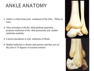
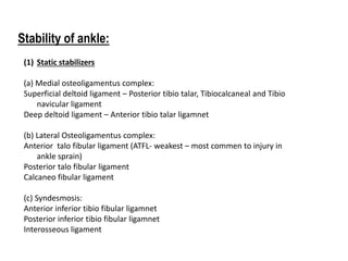
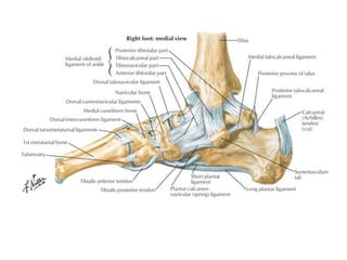
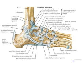
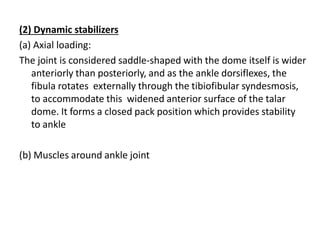
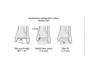
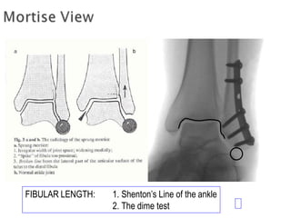
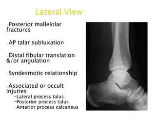
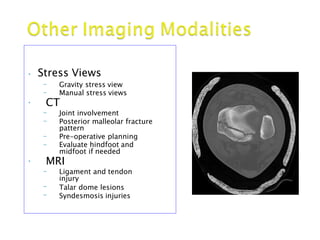
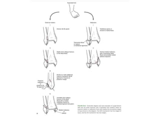
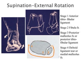
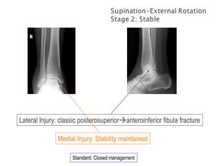
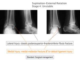
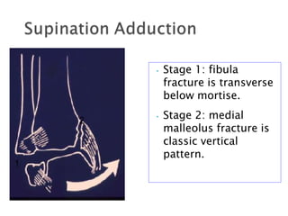
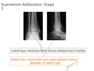
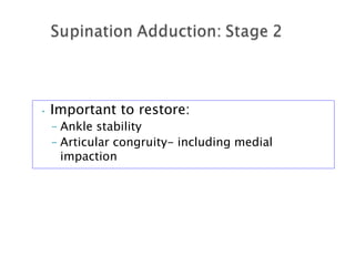
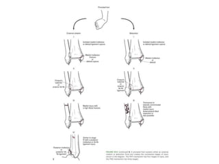
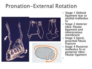
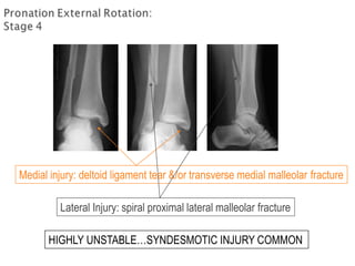
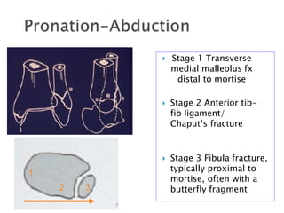
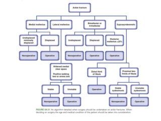
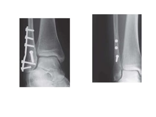
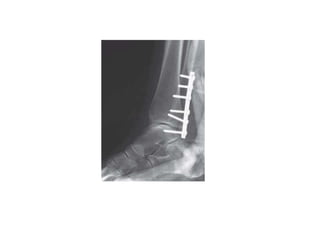
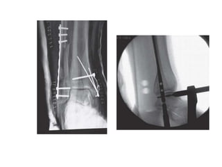
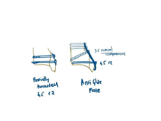
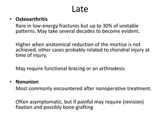
SlideShare a Scribd company logo
DR MRUDEV GANDHI
Resident
Dept of Orthopedics
Pramukh Swami Medical College, Karamsad
 Ankle is a three bone joint composed of the tibia , fibula an
talus.
 Talus articulates with the tibial plafond superiorly ,
posterior malleolus of the tibia posteriorly and medial
malleolus medially
 Lateral articulation is with malleolus of fibula
 Medial malleolus is shorter and anterior and thus axis of
the joint is 15 degrees of external rotation.
Stability of ankle:
(1) Static stabilizers
(a) Medial osteoligamentus complex:
Superficial deltoid ligament – Posterior tibio talar, Tibiocalcaneal and Tibio
navicular ligament
Deep deltoid ligament – Anterior tibio talar ligamnet
(b) Lateral Osteoligamentus complex:
Anterior talo fibular ligament (ATFL- weakest – most commen to injury in
ankle sprain)
Posterior talo fibular ligament
Calcaneo fibular ligament
(c) Syndesmosis:
Anterior inferior tibio fibular ligamnet
Posterior inferior tibio fibular ligamnet
Interosseous ligament
Anterior Colliculus
Posterior Colliculus
Intercollicular Groove
Medial malleolus consists of:
-Anterior Colliculus
-Intercollicular Groove
-Posterior Colliculus
Origi
Chaput tubercle
Wagstaffe
tubercle
Volkman
tubercle
(2) Dynamic stabilizers
(a) Axial loading:
The joint is considered saddle-shaped with the dome itself is wider
anteriorly than posteriorly, and as the ankle dorsiflexes, the
fibula rotates externally through the tibiofibular syndesmosis,
to accommodate this widened anterior surface of the talar
dome. It forms a closed pack position which provides stability
to ankle
(b) Muscles around ankle joint
INTRODUCTION
Ankle fractures are among the most common injuries and
management of these fractures depends upon careful
identification of the extent of bony injury as well as soft tissue
and ligamentous damage.
Once defined, the key to successful outcome following
rotational ankle fractures is anatomic restoration and healing of
ankle mortise.
Low velocity injuries a/w rotational component
Clinical Evaluation:
Detailed History
Detailed Examination
a/w soft tiuuse injuries
Compartment syndrome
IMAGING AND DIAGNOSTIC MODALITIES
OTTAWA ANKLE RULES
To manage the large volume of ankle injuries of patients who
presented to emergency certain criteria has been established for
requiring ankle radiographs.
Pain exists near one or both of the malleoli PLUS one or more of the
following:
•Age > 55 yrs old
•Inability to bear weight
•Bone tenderness over the posterior edge or tip of either malleolus .
•Plain Films
–AP, Mortise, Lateral
views of the ankle
–Image the entire
tibia to knee joint
–Foot films when
tender to palpation
– Common
associated fractures
are:
•5th metatarsal
base fracture
•Calcaneal
fracture
Although the OTTAWA RULES have been validated and found to be both cost
effective and reliable (up to 100% sensitivity their implementation has been
inconsistent in general clinical practice
Quaitative analysis◦Tibiofibular overlap
< 10mm is abnormal - implies
syndesmotic injury
◦Tibiofibular clear space
◦> 5mm is abnormal - implies
syndesmotic injury
◦Talar tilt
◦> 2mm is considered abnormal
Consider a comparison with
radiographs of the normal side if there
are unresolved concerns of injury
 Taken with ankle in
15-25 degrees of
internal rotation
 Useful in
evaluation of
articular surface
between talar
dome and mortise
10 degrees internal rotation of 5th MT with respect to a verticalline
 Medial clear space
◦ Between lateral border of
medial malleous and
medial talus
◦ <4mm is normal
◦ >4mm suggests lateral
shift of talus
FIBULAR LENGTH: 1. Shenton’s Line of the ankle
2. The dime test
•Posterior mallelolar
fractures
•AP talar subluxation
•Distal fibular translation
&/or angulation
•Syndesmotic relationship
•Associated or occult
injuries
–Lateral process talus
–Posterior process talus
–Anterior process calcaneus
• Stress Views
– Gravity stress view
– Manual stress views
• CT
Joint involvement
Posterior malleolar fracture
pattern
Pre-operative planning
–
–
–
– Evaluate hindfoot and
midfoot if needed
• MRI
Ligament and tendon–
–
–
injury
Talar dome lesions
Syndesmosis injuries
Radiography after reduction should be studied with
following requirements in mind:
•Normal relationship of ankle mortise must be restored.
•Weight bearing alignment of ankle must be at right angle to the
longitudinal axis of leg
•Counters of the articular surface must be as smooth as possible
• Classification systems
– Lauge-Hansen
– Weber
– OTA
• Additional Anatomic Evaluation
– Posterior Malleolar Fractures
– Syndesmotic Injuries
– Common Eponyms
 Based on cadaveric study
• First word: position of foot at time of injury
• Second word: force applied to foot relative to
tibia at time of injury
Types:
Supination External Rotation
Supination Adduction
Pronation External Rotation
Pronation Abduction
1
3 2
4
Stage 1 Anterior
tibio- fibular
ligament
Stage 2 Fibula fx
Stage 3 Posterior
malleolus fx or
posterior tibio-
fibular ligament
Stage 4 Deltoid
ligament tear or
medial malleolus
fx
Standard: Closed management
Lateral Injury: classic posterosuperioranteroinferior fibula fracture
Medial Injury: Stability maintained
Lateral Injury: classic posterosuperioranteroinferior fibula fracture
Medial Injury: medial malleolar fracture &*/or deltoid ligament injury
Standard: Surgical management
• Stage 1: fibula
fracture is transverse
below mortise.
• Stage 2: medial
malleolus fracture is
classic vertical
pattern.
1
2
Lateral Injury: transverse fibular fracture at/below level of mortise
Medial injury: vertical shear type medial malleolar fracture
BEWARE OF IMPACTION
• Important to restore:
– Ankle stability
– Articular congruity- including medial
impaction
 Stage 1 Deltoid
ligament tear or
medial malleolus
fx
 Stage 2 Anterior
tibio-fibular
ligament and
interosseous
membrane
 Stage 3 Spiral,
proximal fibula
fracture
 Stage 4 Posterior
malleolus fx or
posterior tibio-
fibular ligament

34
1 2
Medial injury: deltoid ligament tear &/or transverse medial malleolar fracture
Lateral Injury: spiral proximal lateral malleolar fracture
HIGHLY UNSTABLE…SYNDESMOTIC INJURY COMMON
• Must x-ray knee to ankle to assess
injury
• Syndesmosis is disrupted in most cases
– Eponym: Maissoneuve Fracture
• Restore:
– Fibular length and rotation
– Ankle mortise
– Syndesmotic stability
 Stage 1 Transverse
medial malleolus fx
distal to mortise
 Stage 2 Anterior tib-
fib ligament/
Chaput’s fracture
 Stage 3 Fibula fracture,
typically proximal to
mortise, often with a
butterfly fragment
1
2 3
Medial injury: tranverse to short oblique medial malleolar fracture
Lateral Injury: comminuted impaction type distal lateral malleolar fracture
Based on location of fibula
fracture relative to mortise
and appearance
Weber A - fibula distal to
Syndesmosis
Weber B - fibula at the level
of Syndesmosis
Weber C - fibula above the
level of Syndesmosis
Concept - the higher the
fibula the more severe the
injury
SKELETAL TRAUMA
Function:
Stability- prevents posterior translation of talus &
enhances syndesmotic stability
Weight bearing- increases surface area of ankle joint
• Fracture pattern:
– Variable
– Difficult to assess on standard lateral
radiograph
•External rotation lateral view (50
degrees)
•CT scan
Type I- posterolateral oblique type Type II- medial extension type
Type III- small shell type
67% 19%
14%
FUNCTION:
Stability- resists external rotation, axial, & lateral
displacement of talus
Weight bearing- allows for standard loading
• Diastasis requires rupture of three strong ligaments and
iterosseous membrane, hence suggesting a very substantial
insult to ankle.
• Severe abduction forces causes  torsional movement of
talus which  forces the tibia and fibula causing 
syndesmosis injury.
• Pronation type is frequently associated with syndesmosis
injury than Supination injuries.
• PER with deltoid rupture is particularly at high risk.
• Radiographic evaluation of Syndesmosis injury
• On AP view
– Tibio fibular clear space > 5mm (synd-A)
– Tibio fibular overlap <5mm (synd-B)
• On Mortise view
- Tibio fibular overlap <1mm (synd-B)
Comparison radiograph of opposite normal ankle is
more accurate.
• Intra operative stress testing:
(1)Lateral force to heel to displace the fibula
laterally (cotton’s test)
(2)Pulling the fibula laterally with a hook (hook
test) – most popular by the surgeons
(3)External rotation stress test
cotton’s test
Hook test
•
•
•
Maisonneuve Fracture
– Fracture of proximal fibula with
syndesmotic disruption
Volkmann Fracture
– Fracture of tibial attachment of
PITFL
– Posterior malleolar fracture type
Tillaux-Chaput Fracture
– Fracture of tibial attachment of
AITFL
Wagstaffe-LeFort fracture.
In the Wagstaffe-LeFort
fracture, seen here
schematically on the
anteroposterior view, the
medial portion of the fibula is
avulsed at the insertion of the
anterior tibiofibular ligament.
The ligament, however,
remains intact.
Management of
Ankle Fractures
Isolated lateral malleolus fracture:
(with no instability)
- Truly isolated lateral malleolus #- stable
- SER 2 and SAD 1 type
- No tibiotalar incongruence
- Can be managed conservatively with
weight bearing cast, ankle brace, elastic
bandaging, stabilizing shoes, air stirrup
devices.
Lateral malleolus fracture with
associated instability
- A/w deltoid ligament failure (SER 4 )
- It may be Frank instability or Occult instability
Frank instability is diagnosed by
- obvious deformity at time of presentation
- clear displacement of talus on trauma
radiographs
Surgical treatment is required for lateral malleolus
in such cases.
• Occult ankle instability is checked by,
(a) Clinically –
swelling , tenderness, bruising over posterior aspect of medial
malleolus
(b) Stress view radiographs – mortise views
medial clear space >5mm
Surgical fixation of lateral malleolus is required in such cases.
* In many centers pragmatic approach of walking plaster is taken. If no
talar shift is noticed on one week follow up xray the ankle has
proved its stability.
 SER-2
 Negative Stress view
 External rotation of
foot with ankle in
neutral flexion (00)
+ Stress View
Widened Medial Clear Space
SE-4
Isolated medial malleolus fracture:
• This includes
- anterior colliculus # with/without deep deltoid injuty
- posterio colliculus #
- supracollicular #
- chip avulsion fractures
Undisplaced fractures can be treated conservatively but
fractures with below knee cast for 6 weeks f/b
progressive ewight bearing and phyiotherapy
Fractures with significant displacement require fixation.
Bimalleolar fractures:
• By large bimalleolar fractures are unstable and should be
managed operatively.
• Only undisplaced bimalleolar fractures can be treated
conservatively.
Posterior malleolus fractures:
• Fractures involving >25% of joint surface should be managed
operatively ( McDaniel and Wilson et al.)
• pre operative ct scan is required
Simple algorithm for ankle fractures:
Fixation techniques:
Ankle fracture
Fixation of lateral
malleolus
Simple oblique
fracture
(SER 3,4)
Inter frag screw
+/- neutralization plate
Or
Malleolar screw
Simple transverse
fracture
(PER 3,4)
Compression plate
Comminuted fracture
(PAB 3)
Bridge platting
Or
IM nailing
Fixation of medial
malleolus
Vertical fracture
(SAD 2)
2 transverse screws
Or
Antiglide plate
Oblique fracture
Two 4.5 mm partially
threaded cancellous
screws perpendicular
to the fracture line
Transverse fracture
Tension bend wire
Fixation of
posterior
malleolus
<25 % of plafond
Can be conserved
>25% of plafond
Cancellous screws
AP or
Antiglide plate
Complications of
Ankle fracture
Early
• Wound infection/dehiscence
1–10% Superficial infections can be treated with antibiotics and dressings.
Deep infections may respond to suppression antibiotics until the fracture
has united but then usually require surgery to debride the wound and
obtain bacteriologic specimens.
Exposed hardware may require removal and the use of a spanning external
fixator until the infection is eradicated
• Loss of reduction 0–2%.
-Most common in conservatively treated, unstable fractures.
-In surgically treated fractures this may be related to inadequate initial
reduction, inadequate fixation, poor bone stock, peripheral neuropathy or
psychiatric illness.
• Malunion
• Osteoarthritis
• Thromboembolism
Late
• Osteoarthritis
Rare in low-energy fractures but up to 30% of unstable
patterns. May take several decades to become evident.
Higher when anatomical reduction of the mortise is not
achieved, other cases probably related to chondral injury at
time of injury.
May require functional bracing or an arthrodesis
• Nonunion
Most commonly encountered after nonoperative treatment.
Often asymptomatic, but if painful may require (revision)
fixation and possibly bone grafting
• Symptomatic hardware
• Compartment syndrome
Rare, associated with high-energy fractures
• Neuroma
The superficial peroneal, sural, and saphenous nerves are
all at risk in
the subcutaneous layer and injurymay result in a patch of
anesthetic, or worse, dysesthetic skin.
THANK YOU

More Related Content

What's hot

Ankle fractures
Ankle fracturesAnkle fractures
Ankle fractures
Anand Dev
 
TALUS FRACTURE AND MANAGEMENT.
TALUS FRACTURE AND MANAGEMENT.TALUS FRACTURE AND MANAGEMENT.
TALUS FRACTURE AND MANAGEMENT.
Dr. Anshu Sharma
 
Distal radius fracture
Distal radius fractureDistal radius fracture
Distal radius fracture
Subhash Das
 
39. tibial plafond (pilon) fractures
39. tibial plafond (pilon) fractures39. tibial plafond (pilon) fractures
39. tibial plafond (pilon) fractures
Muhammad Abdelghani
 
Poller screw
Poller screwPoller screw
Poller screw
drsiddharthdubey
 
Nonunion ppt
Nonunion pptNonunion ppt
Nonunion ppt
Saurabh Chahar
 
Non union by rv ppt
Non union  by rv ppt Non union  by rv ppt
Non union by rv ppt
ravi varma
 
Distal femur fracture
Distal femur fractureDistal femur fracture
Distal femur fracture
Dr Sharanprasad Hongal
 
Ankle fractures final
Ankle fractures finalAnkle fractures final
Ankle fractures finalAnkur Mittal
 
Perilunate dislocations
Perilunate dislocationsPerilunate dislocations
Perilunate dislocations
Rashik Ismail
 
Masquelet technique for management of large bone defects.
Masquelet technique for management of large bone defects.Masquelet technique for management of large bone defects.
Masquelet technique for management of large bone defects.
Kushi Rithvic
 
DHS vs PFNA for Intertrochanteric fractures - Dr Chintan N Patel
DHS vs PFNA for Intertrochanteric fractures - Dr Chintan N PatelDHS vs PFNA for Intertrochanteric fractures - Dr Chintan N Patel
DHS vs PFNA for Intertrochanteric fractures - Dr Chintan N Patel
DrChintan Patel
 
Tens
TensTens
Ankle & Foot Xray & Surgical Approaches
Ankle & Foot Xray & Surgical ApproachesAnkle & Foot Xray & Surgical Approaches
Ankle & Foot Xray & Surgical Approaches
Mirant Dave
 
CORA (center of rotation of angulation)
CORA (center of rotation of angulation)CORA (center of rotation of angulation)
CORA (center of rotation of angulation)
Morshed Abir
 
Septic arthritis sequelae.
Septic arthritis sequelae.Septic arthritis sequelae.
Septic arthritis sequelae.
sabique mp
 
principles of External fixation
principles of External fixationprinciples of External fixation
principles of External fixation
Ahmad Sulong
 

What's hot (20)

Ankle fractures
Ankle fracturesAnkle fractures
Ankle fractures
 
TALUS FRACTURE AND MANAGEMENT.
TALUS FRACTURE AND MANAGEMENT.TALUS FRACTURE AND MANAGEMENT.
TALUS FRACTURE AND MANAGEMENT.
 
Distal radius fracture
Distal radius fractureDistal radius fracture
Distal radius fracture
 
39. tibial plafond (pilon) fractures
39. tibial plafond (pilon) fractures39. tibial plafond (pilon) fractures
39. tibial plafond (pilon) fractures
 
Poller screw
Poller screwPoller screw
Poller screw
 
Nonunion ppt
Nonunion pptNonunion ppt
Nonunion ppt
 
Ankle injury
Ankle injuryAnkle injury
Ankle injury
 
Non union by rv ppt
Non union  by rv ppt Non union  by rv ppt
Non union by rv ppt
 
Distal femur fracture
Distal femur fractureDistal femur fracture
Distal femur fracture
 
Ankle fractures final
Ankle fractures finalAnkle fractures final
Ankle fractures final
 
Perilunate dislocations
Perilunate dislocationsPerilunate dislocations
Perilunate dislocations
 
ankle fractures
ankle fracturesankle fractures
ankle fractures
 
Masquelet technique for management of large bone defects.
Masquelet technique for management of large bone defects.Masquelet technique for management of large bone defects.
Masquelet technique for management of large bone defects.
 
DHS vs PFNA for Intertrochanteric fractures - Dr Chintan N Patel
DHS vs PFNA for Intertrochanteric fractures - Dr Chintan N PatelDHS vs PFNA for Intertrochanteric fractures - Dr Chintan N Patel
DHS vs PFNA for Intertrochanteric fractures - Dr Chintan N Patel
 
Tens
TensTens
Tens
 
Ankle arthrodesis
Ankle arthrodesisAnkle arthrodesis
Ankle arthrodesis
 
Ankle & Foot Xray & Surgical Approaches
Ankle & Foot Xray & Surgical ApproachesAnkle & Foot Xray & Surgical Approaches
Ankle & Foot Xray & Surgical Approaches
 
CORA (center of rotation of angulation)
CORA (center of rotation of angulation)CORA (center of rotation of angulation)
CORA (center of rotation of angulation)
 
Septic arthritis sequelae.
Septic arthritis sequelae.Septic arthritis sequelae.
Septic arthritis sequelae.
 
principles of External fixation
principles of External fixationprinciples of External fixation
principles of External fixation
 

Similar to Ankle fracture

Anklefracturesfinal 120314092959-phpapp01
Anklefracturesfinal 120314092959-phpapp01Anklefracturesfinal 120314092959-phpapp01
Anklefracturesfinal 120314092959-phpapp01
Drpraveen Kumar
 
Ankle fracture
Ankle fracture Ankle fracture
Ankle fracture
AashishYadav35
 
Ankle fracture
Ankle fractureAnkle fracture
Ankle fracture
Dr Prajith
 
Ankle joint final
Ankle   joint finalAnkle   joint final
Ankle joint final
Sahil Arora
 
ANKLE FRACTURES
ANKLE FRACTURESANKLE FRACTURES
ANKLE FRACTURES
Umesh Yadav
 
L12 ankle fxs
L12 ankle fxsL12 ankle fxs
L12 ankle fxs
Claudiu Cucu
 
Ankle injuries & Trimallelor fracture classification
Ankle injuries & Trimallelor  fracture classification Ankle injuries & Trimallelor  fracture classification
Ankle injuries & Trimallelor fracture classification
Ponnilavan Ponz
 
ankleandfootinjuries-110514115858-phpapp01.pdf
ankleandfootinjuries-110514115858-phpapp01.pdfankleandfootinjuries-110514115858-phpapp01.pdf
ankleandfootinjuries-110514115858-phpapp01.pdf
HarmonyOyiko
 
Orthopedics 5th year, 9th lecture (Dr. Ali A.Nabi)
Orthopedics 5th year, 9th lecture (Dr. Ali A.Nabi)Orthopedics 5th year, 9th lecture (Dr. Ali A.Nabi)
Orthopedics 5th year, 9th lecture (Dr. Ali A.Nabi)
College of Medicine, Sulaymaniyah
 
Ankle injury amanj
Ankle injury amanjAnkle injury amanj
Ankle injury amanjAmanj Gardi
 
AJM Sheet: pes cavus
AJM Sheet: pes cavusAJM Sheet: pes cavus
AJM Sheet: pes cavus
Podiatry Town
 
Ankle seminar
Ankle seminarAnkle seminar
Ankle seminar
Arun Sivaram
 
Ankle fractures
Ankle fracturesAnkle fractures
Ankle fractures
Kunal Arora
 
Closed ankle injuries
Closed ankle injuriesClosed ankle injuries
Closed ankle injuries
Asi-oqua Bassey
 
Injuries around the ankle by Dr Bipul Borthakur ppt
Injuries around the ankle by Dr Bipul Borthakur pptInjuries around the ankle by Dr Bipul Borthakur ppt
Injuries around the ankle by Dr Bipul Borthakur ppt
BipulBorthakur
 
Ankle and foot injuries
Ankle and foot injuriesAnkle and foot injuries
Ankle and foot injuries
Amardeep kaur
 
Mandibular fracture
Mandibular fracture Mandibular fracture
Mandibular fracture
Abhishek PT
 
Ankle fractures
Ankle fractures Ankle fractures
Ankle fractures
AntoJose20
 
Lower Limb Fractures.pptx
Lower Limb Fractures.pptxLower Limb Fractures.pptx
Lower Limb Fractures.pptx
Udit Biswal
 
Ankle Sprain
Ankle SprainAnkle Sprain
Ankle Sprain
Abdulla Kamal
 

Similar to Ankle fracture (20)

Anklefracturesfinal 120314092959-phpapp01
Anklefracturesfinal 120314092959-phpapp01Anklefracturesfinal 120314092959-phpapp01
Anklefracturesfinal 120314092959-phpapp01
 
Ankle fracture
Ankle fracture Ankle fracture
Ankle fracture
 
Ankle fracture
Ankle fractureAnkle fracture
Ankle fracture
 
Ankle joint final
Ankle   joint finalAnkle   joint final
Ankle joint final
 
ANKLE FRACTURES
ANKLE FRACTURESANKLE FRACTURES
ANKLE FRACTURES
 
L12 ankle fxs
L12 ankle fxsL12 ankle fxs
L12 ankle fxs
 
Ankle injuries & Trimallelor fracture classification
Ankle injuries & Trimallelor  fracture classification Ankle injuries & Trimallelor  fracture classification
Ankle injuries & Trimallelor fracture classification
 
ankleandfootinjuries-110514115858-phpapp01.pdf
ankleandfootinjuries-110514115858-phpapp01.pdfankleandfootinjuries-110514115858-phpapp01.pdf
ankleandfootinjuries-110514115858-phpapp01.pdf
 
Orthopedics 5th year, 9th lecture (Dr. Ali A.Nabi)
Orthopedics 5th year, 9th lecture (Dr. Ali A.Nabi)Orthopedics 5th year, 9th lecture (Dr. Ali A.Nabi)
Orthopedics 5th year, 9th lecture (Dr. Ali A.Nabi)
 
Ankle injury amanj
Ankle injury amanjAnkle injury amanj
Ankle injury amanj
 
AJM Sheet: pes cavus
AJM Sheet: pes cavusAJM Sheet: pes cavus
AJM Sheet: pes cavus
 
Ankle seminar
Ankle seminarAnkle seminar
Ankle seminar
 
Ankle fractures
Ankle fracturesAnkle fractures
Ankle fractures
 
Closed ankle injuries
Closed ankle injuriesClosed ankle injuries
Closed ankle injuries
 
Injuries around the ankle by Dr Bipul Borthakur ppt
Injuries around the ankle by Dr Bipul Borthakur pptInjuries around the ankle by Dr Bipul Borthakur ppt
Injuries around the ankle by Dr Bipul Borthakur ppt
 
Ankle and foot injuries
Ankle and foot injuriesAnkle and foot injuries
Ankle and foot injuries
 
Mandibular fracture
Mandibular fracture Mandibular fracture
Mandibular fracture
 
Ankle fractures
Ankle fractures Ankle fractures
Ankle fractures
 
Lower Limb Fractures.pptx
Lower Limb Fractures.pptxLower Limb Fractures.pptx
Lower Limb Fractures.pptx
 
Ankle Sprain
Ankle SprainAnkle Sprain
Ankle Sprain
 

Recently uploaded

The approach at University of Liverpool.pptx
The approach at University of Liverpool.pptxThe approach at University of Liverpool.pptx
The approach at University of Liverpool.pptx
Jisc
 
Overview on Edible Vaccine: Pros & Cons with Mechanism
Overview on Edible Vaccine: Pros & Cons with MechanismOverview on Edible Vaccine: Pros & Cons with Mechanism
Overview on Edible Vaccine: Pros & Cons with Mechanism
DeeptiGupta154
 
Sectors of the Indian Economy - Class 10 Study Notes pdf
Sectors of the Indian Economy - Class 10 Study Notes pdfSectors of the Indian Economy - Class 10 Study Notes pdf
Sectors of the Indian Economy - Class 10 Study Notes pdf
Vivekanand Anglo Vedic Academy
 
TESDA TM1 REVIEWER FOR NATIONAL ASSESSMENT WRITTEN AND ORAL QUESTIONS WITH A...
TESDA TM1 REVIEWER  FOR NATIONAL ASSESSMENT WRITTEN AND ORAL QUESTIONS WITH A...TESDA TM1 REVIEWER  FOR NATIONAL ASSESSMENT WRITTEN AND ORAL QUESTIONS WITH A...
TESDA TM1 REVIEWER FOR NATIONAL ASSESSMENT WRITTEN AND ORAL QUESTIONS WITH A...
EugeneSaldivar
 
Thesis Statement for students diagnonsed withADHD.ppt
Thesis Statement for students diagnonsed withADHD.pptThesis Statement for students diagnonsed withADHD.ppt
Thesis Statement for students diagnonsed withADHD.ppt
EverAndrsGuerraGuerr
 
Operation Blue Star - Saka Neela Tara
Operation Blue Star   -  Saka Neela TaraOperation Blue Star   -  Saka Neela Tara
Operation Blue Star - Saka Neela Tara
Balvir Singh
 
aaaaaaaaaaaaaaaaaaaaaaaaaaaaaaaaaaaaaaaaa
aaaaaaaaaaaaaaaaaaaaaaaaaaaaaaaaaaaaaaaaaaaaaaaaaaaaaaaaaaaaaaaaaaaaaaaaaaaaaaaaaa
aaaaaaaaaaaaaaaaaaaaaaaaaaaaaaaaaaaaaaaaa
siemaillard
 
Instructions for Submissions thorugh G- Classroom.pptx
Instructions for Submissions thorugh G- Classroom.pptxInstructions for Submissions thorugh G- Classroom.pptx
Instructions for Submissions thorugh G- Classroom.pptx
Jheel Barad
 
Polish students' mobility in the Czech Republic
Polish students' mobility in the Czech RepublicPolish students' mobility in the Czech Republic
Polish students' mobility in the Czech Republic
Anna Sz.
 
The French Revolution Class 9 Study Material pdf free download
The French Revolution Class 9 Study Material pdf free downloadThe French Revolution Class 9 Study Material pdf free download
The French Revolution Class 9 Study Material pdf free download
Vivekanand Anglo Vedic Academy
 
Language Across the Curriculm LAC B.Ed.
Language Across the  Curriculm LAC B.Ed.Language Across the  Curriculm LAC B.Ed.
Language Across the Curriculm LAC B.Ed.
Atul Kumar Singh
 
aaaaaaaaaaaaaaaaaaaaaaaaaaaaaaaaaaaaaaaaaaaaaaaaaaaaaaa
aaaaaaaaaaaaaaaaaaaaaaaaaaaaaaaaaaaaaaaaaaaaaaaaaaaaaaaaaaaaaaaaaaaaaaaaaaaaaaaaaaaaaaaaaaaaaaaaaaaaaaaaaaaaaa
aaaaaaaaaaaaaaaaaaaaaaaaaaaaaaaaaaaaaaaaaaaaaaaaaaaaaaa
siemaillard
 
Unit 8 - Information and Communication Technology (Paper I).pdf
Unit 8 - Information and Communication Technology (Paper I).pdfUnit 8 - Information and Communication Technology (Paper I).pdf
Unit 8 - Information and Communication Technology (Paper I).pdf
Thiyagu K
 
How to Create Map Views in the Odoo 17 ERP
How to Create Map Views in the Odoo 17 ERPHow to Create Map Views in the Odoo 17 ERP
How to Create Map Views in the Odoo 17 ERP
Celine George
 
Fish and Chips - have they had their chips
Fish and Chips - have they had their chipsFish and Chips - have they had their chips
Fish and Chips - have they had their chips
GeoBlogs
 
PART A. Introduction to Costumer Service
PART A. Introduction to Costumer ServicePART A. Introduction to Costumer Service
PART A. Introduction to Costumer Service
PedroFerreira53928
 
MARUTI SUZUKI- A Successful Joint Venture in India.pptx
MARUTI SUZUKI- A Successful Joint Venture in India.pptxMARUTI SUZUKI- A Successful Joint Venture in India.pptx
MARUTI SUZUKI- A Successful Joint Venture in India.pptx
bennyroshan06
 
How to Break the cycle of negative Thoughts
How to Break the cycle of negative ThoughtsHow to Break the cycle of negative Thoughts
How to Break the cycle of negative Thoughts
Col Mukteshwar Prasad
 
The Roman Empire A Historical Colossus.pdf
The Roman Empire A Historical Colossus.pdfThe Roman Empire A Historical Colossus.pdf
The Roman Empire A Historical Colossus.pdf
kaushalkr1407
 
How to Make a Field invisible in Odoo 17
How to Make a Field invisible in Odoo 17How to Make a Field invisible in Odoo 17
How to Make a Field invisible in Odoo 17
Celine George
 

Recently uploaded (20)

The approach at University of Liverpool.pptx
The approach at University of Liverpool.pptxThe approach at University of Liverpool.pptx
The approach at University of Liverpool.pptx
 
Overview on Edible Vaccine: Pros & Cons with Mechanism
Overview on Edible Vaccine: Pros & Cons with MechanismOverview on Edible Vaccine: Pros & Cons with Mechanism
Overview on Edible Vaccine: Pros & Cons with Mechanism
 
Sectors of the Indian Economy - Class 10 Study Notes pdf
Sectors of the Indian Economy - Class 10 Study Notes pdfSectors of the Indian Economy - Class 10 Study Notes pdf
Sectors of the Indian Economy - Class 10 Study Notes pdf
 
TESDA TM1 REVIEWER FOR NATIONAL ASSESSMENT WRITTEN AND ORAL QUESTIONS WITH A...
TESDA TM1 REVIEWER  FOR NATIONAL ASSESSMENT WRITTEN AND ORAL QUESTIONS WITH A...TESDA TM1 REVIEWER  FOR NATIONAL ASSESSMENT WRITTEN AND ORAL QUESTIONS WITH A...
TESDA TM1 REVIEWER FOR NATIONAL ASSESSMENT WRITTEN AND ORAL QUESTIONS WITH A...
 
Thesis Statement for students diagnonsed withADHD.ppt
Thesis Statement for students diagnonsed withADHD.pptThesis Statement for students diagnonsed withADHD.ppt
Thesis Statement for students diagnonsed withADHD.ppt
 
Operation Blue Star - Saka Neela Tara
Operation Blue Star   -  Saka Neela TaraOperation Blue Star   -  Saka Neela Tara
Operation Blue Star - Saka Neela Tara
 
aaaaaaaaaaaaaaaaaaaaaaaaaaaaaaaaaaaaaaaaa
aaaaaaaaaaaaaaaaaaaaaaaaaaaaaaaaaaaaaaaaaaaaaaaaaaaaaaaaaaaaaaaaaaaaaaaaaaaaaaaaaa
aaaaaaaaaaaaaaaaaaaaaaaaaaaaaaaaaaaaaaaaa
 
Instructions for Submissions thorugh G- Classroom.pptx
Instructions for Submissions thorugh G- Classroom.pptxInstructions for Submissions thorugh G- Classroom.pptx
Instructions for Submissions thorugh G- Classroom.pptx
 
Polish students' mobility in the Czech Republic
Polish students' mobility in the Czech RepublicPolish students' mobility in the Czech Republic
Polish students' mobility in the Czech Republic
 
The French Revolution Class 9 Study Material pdf free download
The French Revolution Class 9 Study Material pdf free downloadThe French Revolution Class 9 Study Material pdf free download
The French Revolution Class 9 Study Material pdf free download
 
Language Across the Curriculm LAC B.Ed.
Language Across the  Curriculm LAC B.Ed.Language Across the  Curriculm LAC B.Ed.
Language Across the Curriculm LAC B.Ed.
 
aaaaaaaaaaaaaaaaaaaaaaaaaaaaaaaaaaaaaaaaaaaaaaaaaaaaaaa
aaaaaaaaaaaaaaaaaaaaaaaaaaaaaaaaaaaaaaaaaaaaaaaaaaaaaaaaaaaaaaaaaaaaaaaaaaaaaaaaaaaaaaaaaaaaaaaaaaaaaaaaaaaaaa
aaaaaaaaaaaaaaaaaaaaaaaaaaaaaaaaaaaaaaaaaaaaaaaaaaaaaaa
 
Unit 8 - Information and Communication Technology (Paper I).pdf
Unit 8 - Information and Communication Technology (Paper I).pdfUnit 8 - Information and Communication Technology (Paper I).pdf
Unit 8 - Information and Communication Technology (Paper I).pdf
 
How to Create Map Views in the Odoo 17 ERP
How to Create Map Views in the Odoo 17 ERPHow to Create Map Views in the Odoo 17 ERP
How to Create Map Views in the Odoo 17 ERP
 
Fish and Chips - have they had their chips
Fish and Chips - have they had their chipsFish and Chips - have they had their chips
Fish and Chips - have they had their chips
 
PART A. Introduction to Costumer Service
PART A. Introduction to Costumer ServicePART A. Introduction to Costumer Service
PART A. Introduction to Costumer Service
 
MARUTI SUZUKI- A Successful Joint Venture in India.pptx
MARUTI SUZUKI- A Successful Joint Venture in India.pptxMARUTI SUZUKI- A Successful Joint Venture in India.pptx
MARUTI SUZUKI- A Successful Joint Venture in India.pptx
 
How to Break the cycle of negative Thoughts
How to Break the cycle of negative ThoughtsHow to Break the cycle of negative Thoughts
How to Break the cycle of negative Thoughts
 
The Roman Empire A Historical Colossus.pdf
The Roman Empire A Historical Colossus.pdfThe Roman Empire A Historical Colossus.pdf
The Roman Empire A Historical Colossus.pdf
 
How to Make a Field invisible in Odoo 17
How to Make a Field invisible in Odoo 17How to Make a Field invisible in Odoo 17
How to Make a Field invisible in Odoo 17
 

Ankle fracture

  • 1. DR MRUDEV GANDHI Resident Dept of Orthopedics Pramukh Swami Medical College, Karamsad
  • 2.  Ankle is a three bone joint composed of the tibia , fibula an talus.  Talus articulates with the tibial plafond superiorly , posterior malleolus of the tibia posteriorly and medial malleolus medially  Lateral articulation is with malleolus of fibula  Medial malleolus is shorter and anterior and thus axis of the joint is 15 degrees of external rotation.
  • 3. Stability of ankle: (1) Static stabilizers (a) Medial osteoligamentus complex: Superficial deltoid ligament – Posterior tibio talar, Tibiocalcaneal and Tibio navicular ligament Deep deltoid ligament – Anterior tibio talar ligamnet (b) Lateral Osteoligamentus complex: Anterior talo fibular ligament (ATFL- weakest – most commen to injury in ankle sprain) Posterior talo fibular ligament Calcaneo fibular ligament (c) Syndesmosis: Anterior inferior tibio fibular ligamnet Posterior inferior tibio fibular ligamnet Interosseous ligament
  • 4.
  • 5. Anterior Colliculus Posterior Colliculus Intercollicular Groove Medial malleolus consists of: -Anterior Colliculus -Intercollicular Groove -Posterior Colliculus Origi
  • 7. (2) Dynamic stabilizers (a) Axial loading: The joint is considered saddle-shaped with the dome itself is wider anteriorly than posteriorly, and as the ankle dorsiflexes, the fibula rotates externally through the tibiofibular syndesmosis, to accommodate this widened anterior surface of the talar dome. It forms a closed pack position which provides stability to ankle (b) Muscles around ankle joint
  • 8. INTRODUCTION Ankle fractures are among the most common injuries and management of these fractures depends upon careful identification of the extent of bony injury as well as soft tissue and ligamentous damage. Once defined, the key to successful outcome following rotational ankle fractures is anatomic restoration and healing of ankle mortise. Low velocity injuries a/w rotational component
  • 9. Clinical Evaluation: Detailed History Detailed Examination a/w soft tiuuse injuries Compartment syndrome
  • 10. IMAGING AND DIAGNOSTIC MODALITIES OTTAWA ANKLE RULES To manage the large volume of ankle injuries of patients who presented to emergency certain criteria has been established for requiring ankle radiographs. Pain exists near one or both of the malleoli PLUS one or more of the following: •Age > 55 yrs old •Inability to bear weight •Bone tenderness over the posterior edge or tip of either malleolus .
  • 11. •Plain Films –AP, Mortise, Lateral views of the ankle –Image the entire tibia to knee joint –Foot films when tender to palpation – Common associated fractures are: •5th metatarsal base fracture •Calcaneal fracture Although the OTTAWA RULES have been validated and found to be both cost effective and reliable (up to 100% sensitivity their implementation has been inconsistent in general clinical practice
  • 12. Quaitative analysis◦Tibiofibular overlap < 10mm is abnormal - implies syndesmotic injury ◦Tibiofibular clear space ◦> 5mm is abnormal - implies syndesmotic injury ◦Talar tilt ◦> 2mm is considered abnormal Consider a comparison with radiographs of the normal side if there are unresolved concerns of injury
  • 13.  Taken with ankle in 15-25 degrees of internal rotation  Useful in evaluation of articular surface between talar dome and mortise
  • 14. 10 degrees internal rotation of 5th MT with respect to a verticalline
  • 15.  Medial clear space ◦ Between lateral border of medial malleous and medial talus ◦ <4mm is normal ◦ >4mm suggests lateral shift of talus
  • 16.
  • 17. FIBULAR LENGTH: 1. Shenton’s Line of the ankle 2. The dime test
  • 18. •Posterior mallelolar fractures •AP talar subluxation •Distal fibular translation &/or angulation •Syndesmotic relationship •Associated or occult injuries –Lateral process talus –Posterior process talus –Anterior process calcaneus
  • 19. • Stress Views – Gravity stress view – Manual stress views • CT Joint involvement Posterior malleolar fracture pattern Pre-operative planning – – – – Evaluate hindfoot and midfoot if needed • MRI Ligament and tendon– – – injury Talar dome lesions Syndesmosis injuries
  • 20.
  • 21. Radiography after reduction should be studied with following requirements in mind: •Normal relationship of ankle mortise must be restored. •Weight bearing alignment of ankle must be at right angle to the longitudinal axis of leg •Counters of the articular surface must be as smooth as possible
  • 22. • Classification systems – Lauge-Hansen – Weber – OTA • Additional Anatomic Evaluation – Posterior Malleolar Fractures – Syndesmotic Injuries – Common Eponyms
  • 23.  Based on cadaveric study • First word: position of foot at time of injury • Second word: force applied to foot relative to tibia at time of injury Types: Supination External Rotation Supination Adduction Pronation External Rotation Pronation Abduction
  • 24.
  • 25. 1 3 2 4 Stage 1 Anterior tibio- fibular ligament Stage 2 Fibula fx Stage 3 Posterior malleolus fx or posterior tibio- fibular ligament Stage 4 Deltoid ligament tear or medial malleolus fx
  • 26. Standard: Closed management Lateral Injury: classic posterosuperioranteroinferior fibula fracture Medial Injury: Stability maintained
  • 27. Lateral Injury: classic posterosuperioranteroinferior fibula fracture Medial Injury: medial malleolar fracture &*/or deltoid ligament injury Standard: Surgical management
  • 28. • Stage 1: fibula fracture is transverse below mortise. • Stage 2: medial malleolus fracture is classic vertical pattern. 1 2
  • 29. Lateral Injury: transverse fibular fracture at/below level of mortise Medial injury: vertical shear type medial malleolar fracture BEWARE OF IMPACTION
  • 30. • Important to restore: – Ankle stability – Articular congruity- including medial impaction
  • 31.
  • 32.  Stage 1 Deltoid ligament tear or medial malleolus fx  Stage 2 Anterior tibio-fibular ligament and interosseous membrane  Stage 3 Spiral, proximal fibula fracture  Stage 4 Posterior malleolus fx or posterior tibio- fibular ligament  34 1 2
  • 33. Medial injury: deltoid ligament tear &/or transverse medial malleolar fracture Lateral Injury: spiral proximal lateral malleolar fracture HIGHLY UNSTABLE…SYNDESMOTIC INJURY COMMON
  • 34. • Must x-ray knee to ankle to assess injury • Syndesmosis is disrupted in most cases – Eponym: Maissoneuve Fracture • Restore: – Fibular length and rotation – Ankle mortise – Syndesmotic stability
  • 35.  Stage 1 Transverse medial malleolus fx distal to mortise  Stage 2 Anterior tib- fib ligament/ Chaput’s fracture  Stage 3 Fibula fracture, typically proximal to mortise, often with a butterfly fragment 1 2 3
  • 36. Medial injury: tranverse to short oblique medial malleolar fracture Lateral Injury: comminuted impaction type distal lateral malleolar fracture
  • 37. Based on location of fibula fracture relative to mortise and appearance Weber A - fibula distal to Syndesmosis Weber B - fibula at the level of Syndesmosis Weber C - fibula above the level of Syndesmosis Concept - the higher the fibula the more severe the injury
  • 39. Function: Stability- prevents posterior translation of talus & enhances syndesmotic stability Weight bearing- increases surface area of ankle joint
  • 40. • Fracture pattern: – Variable – Difficult to assess on standard lateral radiograph •External rotation lateral view (50 degrees) •CT scan
  • 41. Type I- posterolateral oblique type Type II- medial extension type Type III- small shell type 67% 19% 14%
  • 42. FUNCTION: Stability- resists external rotation, axial, & lateral displacement of talus Weight bearing- allows for standard loading
  • 43. • Diastasis requires rupture of three strong ligaments and iterosseous membrane, hence suggesting a very substantial insult to ankle. • Severe abduction forces causes  torsional movement of talus which  forces the tibia and fibula causing  syndesmosis injury. • Pronation type is frequently associated with syndesmosis injury than Supination injuries. • PER with deltoid rupture is particularly at high risk.
  • 44. • Radiographic evaluation of Syndesmosis injury • On AP view – Tibio fibular clear space > 5mm (synd-A) – Tibio fibular overlap <5mm (synd-B) • On Mortise view - Tibio fibular overlap <1mm (synd-B) Comparison radiograph of opposite normal ankle is more accurate.
  • 45. • Intra operative stress testing: (1)Lateral force to heel to displace the fibula laterally (cotton’s test) (2)Pulling the fibula laterally with a hook (hook test) – most popular by the surgeons (3)External rotation stress test
  • 48. • • • Maisonneuve Fracture – Fracture of proximal fibula with syndesmotic disruption Volkmann Fracture – Fracture of tibial attachment of PITFL – Posterior malleolar fracture type Tillaux-Chaput Fracture – Fracture of tibial attachment of AITFL
  • 49. Wagstaffe-LeFort fracture. In the Wagstaffe-LeFort fracture, seen here schematically on the anteroposterior view, the medial portion of the fibula is avulsed at the insertion of the anterior tibiofibular ligament. The ligament, however, remains intact.
  • 51. Isolated lateral malleolus fracture: (with no instability) - Truly isolated lateral malleolus #- stable - SER 2 and SAD 1 type - No tibiotalar incongruence - Can be managed conservatively with weight bearing cast, ankle brace, elastic bandaging, stabilizing shoes, air stirrup devices.
  • 52. Lateral malleolus fracture with associated instability - A/w deltoid ligament failure (SER 4 ) - It may be Frank instability or Occult instability Frank instability is diagnosed by - obvious deformity at time of presentation - clear displacement of talus on trauma radiographs Surgical treatment is required for lateral malleolus in such cases.
  • 53. • Occult ankle instability is checked by, (a) Clinically – swelling , tenderness, bruising over posterior aspect of medial malleolus (b) Stress view radiographs – mortise views medial clear space >5mm Surgical fixation of lateral malleolus is required in such cases. * In many centers pragmatic approach of walking plaster is taken. If no talar shift is noticed on one week follow up xray the ankle has proved its stability.
  • 54.  SER-2  Negative Stress view  External rotation of foot with ankle in neutral flexion (00) + Stress View Widened Medial Clear Space SE-4
  • 55. Isolated medial malleolus fracture: • This includes - anterior colliculus # with/without deep deltoid injuty - posterio colliculus # - supracollicular # - chip avulsion fractures Undisplaced fractures can be treated conservatively but fractures with below knee cast for 6 weeks f/b progressive ewight bearing and phyiotherapy Fractures with significant displacement require fixation.
  • 56. Bimalleolar fractures: • By large bimalleolar fractures are unstable and should be managed operatively. • Only undisplaced bimalleolar fractures can be treated conservatively. Posterior malleolus fractures: • Fractures involving >25% of joint surface should be managed operatively ( McDaniel and Wilson et al.) • pre operative ct scan is required
  • 57. Simple algorithm for ankle fractures:
  • 59. Fixation of lateral malleolus Simple oblique fracture (SER 3,4) Inter frag screw +/- neutralization plate Or Malleolar screw Simple transverse fracture (PER 3,4) Compression plate Comminuted fracture (PAB 3) Bridge platting Or IM nailing
  • 60.
  • 61.
  • 62.
  • 63. Fixation of medial malleolus Vertical fracture (SAD 2) 2 transverse screws Or Antiglide plate Oblique fracture Two 4.5 mm partially threaded cancellous screws perpendicular to the fracture line Transverse fracture Tension bend wire
  • 64.
  • 65.
  • 66. Fixation of posterior malleolus <25 % of plafond Can be conserved >25% of plafond Cancellous screws AP or Antiglide plate
  • 68. Early • Wound infection/dehiscence 1–10% Superficial infections can be treated with antibiotics and dressings. Deep infections may respond to suppression antibiotics until the fracture has united but then usually require surgery to debride the wound and obtain bacteriologic specimens. Exposed hardware may require removal and the use of a spanning external fixator until the infection is eradicated • Loss of reduction 0–2%. -Most common in conservatively treated, unstable fractures. -In surgically treated fractures this may be related to inadequate initial reduction, inadequate fixation, poor bone stock, peripheral neuropathy or psychiatric illness. • Malunion • Osteoarthritis • Thromboembolism
  • 69. Late • Osteoarthritis Rare in low-energy fractures but up to 30% of unstable patterns. May take several decades to become evident. Higher when anatomical reduction of the mortise is not achieved, other cases probably related to chondral injury at time of injury. May require functional bracing or an arthrodesis • Nonunion Most commonly encountered after nonoperative treatment. Often asymptomatic, but if painful may require (revision) fixation and possibly bone grafting
  • 70. • Symptomatic hardware • Compartment syndrome Rare, associated with high-energy fractures • Neuroma The superficial peroneal, sural, and saphenous nerves are all at risk in the subcutaneous layer and injurymay result in a patch of anesthetic, or worse, dysesthetic skin.