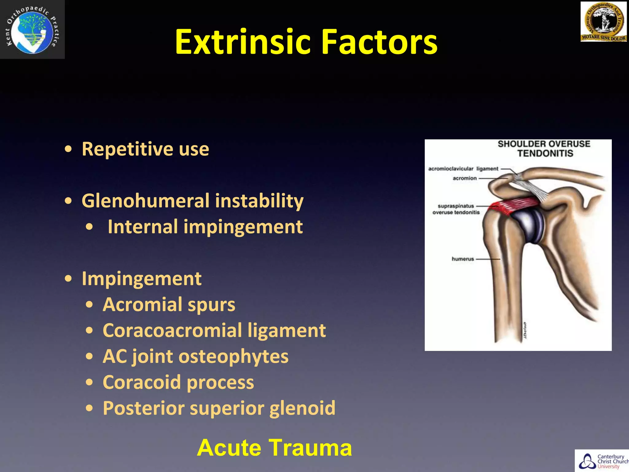
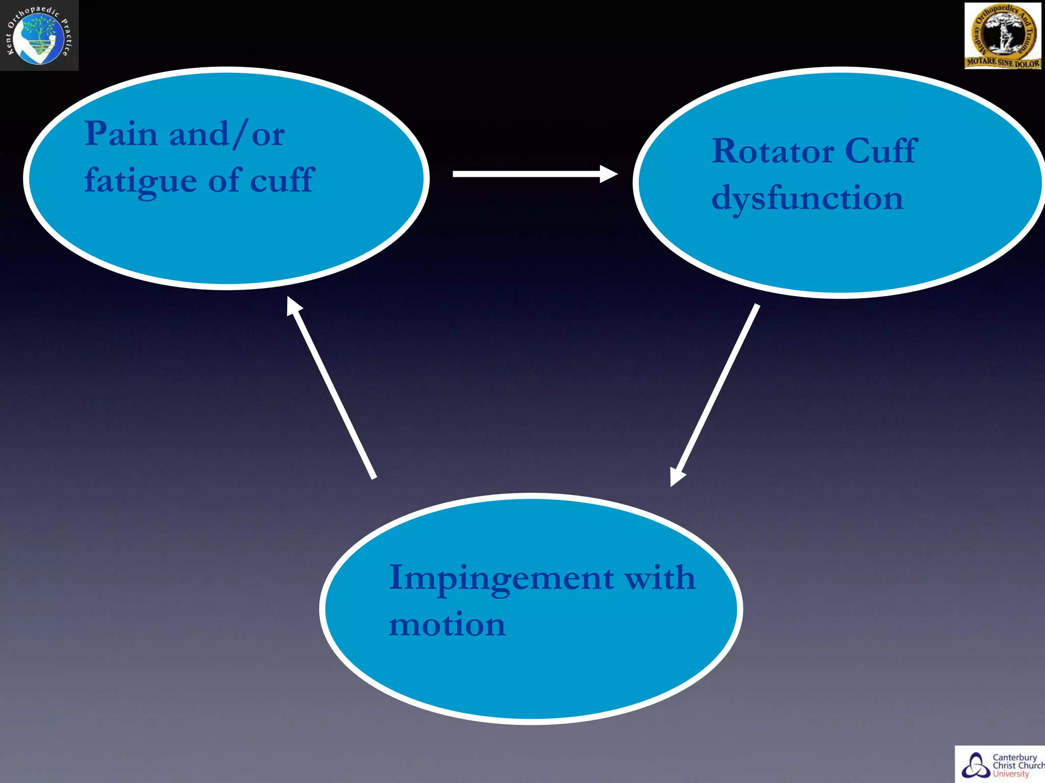
Prof. Bijayendra Singh
Consultant Trauma & Orthopaedic Surgeon
Medway NHS Foundation Trust
Visiting Professor, Canterbury Christ Church University
Cuff Repair
What is Rotator cuff ?
Extrinsic Factors
• Repetitive use
• Glenohumeral instability
• Internal impingement
• Impingement
• Acromial spurs
• Coracoacromial ligament
• AC joint osteophytes
• Coracoid process
• Posterior superior glenoid
Acute Trauma
Intrinsic Factors
• Vascular supply (? significance)
• Distal 1cm of supraspinatus tendon (early studies)
• Hypervascularity with tendonitis
• Codman (1934) described critical zone
• Rathburn (1970) position related to blood supply
• Lohr (1990) bursal side better blood supply :
Increased incidence of articular surface tears?
• Degenerative changes
• Age related
• Change in proteoglycan and collagen content in
symptomatic tendons
Physical Examination
• Inspection: Atrophy, symmetry
• Palpation: AC joint, cuff tenderness
• Range of motion: Active, passive
• Muscle strength
• Special tests
Investigations
• Ultrasound:
• High Accuracy for Full
Thickness Tear
• Poor info on other
pathologies
• Static images for
dynamic investigation
• Operator Dependent
• MRI:
• Gold Standard
• Easier to explain to patient
• Other Shoulder Pathologies
• Muscle Atrophy
• Expensive/Cumbersome
• May find pathologies of no
clinical relevnace..
Natural History
• Asymptomatic: 5 - 40%
• Increases with age
• Sher et al, J Bone Joint Surg Am. 1995 Jan;77(1):10-5:
Abnormal MRI Findings in asymptomatic shoulders
• 96 asymptomatic shoulders MRI
• 14 FT & 19 PT
• > 60: 28% FT, 26% PT (54%)
• 40 - 60: 4% FT, 24% PT (28%)
• < 40: no FT, 4% PT
Pain and/or
fatigue of cuff
Rotator Cuff
dysfunction
Impingement with
motion
Indications for Surgery
• Failed conservative management
• Significant or progressive weakness
• Young, active
• Acute tear
• Early repair if age<50 years and full-thickness tear
• Do we need to repair
Open vs Arthroscopic
Do we need to Repair Cuffs?
Natural History of Non operatively Treated Symptomatic Rotator Cuff
Tears in <60 yrs. (5mm or more)
Safran et al: Am Jr. Sports Medicine, 39(4), 710 - 714
• F/U: 25 - 39 months
• Ultrasonography by same sonographer
• 51/61 evaluated
–30 (49%) tears increased in size
–26 (41%) no change
–5 (8%) reduced
–10(25%) found to have new tears
• No correlation between change in tear size,
–patient age
–prior trauma
–size of tear at index
• Co-relation between considerable pain & increase in tear size
Open Vs Arthroscopic
Open Repair
• Advantages:
• Easy
• No special equipment required
• Direct visualization of cuff repair and acromioplasty
• Good long term follow-up. Several studies with >10
year follow-up show generally stable results with
time
• UKCUFF Trial
Disadvantages
• Deltoid detachment required
• Increased perioperative morbidity
• Unrepairable tear will be opened
• Significant intraarticular pathology can be missed
• Increased blood loss
• Increased rehabilitation time
• Large scar
Arthroscopic Cuff Repair
• Deltoid preservation
• Diagnosis and treatment of any concomitant shoulder
pathology
• Decreased postoperative pain
• Decreased blood loss
• Small surgical scar
• Shorter hospital stay
• Earlier rehabilitation
• Decreased postoperative stiffness
Disadvantages
• Special Instruments
• Cannula
• Suture Passer
• Suture manipulator
• Appropriate Anchors
• Suture Cutter
• Suture Management
Learning Curve
Principles of Repair
• Neer JBJS-A 1972
• Adequate subacromial decompression
• Repair tendon to bone
• Secure fixation of tendon to tuberosity
• Mobilization of muscle-tendon units
• Closely supervised rehabilitation
Single Row vs Double Row
Biomechanics
• Single Row - 220 N
• Double Row - 320 N
• Suture Bridge - 20 - 50% higher
• Almost all biomechanical studies show lower re-tear
rates for double row / Suture Bridge
Clinical Outcomes
• Franceschi et al:
• 30 in each group, UCLA 32.9 vs 33.3 post op
• MRI retear = 12/16 single, 8/26
• Burks et al:
• 20 in each group, No difference in UCLA, ASES, Constant
• Retear 2 in each on MRI
• Grasso et al
• 40 in each group
• No significant difference in DASH, Constant & Muscle Strength
• No post op imaging
Level 1 studies
Rehabilitation
• Four Phases
• Inflammatory: 7 days
• Proliferative: 2 - 3weeks
• Healing Phase: 3 - 12 weeks
• Maturation: 12 - 26 weeks
Ross et al: Rehabilitaiton Following Arthroscopic Rotator Cuff Repair - Review of
Current Literature. JAAOS, 2014, 22(1), 1 - 9
• ROM:
• Some studies have shown better elevation in early stages
• Preop ROM important factor
• NO difference at one year
• Pain:
• No significant difference in early vs late mobilisation
• Muscle Strength:
• No difference, significantly lower than other side
• Re Tear Rates:
• 0 - 94%
• Variable results on radiological re-tears
• No functional difference
Enhancement
• Biology of patient & tendon (can’t be altered)
• Techniques:
• Microfracture of healing bed
• Use of vented anchors
• Doxycycline (reduces effects of MMP)
• PRP
• Mesenchymal stem cells
• No definite evidence at present
Conclusion
• Keys to success:
• Pick a winner
• Good anaesthesia
• Tension-free reduction
• Thorough bursectomy for visualisation
• Work to a system
• Variety of equipment invaluable
My choice
• Small Tears = Single Row - Mattress Repair
• Large > 3 cm = Double Row - Suture-bridge technique
Thank You

Arthroscopic Rotator Cuff Repair

  • 1.
    Prof. Bijayendra Singh ConsultantTrauma & Orthopaedic Surgeon Medway NHS Foundation Trust Visiting Professor, Canterbury Christ Church University Cuff Repair
  • 2.
  • 3.
    Extrinsic Factors • Repetitiveuse • Glenohumeral instability • Internal impingement • Impingement • Acromial spurs • Coracoacromial ligament • AC joint osteophytes • Coracoid process • Posterior superior glenoid Acute Trauma
  • 4.
    Intrinsic Factors • Vascularsupply (? significance) • Distal 1cm of supraspinatus tendon (early studies) • Hypervascularity with tendonitis • Codman (1934) described critical zone • Rathburn (1970) position related to blood supply • Lohr (1990) bursal side better blood supply : Increased incidence of articular surface tears? • Degenerative changes • Age related • Change in proteoglycan and collagen content in symptomatic tendons
  • 5.
    Physical Examination • Inspection:Atrophy, symmetry • Palpation: AC joint, cuff tenderness • Range of motion: Active, passive • Muscle strength • Special tests
  • 6.
  • 7.
    • Ultrasound: • HighAccuracy for Full Thickness Tear • Poor info on other pathologies • Static images for dynamic investigation • Operator Dependent • MRI: • Gold Standard • Easier to explain to patient • Other Shoulder Pathologies • Muscle Atrophy • Expensive/Cumbersome • May find pathologies of no clinical relevnace..
  • 8.
    Natural History • Asymptomatic:5 - 40% • Increases with age
  • 9.
    • Sher etal, J Bone Joint Surg Am. 1995 Jan;77(1):10-5: Abnormal MRI Findings in asymptomatic shoulders • 96 asymptomatic shoulders MRI • 14 FT & 19 PT • > 60: 28% FT, 26% PT (54%) • 40 - 60: 4% FT, 24% PT (28%) • < 40: no FT, 4% PT
  • 10.
    Pain and/or fatigue ofcuff Rotator Cuff dysfunction Impingement with motion
  • 11.
    Indications for Surgery •Failed conservative management • Significant or progressive weakness • Young, active • Acute tear • Early repair if age<50 years and full-thickness tear
  • 12.
    • Do weneed to repair Open vs Arthroscopic
  • 13.
    Do we needto Repair Cuffs?
  • 14.
    Natural History ofNon operatively Treated Symptomatic Rotator Cuff Tears in <60 yrs. (5mm or more) Safran et al: Am Jr. Sports Medicine, 39(4), 710 - 714 • F/U: 25 - 39 months • Ultrasonography by same sonographer • 51/61 evaluated –30 (49%) tears increased in size –26 (41%) no change –5 (8%) reduced –10(25%) found to have new tears • No correlation between change in tear size, –patient age –prior trauma –size of tear at index • Co-relation between considerable pain & increase in tear size
  • 15.
  • 16.
    Open Repair • Advantages: •Easy • No special equipment required • Direct visualization of cuff repair and acromioplasty • Good long term follow-up. Several studies with >10 year follow-up show generally stable results with time • UKCUFF Trial
  • 17.
    Disadvantages • Deltoid detachmentrequired • Increased perioperative morbidity • Unrepairable tear will be opened • Significant intraarticular pathology can be missed • Increased blood loss • Increased rehabilitation time • Large scar
  • 18.
    Arthroscopic Cuff Repair •Deltoid preservation • Diagnosis and treatment of any concomitant shoulder pathology • Decreased postoperative pain • Decreased blood loss • Small surgical scar • Shorter hospital stay • Earlier rehabilitation • Decreased postoperative stiffness
  • 19.
    Disadvantages • Special Instruments •Cannula • Suture Passer • Suture manipulator • Appropriate Anchors • Suture Cutter • Suture Management Learning Curve
  • 21.
    Principles of Repair •Neer JBJS-A 1972 • Adequate subacromial decompression • Repair tendon to bone • Secure fixation of tendon to tuberosity • Mobilization of muscle-tendon units • Closely supervised rehabilitation
  • 22.
    Single Row vsDouble Row
  • 26.
    Biomechanics • Single Row- 220 N • Double Row - 320 N • Suture Bridge - 20 - 50% higher • Almost all biomechanical studies show lower re-tear rates for double row / Suture Bridge
  • 27.
    Clinical Outcomes • Franceschiet al: • 30 in each group, UCLA 32.9 vs 33.3 post op • MRI retear = 12/16 single, 8/26 • Burks et al: • 20 in each group, No difference in UCLA, ASES, Constant • Retear 2 in each on MRI • Grasso et al • 40 in each group • No significant difference in DASH, Constant & Muscle Strength • No post op imaging Level 1 studies
  • 28.
    Rehabilitation • Four Phases •Inflammatory: 7 days • Proliferative: 2 - 3weeks • Healing Phase: 3 - 12 weeks • Maturation: 12 - 26 weeks
  • 32.
    Ross et al:Rehabilitaiton Following Arthroscopic Rotator Cuff Repair - Review of Current Literature. JAAOS, 2014, 22(1), 1 - 9 • ROM: • Some studies have shown better elevation in early stages • Preop ROM important factor • NO difference at one year • Pain: • No significant difference in early vs late mobilisation • Muscle Strength: • No difference, significantly lower than other side • Re Tear Rates: • 0 - 94% • Variable results on radiological re-tears • No functional difference
  • 33.
    Enhancement • Biology ofpatient & tendon (can’t be altered) • Techniques: • Microfracture of healing bed • Use of vented anchors • Doxycycline (reduces effects of MMP) • PRP • Mesenchymal stem cells • No definite evidence at present
  • 34.
    Conclusion • Keys tosuccess: • Pick a winner • Good anaesthesia • Tension-free reduction • Thorough bursectomy for visualisation • Work to a system • Variety of equipment invaluable My choice • Small Tears = Single Row - Mattress Repair • Large > 3 cm = Double Row - Suture-bridge technique
  • 35.

Editor's Notes

  • #2 Thank you for asking me to deliver the Dr. S.K.Lokhare Oration at the 33rd Annual Congress of MOA. Its indeed a great honour and privilege to be able to deliver this lecture. My heartfelt gratitude to the organising committee and the executive at MOA. A special thanks to Ajis & Shiva.
  • #3 Fine-tuning” muscles Keep the humeral head centered on the Generally work to depress the humeral head while powerful deltoid contracts
  • #12 Failed conservative management 3 to 12 month course of NSAIDs, physio, corticosteroid injections, activity modification Significant or progressive weakness, esp. acute Early repair if <50 y.o. and full-thickness tear Differential diagnosis confirms weakness is from rotator cuff tear (i.e. MRI findings correlate with exam, rule out other causes)