SlideShare a Scribd company logo
1 of 57
Application of Ultrasound and
Biological effect
Presenter :- Yashawant Kumar Yadav
BSc. MIT 3rd yr
NAMS , Bir Hospital
1
OUTLINE
• Introduction of Ultrasound
• Modes of Ultrasound with application
• Doppler US and types with application
• Different kinds of probes with medical application
• Biological effect
• References
2
Intro…..
• Any sound with a frequency above 20,000 Hz (or 20 kHz)that is, above the
highest audible frequency—is defined to be ultrasound.
• 1Hz = 1 cycle / vibration
• 1kHz = 1000 cycle per second
• 1MHz = 1 million vibrations per second.
3
Contd….
• A sound wave can be described as a mechanical, longitudinal wave composed of
cyclic compressions and rarefactions of molecules in a medium.
4
Interaction
• Reflection
• Refraction
• Absorption
• Scattering
• Acoustic impedance
• Z = ρv, The units for Z are therefore kg/(m2 · s).
• Matching layer ???
• Z of PZC is 15 times higher than skin
5
Parameters
• Amplitude:
• Power :- (joules /s) (A2)
• Intensity :- The energy per unit cross-sectional area in a sound beam,
• Intensity is expressed in joules/second/square centimeter.
• Joule/second = watt
• (w/cm2)
6
Contd…
Relative scale intensity is defined in units of the decibel (dB)
In common practice, the lowest-intensity audible sound (10-12 W /M2) is assigned
the value of 0 dB.
10dB = 102 Times of 0dB
So 120 dB = 1012 times
i.e 1 trillion times of 0 dB
7
Production of US in Medicine
Piezoelectric US generation.
Natural --
- (quartz, and lead titanate)
Piezoelectric crystals
I. Barium titanate,(BaTiO3) (1st discovered )
II. Rochelle salts, (KNaC4H4O6·4H2O)
III. Potassium niobate (KNbO3)
IV. Ammonium dihydrogen phosphate (ADP)
V. lead zirconate titanate . (Pb[ZRxTi]O3)
VI. Sodium tungstate (N2WO3)
VII. Sodium potassium niobate (NKN) lead free 8
Pulse acoustic phenomenon
• Continuous waves are not useful for structural imaging.
• One pulse typically consists of three to five cycles.
Pulse US parameters
• Pulse duration (0.5 to 3 µs).
• Pulse repetition period: ( 0.1 to 1 ms.)
• Spatial pulse length: (0.1 to 1 mm).
• Duty factor (0.1 % to 1 %. )
• Pulse repetition frequency (PRF): (1 OOO to 10,000 Hz )
PRF is inversely proportional to imaging depth.
9
Tissues attenuation
• Bone is represented as a very bright
structure and appears ‘hyperechoic’.
• Muscle presents as hypoechoic, with
some internal signals as a result of
collagen fibers.
• Tendon show tightly packed
hyperechoic lines representing the
fibrils of the tendon.
10
Contd….
• Nerves as ‘honeycomb’ like structures composed of
hypoechoic spots embedded in a hyperechoic
background.
• Fluid presents has an anechoic appearance on
ultrasound.
• Air bubbles reflect much of the US that engages them and
appear very echo dense (bright).
• Ligament
11
Medical application
• Ultrasound is used in medicine for therapy and diagnosis.
In diagnosis :
• Ultrasound have 4 basic mode :-
1. A – amplitude mode
2. B – brightness mode
3. M- motion mode
4. Doppler
12
A-amplitude mode (10–20 MHz)
• Mode A is a one-dimensional process, and simplest
type of US .
• Basically used to determine depth of abnormality .
(function of depth )
• Therapeutic ultrasound aimed at a specific tumor or calculus is
also A-mode, to allow for pinpoint accurate focus of the
destructive wave energy.
• As an example on how Mode A can be applied,
• US encephalography, and US ophthalmology
13
14
Contd…
In a case where the middle brain structures have been displaced (for
example by the presence of an odd mass in a lobe that pushes them into the
other lobe)
15
Contd….
16
B – Brightness mode
• The B-mode is a two-dimensional (2D) image
of the area that is simultaneously scanned by a
linear array of 100–300 piezoelectric elements
rather than a single one as in A-mode
• B-Mode composed of bright dots representing
the ultrasound echoes.
• The brightness of each dot is determined by
the amplitude of the returned echo signal.
17
18
M- Motion Mode
• A continuous B-mode display. Displays one
scan line versus time.
• Allows for a high frame rate, accuracy of
linear measurements, and tracking of motion
of reflectors
(x-axis: time; y-axis: reflector depth)..
• M-mode is used extensively in cardiac and
fetal cardiac imaging.
• Its an application of B - mode imaging
19
Doppler mode
• The Doppler effect is defined as the change in
the frequency of sound emitted or reflected by a
moving object. The amount of change is termed
the Doppler shift.
20
Th is angle is known as the angle of
insonation.
21
Contd…
• The highest Doppler frequency shift from any specific vessel occurs
when the vessel and the beam are aligned; that is, when the angle of
insonation is zero and the cosine function has the value of 1.0.
• The least desirable situation occurs when the angle approaches 90° as
then the Doppler frequency shift will be minimum.
• In clinical practice it is often not possible to align the beam and the
vessel.
• As a rule, provided the angle is less than about 60°, good-quality
spectral waveforms can be obtained. 22
Application of doppler
1. Peripheral vascular system - Arteries and veins of the extremities
2. Central arteries / veins - Abdominal aorta, IVC, mesenteric vessels
3. Cerebrovascular system -
4. Cardiology
5. Obstetrics and Gynecology - IUGR, Uterine arteries, Ovarian torsion
6. Renovascular hypertension
7. Male infertility / erectile dysfunction, testicular torsion
8. Tumors, AV malformations.
23
Contd…
There are five methods of imaging by employing Doppler principle.
a) Continuous wave doppler
b) Pulsed wave Doppler
c) Color Doppler
d) Power Doppler
e) Spectral Doppler or Duplex scanning
24
CONTINIOUS WAVE DOPPLER
• This employs two piezoelectric crystals, both contained
in a single head. One crystal transmits a continuous
sonic signal at a known frequency (3-8 MHz).
• The other crystal receives the returning echoes and
records their frequency.
• It is employed in detection of blood flow but does not
give information of depth, direction and velocity of
flow.
• From a practical standpoint, continuous wave Doppler
should be used when measuring velocities > 1.2 m/s.
(e.g., regurgitant jets, stenotic valves).
• No depth resolution
25
26
Contd…
Pulsed wave Doppler (PW):-
Provide depth information , variable depth can be acquired by changing beam
frequency (low for high penetration and high for low penetration)
 Three factors affects PWD To evaluate mobile structure
I. High pulse repetition frequency
II. Optimum transducer frequency
III. Insonation angle (less than 600)
It is customarily combined with a B mode / 2-Dimensional mode imaging, which is
then called duplex scanning.
measuring relatively low-flow velocities ( < ~ 1.2 m/s) in specific areas of interest
(e.g., pulmonary vein flow, mitral valve inflow). 27
Contd..
• Because sampling is intermittent, the pulse repetition frequency limits the
maximum Doppler shift (and thus maximum velocity) that can be measured
accurately.
• Velocities higher than this maximum velocity will appear to wrap around on the
display, a phenomenon known as aliasing.
• The Doppler frequency shift at which aliasing occurs, equal to PRF divided by 2,
is termed the Nyquist limit.
28
Contd..
• Doppler shift (expressed in Hz) = (2 · v · Fi cosine ϴ)/c (if ϴ= 0/180 =1/-1)
Doppler shift= (v· Fi)/770
v = 770· (Doppler shift/Fi)
• if a 5-MHz transducer can only send out about 15,000 pulses per second, the
Nyquist limit is 7500 Hz (15,000/2).
• Using the velocity equation shown earlier, the maximum velocity that can be
measured without aliasing is about 1.15m/s (770 X (7500/5,000,000)).
29
30
Color doppler(Also called color flow imaging (CFI) or color velocity imaging (CVI) )
• Ten or more pulse packets are used to
produce an estimate of mean velocity of all
reflectors.
• This data is assigned color by the machine
and is superimposed on B mode data from
stationary structures.
31
Contd…
• Flow that travels away from the transducer (negative Doppler shift) is depicted in
blue,
• Flow that is traveling toward the transducer (positive Doppler shift) is depicted in
red, with lighter shades of each color denoting higher velocities.
• A third color, usually green or yellow, is often used to denote areas of high flow
turbulence.
Clinical use :-
• overall blood flow to a region of interest. Depiction of the general velocity and
direction of blood flow within the heart and blood vessels.
• It also allows the. generation of unique phenomena such as the fluid color sign
32
Contd …
• The fluid color sign is a diagnostic sign to differentiate a pleural
effusion from pleural thickening by means of color Doppler ultrasound.
• In the case of pleural effusion a color signal is seen in the pleural fluid during
respiratory and cardiac movement, whereas this color signal is not seen in the case
of pleural thickening.
33
Power Doppler or Energy mode imaging
• An alternative processing method.
which ignores the velocity and simply
estimates the strength/amplitude of
the Doppler signal detected from each
location. And encoded as a color .
• The Energy mode image is
continuous and hence not sensitive to
relative flow direction. It does not
give the absolute values of velocity
but only gives the strength of the
Doppler signal.
34
Contd….
• The higher sensitivity to slow
flow.
• Power Doppler is particularly
useful when examining superficial
structures, like thyroid, testis,
renal grafts and subcutaneous
lesions. It may be used to look for
tumor vessels, to evaluate tiny
low-flow vessels and detect subtle
ischemic areas.
35
Contd…
Advantages of Power Doppler over Color flow imaging
I. More sensitive to flow states (3-5% more sensitive than
CFD)
II. Angle effects are ignored unless the angle is close to 90°
III. Aliasing is not applicable.
There are disadvantages also
(i) Values of velocity and the direction of blood flow cannot
be assessed.
(ii) Because of more averaging of information at slower
frame rates. slow moving soft tissue signals appear as
flash artifacts
36
Spectral doppler
• Spectral Doppler displays the blood flow
measurements graphically, displaying
flow velocities recorded over time.
• For spectral Doppler , the machine
represents the movement of blood via an
audible flow sound, and a graph
depicting flow velocities in relation to
time.
• This mode is ideal for measuring lower
velocities at chosen anatomic locations,
37
ULTRASOUND PROBES
• Linear array
• Curvilinear array
• Phased array
• linear array, “L”
• electronic linear scanning, “E”
in the xz plane
• Curve scan “C”
• “E<xz” electronically focus
38
Acoustic window’s
An acoustic window is the
location from which
an ultrasound probe makes it's
scan.
39
Linear (5-15MHz)
• Designed to make contact with flat surfaces, with the
footprint decreasing in size with increasing frequency.
• Here rectangular and trapezoidal formats provide appropriate
viewing areas. With abdominal imaging, to increase the
viewing area with minimal increases of the contact area
• Use for high-resolution imaging of superficial structures,
40
Contd..
• This probe is ideal for ultrasound-guided needle entry (particularly vascular
access),
• evaluation of deep venous thrombosis,
• chest imaging for pleural lines (pneumothorax),
• skin and soft tissue imaging for abscesses.
• Nerve blocks
• Ophthalmologic
• Joint /tendon
• Thyroid/breast
• Evaluation of superficial structure
41
Curve linear array (1-8MHz)
• convex arrays designed to make surface contact in
deformable soft areas of the body.
• The curvilinear (convex) array probe is used for scanning
deeper structures,
• The crystal alignment along a curved surface generates a
beam that fans outward. The result is a field of view wider
than the probe’s footprint,
• These probes are most often used in abdominal and pelvic
diagnostic applications. 42
Contd….
• These probes are most often used in abdominal and pelvic diagnostic applications.
• Hepatobiliary
• Renal
• Appendix (pediatric )
• Aorta
• Focus assessment in case of trauma
• Fetal heart tones and obstetric complication (pregnancy )
• Emergency lung examination
• Paracentesis and thoracentesis
43
Phase array (2-7.5MHz)
• Consisting of 64-128 elements. It is a smaller
assembly .
• The phased array probe is a lower frequency probe.
• Beam can be steer electronically , variable field
shape can be acquired .
• The smaller footprint allows for image acquisition
between the ribs, and the slightly convex surface
produces a fanning beam with a sector-shaped image.
44
Contd…..
• This probe is used for echocardiography and thoracic imaging but can be applied
to abdominal and pelvic imaging as well.
• Procedure guide
• Alternative to curvilinear for abdominal structure evaluation
• Pericardiocentesis
• Thoracentesis
45
46
Biological effect
• Energy deposition in biological tissue.
• With CW ultrasound, the tissues are irradiated throughout the duration of the
exposure, and the total energy deposited in tissue will be much more in
comparison to PW irradiation over the same total exposure duration,
• Consequences of high intensity, prolonged exposures
1. Elevation of tissue temperature (thermal effect )
2. Cavitation in liquids (non thermal effect )
Interlinked
47
Elevation of temperature
• The mechanical energy of ultrasound can be transformed into heat through
absorption in tissue, resulting in the elevation of tissue temperature.
• Temperatures exceeding 4°C above normal body temperature, maintained for
long periods of time, may be harmful to the embryo and fetus, leading to a variety
of biological changes which can result in abortion, fetal death, or developmental
abnormalities,(teratogenic effect )
• These effects have been seen following whole body heating of both the mother
and the offspring, (approx. 0.5°C min−1 )
48
Contd..
The World Federation for Ultrasound in Medicine and Biology (WFUMB)
recommends that diagnostic exposures that produce a maximum temperature rise of
1.50 c above the normal physiological level of 37°C
• Ultrasound heating is very rapid . In addition, ultrasound exposures in pregnancy
only subject a small fraction of the maternal volume to a potential temperature
rise, and thus any heating of the embryo is likely to be rapidly dissipated.
49
Cavitation in liquids( non thermal effect )
• Cavitation is a phenomenon which takes place in
liquid cavities exposed to very high intensities of
ultrasound
• The presence of gas within the volume of the
liquid may lead to the formation of micro bubbles
during the low pressure phase (rarefaction) of the
ultrasound wave cycle. With changes in pressure,
the bubbles may grow in size. During the high
pressure phase (compression),
• The micro bubbles may collapse a with the
release of large quantities of mechanical energy
which can cause biological damage.
50
Contd..
• cavitation can also be the cause of free radical formation.
• Free radicals are highly reactive chemical species which can attack bio molecules
through a chain of chemical reactions, producing toxic bio products.
• This leads to damage of biological matter not only locally, but also remotely
through diffusion of toxic products.
• cavitation is unlikely to occur with PW ultrasound
51
Drug delivery (Sonoporation )
• Refers to a technique capable of
controlling drug delivery efficiency by
maximizing the drug permeability of
surrounding tissues or cells using a
reaction mechanism between
microbubbles and ultrasonic waves.
• Injecting microbubbles intravenously can minimize drug side effects that may
cause nonspecific delivery of drugs into healthy organs because the microbubbles
only collapse in specific diseased areas due to focused ultrasound irradiation.
52
53
Contd …
• ultrasound-mediated microbubble destruction (UMMD)
• ultrasound-targeted microbubble destruction (UTMD).
54
THANK
YOU
55
References
• https://theultrasoundsite.co.uk/ultrasound-case-studies/starting-article-2-
different-tissues-ultrasound-look-like/
• https://www.nysora.com/foundations-of-regional-
anesthesia/equipment/physics-of-ultrasound/
• https://radiologykey.com/doppler-imaging-2/
• https://radiopaedia.org/articles/phased-array
• https://www.ncbi.nlm.nih.gov/pmc/articles/PMC6208473/
• Martinoli, C., Pretolesi, F., Crespi, G., Bianchi, S., Gandolfo, N., Valle, M.,
& Derchi, L. E. (1998). Power Doppler sonography: clinical
applications. European journal of radiology, 27 Suppl 2, S133–S140.
https://doi.org/10.1016/s0720-048x(98)00054-0
• https://echocardiographer.org/Echo%20Physics/spectral%20doppler.html
56
1. Cavitation effect in microbubble contrast agent ??
57

More Related Content

What's hot

Ultrasound imaging system
Ultrasound imaging system Ultrasound imaging system
Ultrasound imaging system Riadh Al-Haidari
 
Design of ultrasound transducer
Design of ultrasound transducer Design of ultrasound transducer
Design of ultrasound transducer SudarshanKanse1
 
Ultrasound physics and image optimization1 (1)
Ultrasound physics and image optimization1 (1)Ultrasound physics and image optimization1 (1)
Ultrasound physics and image optimization1 (1)Prajwith Rai
 
Ultrasound instrumentation
Ultrasound instrumentationUltrasound instrumentation
Ultrasound instrumentationVnAy Kris
 
Interactions of ultrasound with matter
Interactions of ultrasound with matterInteractions of ultrasound with matter
Interactions of ultrasound with matterSwapnil Shetty
 
Ultrasound Physics
Ultrasound PhysicsUltrasound Physics
Ultrasound Physicsu.surgery
 
Types of Ultrasound Probes
Types of Ultrasound ProbesTypes of Ultrasound Probes
Types of Ultrasound Probeshafsatalat1
 
Principle of usg imaging, construction of transducers
Principle of usg imaging, construction of transducersPrinciple of usg imaging, construction of transducers
Principle of usg imaging, construction of transducersDev Lakhera
 
Ultrasound Physics Made easy - By Dr Chandni Wadhwani
Ultrasound Physics Made easy - By Dr Chandni WadhwaniUltrasound Physics Made easy - By Dr Chandni Wadhwani
Ultrasound Physics Made easy - By Dr Chandni WadhwaniChandni Wadhwani
 
CT Scan Image reconstruction
CT Scan Image reconstructionCT Scan Image reconstruction
CT Scan Image reconstructionGunjan Patel
 
INTRODUCTION TO ULTRASOUND IMAGING ( ULTRASONGRAPHY ) - ANAND GURMAITA (BSc. ...
INTRODUCTION TO ULTRASOUND IMAGING ( ULTRASONGRAPHY ) - ANAND GURMAITA (BSc. ...INTRODUCTION TO ULTRASOUND IMAGING ( ULTRASONGRAPHY ) - ANAND GURMAITA (BSc. ...
INTRODUCTION TO ULTRASOUND IMAGING ( ULTRASONGRAPHY ) - ANAND GURMAITA (BSc. ...ANAND GURMAITA
 
Basic principle of ct and ct generations
Basic principle of ct and ct generationsBasic principle of ct and ct generations
Basic principle of ct and ct generationsTarun Goyal
 
Ultrasound Transducer Constriction And It’s Physics.pptx
Ultrasound Transducer Constriction And It’s Physics.pptxUltrasound Transducer Constriction And It’s Physics.pptx
Ultrasound Transducer Constriction And It’s Physics.pptxDr. Dheeraj Kumar
 
ULTRASOUND PHYSICS
ULTRASOUND PHYSICSULTRASOUND PHYSICS
ULTRASOUND PHYSICSNavni Garg
 
Doppler Ultrasonography And Advancements in USG
Doppler Ultrasonography And Advancements in USGDoppler Ultrasonography And Advancements in USG
Doppler Ultrasonography And Advancements in USGSudil Paudyal
 
Mammographic equipment
Mammographic equipmentMammographic equipment
Mammographic equipmentSharmaRajan4
 
Doppler Effect - Ultrasound
Doppler Effect - UltrasoundDoppler Effect - Ultrasound
Doppler Effect - UltrasoundVictor Ekpo
 

What's hot (20)

Ultrasound imaging system
Ultrasound imaging system Ultrasound imaging system
Ultrasound imaging system
 
Design of ultrasound transducer
Design of ultrasound transducer Design of ultrasound transducer
Design of ultrasound transducer
 
Computed tomography
Computed tomographyComputed tomography
Computed tomography
 
Ultrasound physics and image optimization1 (1)
Ultrasound physics and image optimization1 (1)Ultrasound physics and image optimization1 (1)
Ultrasound physics and image optimization1 (1)
 
Ultrasound instrumentation
Ultrasound instrumentationUltrasound instrumentation
Ultrasound instrumentation
 
Interactions of ultrasound with matter
Interactions of ultrasound with matterInteractions of ultrasound with matter
Interactions of ultrasound with matter
 
Ultrasound Physics
Ultrasound PhysicsUltrasound Physics
Ultrasound Physics
 
Types of Ultrasound Probes
Types of Ultrasound ProbesTypes of Ultrasound Probes
Types of Ultrasound Probes
 
Principle of usg imaging, construction of transducers
Principle of usg imaging, construction of transducersPrinciple of usg imaging, construction of transducers
Principle of usg imaging, construction of transducers
 
Ultrasound Physics Made easy - By Dr Chandni Wadhwani
Ultrasound Physics Made easy - By Dr Chandni WadhwaniUltrasound Physics Made easy - By Dr Chandni Wadhwani
Ultrasound Physics Made easy - By Dr Chandni Wadhwani
 
Ultrasound physics
Ultrasound physicsUltrasound physics
Ultrasound physics
 
CT Scan Image reconstruction
CT Scan Image reconstructionCT Scan Image reconstruction
CT Scan Image reconstruction
 
INTRODUCTION TO ULTRASOUND IMAGING ( ULTRASONGRAPHY ) - ANAND GURMAITA (BSc. ...
INTRODUCTION TO ULTRASOUND IMAGING ( ULTRASONGRAPHY ) - ANAND GURMAITA (BSc. ...INTRODUCTION TO ULTRASOUND IMAGING ( ULTRASONGRAPHY ) - ANAND GURMAITA (BSc. ...
INTRODUCTION TO ULTRASOUND IMAGING ( ULTRASONGRAPHY ) - ANAND GURMAITA (BSc. ...
 
Basic principle of ct and ct generations
Basic principle of ct and ct generationsBasic principle of ct and ct generations
Basic principle of ct and ct generations
 
Ultrasound Transducer Constriction And It’s Physics.pptx
Ultrasound Transducer Constriction And It’s Physics.pptxUltrasound Transducer Constriction And It’s Physics.pptx
Ultrasound Transducer Constriction And It’s Physics.pptx
 
Doppler ultrasonography
Doppler ultrasonographyDoppler ultrasonography
Doppler ultrasonography
 
ULTRASOUND PHYSICS
ULTRASOUND PHYSICSULTRASOUND PHYSICS
ULTRASOUND PHYSICS
 
Doppler Ultrasonography And Advancements in USG
Doppler Ultrasonography And Advancements in USGDoppler Ultrasonography And Advancements in USG
Doppler Ultrasonography And Advancements in USG
 
Mammographic equipment
Mammographic equipmentMammographic equipment
Mammographic equipment
 
Doppler Effect - Ultrasound
Doppler Effect - UltrasoundDoppler Effect - Ultrasound
Doppler Effect - Ultrasound
 

Similar to Application of ultrasound

ULTRASOUND IMAGING PRINCIPLES
ULTRASOUND IMAGING PRINCIPLESULTRASOUND IMAGING PRINCIPLES
ULTRASOUND IMAGING PRINCIPLESINDIA ULTRASOUND
 
ultra sound.pptx
ultra sound.pptxultra sound.pptx
ultra sound.pptxAliMRiyath
 
Introduction to ultrasound & regional anesthesia
Introduction to ultrasound & regional anesthesiaIntroduction to ultrasound & regional anesthesia
Introduction to ultrasound & regional anesthesiaSaad Al-Shamma
 
Ultrasound assignment 3 mark question and answer
Ultrasound assignment 3 mark question and answer Ultrasound assignment 3 mark question and answer
Ultrasound assignment 3 mark question and answer Ganesan Yogananthem
 
Ultrasonography
UltrasonographyUltrasonography
UltrasonographyAnu V
 
Emergency us physics and instrumentation
Emergency us physics  and instrumentationEmergency us physics  and instrumentation
Emergency us physics and instrumentationdrnikahmad
 
BASICS OF EUS PHYSICS.pptx
BASICS OF EUS PHYSICS.pptxBASICS OF EUS PHYSICS.pptx
BASICS OF EUS PHYSICS.pptxPriyaranjanDas22
 
Doppler ultrasounds (1)
Doppler ultrasounds (1)Doppler ultrasounds (1)
Doppler ultrasounds (1)KamalEldirawi
 
1.Introduction to Ultrasound - Adamu.pptx
1.Introduction to Ultrasound - Adamu.pptx1.Introduction to Ultrasound - Adamu.pptx
1.Introduction to Ultrasound - Adamu.pptxabelllll
 
Echo Physics and Doppler
Echo Physics and Doppler Echo Physics and Doppler
Echo Physics and Doppler George Abraham
 
Ultrasound Imaging_2023.pdf
Ultrasound Imaging_2023.pdfUltrasound Imaging_2023.pdf
Ultrasound Imaging_2023.pdfghadaElbanby1
 

Similar to Application of ultrasound (20)

ULTRASOUND IMAGING PRINCIPLES
ULTRASOUND IMAGING PRINCIPLESULTRASOUND IMAGING PRINCIPLES
ULTRASOUND IMAGING PRINCIPLES
 
ultra sound.pptx
ultra sound.pptxultra sound.pptx
ultra sound.pptx
 
Introduction to ultrasound & regional anesthesia
Introduction to ultrasound & regional anesthesiaIntroduction to ultrasound & regional anesthesia
Introduction to ultrasound & regional anesthesia
 
Doppler phys (2)
Doppler phys (2)Doppler phys (2)
Doppler phys (2)
 
Ultrasound assignment 3 mark question and answer
Ultrasound assignment 3 mark question and answer Ultrasound assignment 3 mark question and answer
Ultrasound assignment 3 mark question and answer
 
Doppler us (3)
Doppler us (3)Doppler us (3)
Doppler us (3)
 
Doppler physics.pptx
Doppler physics.pptxDoppler physics.pptx
Doppler physics.pptx
 
Ultrasonography
UltrasonographyUltrasonography
Ultrasonography
 
Doppler Effect
Doppler EffectDoppler Effect
Doppler Effect
 
Emergency us physics and instrumentation
Emergency us physics  and instrumentationEmergency us physics  and instrumentation
Emergency us physics and instrumentation
 
Doppler echocardiography
Doppler echocardiographyDoppler echocardiography
Doppler echocardiography
 
Doppler echocardiography
Doppler echocardiographyDoppler echocardiography
Doppler echocardiography
 
Doppler physics ppt
Doppler physics pptDoppler physics ppt
Doppler physics ppt
 
BASICS OF EUS PHYSICS.pptx
BASICS OF EUS PHYSICS.pptxBASICS OF EUS PHYSICS.pptx
BASICS OF EUS PHYSICS.pptx
 
ULTRA.pptx
ULTRA.pptxULTRA.pptx
ULTRA.pptx
 
PRINCIPLES OF ULTRASONOGRAPHY
PRINCIPLES OF ULTRASONOGRAPHYPRINCIPLES OF ULTRASONOGRAPHY
PRINCIPLES OF ULTRASONOGRAPHY
 
Doppler ultrasounds (1)
Doppler ultrasounds (1)Doppler ultrasounds (1)
Doppler ultrasounds (1)
 
1.Introduction to Ultrasound - Adamu.pptx
1.Introduction to Ultrasound - Adamu.pptx1.Introduction to Ultrasound - Adamu.pptx
1.Introduction to Ultrasound - Adamu.pptx
 
Echo Physics and Doppler
Echo Physics and Doppler Echo Physics and Doppler
Echo Physics and Doppler
 
Ultrasound Imaging_2023.pdf
Ultrasound Imaging_2023.pdfUltrasound Imaging_2023.pdf
Ultrasound Imaging_2023.pdf
 

More from Yashawant Yadav

Mr procedure of pelvis and hip joint
Mr procedure of pelvis and hip jointMr procedure of pelvis and hip joint
Mr procedure of pelvis and hip jointYashawant Yadav
 
Positron emission tomography pet scan and its applications
Positron emission tomography pet scan and its applicationsPositron emission tomography pet scan and its applications
Positron emission tomography pet scan and its applicationsYashawant Yadav
 
Ptc )percutaneous transhepatic cholangiography
Ptc )percutaneous transhepatic cholangiographyPtc )percutaneous transhepatic cholangiography
Ptc )percutaneous transhepatic cholangiographyYashawant Yadav
 
Ct coronary angiography edited 1st
Ct   coronary  angiography edited 1stCt   coronary  angiography edited 1st
Ct coronary angiography edited 1stYashawant Yadav
 
Mri spin echo pulse sequences its variations and
Mri spin echo pulse sequences its variations andMri spin echo pulse sequences its variations and
Mri spin echo pulse sequences its variations andYashawant Yadav
 
Technique of thoracic and lumber spine radiography
Technique  of thoracic and lumber spine radiographyTechnique  of thoracic and lumber spine radiography
Technique of thoracic and lumber spine radiographyYashawant Yadav
 
Digital breast tomosynthesis
Digital breast tomosynthesisDigital breast tomosynthesis
Digital breast tomosynthesisYashawant Yadav
 
Ptc )percutaneous transhepatic cholangiography
Ptc )percutaneous transhepatic cholangiographyPtc )percutaneous transhepatic cholangiography
Ptc )percutaneous transhepatic cholangiographyYashawant Yadav
 
High voltage transformer and high frequency generator
High voltage transformer and high frequency generatorHigh voltage transformer and high frequency generator
High voltage transformer and high frequency generatorYashawant Yadav
 
Radiological contrast media
Radiological contrast mediaRadiological contrast media
Radiological contrast mediaYashawant Yadav
 
Procedure of ercp and t tube cholangiography
Procedure of ercp and t tube cholangiographyProcedure of ercp and t tube cholangiography
Procedure of ercp and t tube cholangiographyYashawant Yadav
 
Presentation1.pptx thoraccic and lumber spine
Presentation1.pptx thoraccic and lumber spinePresentation1.pptx thoraccic and lumber spine
Presentation1.pptx thoraccic and lumber spineYashawant Yadav
 
Technique of dental radiographic
Technique of dental radiographicTechnique of dental radiographic
Technique of dental radiographicYashawant Yadav
 
Presentation2.pptx technique chest 2
Presentation2.pptx technique chest 2Presentation2.pptx technique chest 2
Presentation2.pptx technique chest 2Yashawant Yadav
 
Image characteristics of x ray film
Image characteristics of x ray filmImage characteristics of x ray film
Image characteristics of x ray filmYashawant Yadav
 
Mammographic equipment and its advancement
Mammographic equipment and its advancementMammographic equipment and its advancement
Mammographic equipment and its advancementYashawant Yadav
 
Dacrocystography and sialography
Dacrocystography and sialographyDacrocystography and sialography
Dacrocystography and sialographyYashawant Yadav
 
Ct image quality artifacts and it remedy
Ct image quality artifacts and it remedyCt image quality artifacts and it remedy
Ct image quality artifacts and it remedyYashawant Yadav
 

More from Yashawant Yadav (20)

Mr procedure of pelvis and hip joint
Mr procedure of pelvis and hip jointMr procedure of pelvis and hip joint
Mr procedure of pelvis and hip joint
 
Positron emission tomography pet scan and its applications
Positron emission tomography pet scan and its applicationsPositron emission tomography pet scan and its applications
Positron emission tomography pet scan and its applications
 
Ptc )percutaneous transhepatic cholangiography
Ptc )percutaneous transhepatic cholangiographyPtc )percutaneous transhepatic cholangiography
Ptc )percutaneous transhepatic cholangiography
 
Ct coronary angiography edited 1st
Ct   coronary  angiography edited 1stCt   coronary  angiography edited 1st
Ct coronary angiography edited 1st
 
Ct protocol for ivu
Ct protocol for ivuCt protocol for ivu
Ct protocol for ivu
 
Mri spin echo pulse sequences its variations and
Mri spin echo pulse sequences its variations andMri spin echo pulse sequences its variations and
Mri spin echo pulse sequences its variations and
 
Technique of thoracic and lumber spine radiography
Technique  of thoracic and lumber spine radiographyTechnique  of thoracic and lumber spine radiography
Technique of thoracic and lumber spine radiography
 
Digital breast tomosynthesis
Digital breast tomosynthesisDigital breast tomosynthesis
Digital breast tomosynthesis
 
Ptc )percutaneous transhepatic cholangiography
Ptc )percutaneous transhepatic cholangiographyPtc )percutaneous transhepatic cholangiography
Ptc )percutaneous transhepatic cholangiography
 
High voltage transformer and high frequency generator
High voltage transformer and high frequency generatorHigh voltage transformer and high frequency generator
High voltage transformer and high frequency generator
 
Radiological contrast media
Radiological contrast mediaRadiological contrast media
Radiological contrast media
 
Procedure of ercp and t tube cholangiography
Procedure of ercp and t tube cholangiographyProcedure of ercp and t tube cholangiography
Procedure of ercp and t tube cholangiography
 
Presentation1.pptx thoraccic and lumber spine
Presentation1.pptx thoraccic and lumber spinePresentation1.pptx thoraccic and lumber spine
Presentation1.pptx thoraccic and lumber spine
 
Technique of dental radiographic
Technique of dental radiographicTechnique of dental radiographic
Technique of dental radiographic
 
Presentation2.pptx technique chest 2
Presentation2.pptx technique chest 2Presentation2.pptx technique chest 2
Presentation2.pptx technique chest 2
 
Image characteristics of x ray film
Image characteristics of x ray filmImage characteristics of x ray film
Image characteristics of x ray film
 
Procedure of upper gi
Procedure of upper giProcedure of upper gi
Procedure of upper gi
 
Mammographic equipment and its advancement
Mammographic equipment and its advancementMammographic equipment and its advancement
Mammographic equipment and its advancement
 
Dacrocystography and sialography
Dacrocystography and sialographyDacrocystography and sialography
Dacrocystography and sialography
 
Ct image quality artifacts and it remedy
Ct image quality artifacts and it remedyCt image quality artifacts and it remedy
Ct image quality artifacts and it remedy
 

Recently uploaded

Computed Fields and api Depends in the Odoo 17
Computed Fields and api Depends in the Odoo 17Computed Fields and api Depends in the Odoo 17
Computed Fields and api Depends in the Odoo 17Celine George
 
Roles & Responsibilities in Pharmacovigilance
Roles & Responsibilities in PharmacovigilanceRoles & Responsibilities in Pharmacovigilance
Roles & Responsibilities in PharmacovigilanceSamikshaHamane
 
Final demo Grade 9 for demo Plan dessert.pptx
Final demo Grade 9 for demo Plan dessert.pptxFinal demo Grade 9 for demo Plan dessert.pptx
Final demo Grade 9 for demo Plan dessert.pptxAvyJaneVismanos
 
KSHARA STURA .pptx---KSHARA KARMA THERAPY (CAUSTIC THERAPY)————IMP.OF KSHARA ...
KSHARA STURA .pptx---KSHARA KARMA THERAPY (CAUSTIC THERAPY)————IMP.OF KSHARA ...KSHARA STURA .pptx---KSHARA KARMA THERAPY (CAUSTIC THERAPY)————IMP.OF KSHARA ...
KSHARA STURA .pptx---KSHARA KARMA THERAPY (CAUSTIC THERAPY)————IMP.OF KSHARA ...M56BOOKSTORE PRODUCT/SERVICE
 
Solving Puzzles Benefits Everyone (English).pptx
Solving Puzzles Benefits Everyone (English).pptxSolving Puzzles Benefits Everyone (English).pptx
Solving Puzzles Benefits Everyone (English).pptxOH TEIK BIN
 
“Oh GOSH! Reflecting on Hackteria's Collaborative Practices in a Global Do-It...
“Oh GOSH! Reflecting on Hackteria's Collaborative Practices in a Global Do-It...“Oh GOSH! Reflecting on Hackteria's Collaborative Practices in a Global Do-It...
“Oh GOSH! Reflecting on Hackteria's Collaborative Practices in a Global Do-It...Marc Dusseiller Dusjagr
 
Types of Journalistic Writing Grade 8.pptx
Types of Journalistic Writing Grade 8.pptxTypes of Journalistic Writing Grade 8.pptx
Types of Journalistic Writing Grade 8.pptxEyham Joco
 
भारत-रोम व्यापार.pptx, Indo-Roman Trade,
भारत-रोम व्यापार.pptx, Indo-Roman Trade,भारत-रोम व्यापार.pptx, Indo-Roman Trade,
भारत-रोम व्यापार.pptx, Indo-Roman Trade,Virag Sontakke
 
Painted Grey Ware.pptx, PGW Culture of India
Painted Grey Ware.pptx, PGW Culture of IndiaPainted Grey Ware.pptx, PGW Culture of India
Painted Grey Ware.pptx, PGW Culture of IndiaVirag Sontakke
 
Biting mechanism of poisonous snakes.pdf
Biting mechanism of poisonous snakes.pdfBiting mechanism of poisonous snakes.pdf
Biting mechanism of poisonous snakes.pdfadityarao40181
 
Historical philosophical, theoretical, and legal foundations of special and i...
Historical philosophical, theoretical, and legal foundations of special and i...Historical philosophical, theoretical, and legal foundations of special and i...
Historical philosophical, theoretical, and legal foundations of special and i...jaredbarbolino94
 
POINT- BIOCHEMISTRY SEM 2 ENZYMES UNIT 5.pptx
POINT- BIOCHEMISTRY SEM 2 ENZYMES UNIT 5.pptxPOINT- BIOCHEMISTRY SEM 2 ENZYMES UNIT 5.pptx
POINT- BIOCHEMISTRY SEM 2 ENZYMES UNIT 5.pptxSayali Powar
 
ECONOMIC CONTEXT - PAPER 1 Q3: NEWSPAPERS.pptx
ECONOMIC CONTEXT - PAPER 1 Q3: NEWSPAPERS.pptxECONOMIC CONTEXT - PAPER 1 Q3: NEWSPAPERS.pptx
ECONOMIC CONTEXT - PAPER 1 Q3: NEWSPAPERS.pptxiammrhaywood
 
Software Engineering Methodologies (overview)
Software Engineering Methodologies (overview)Software Engineering Methodologies (overview)
Software Engineering Methodologies (overview)eniolaolutunde
 
Interactive Powerpoint_How to Master effective communication
Interactive Powerpoint_How to Master effective communicationInteractive Powerpoint_How to Master effective communication
Interactive Powerpoint_How to Master effective communicationnomboosow
 
Crayon Activity Handout For the Crayon A
Crayon Activity Handout For the Crayon ACrayon Activity Handout For the Crayon A
Crayon Activity Handout For the Crayon AUnboundStockton
 
Introduction to AI in Higher Education_draft.pptx
Introduction to AI in Higher Education_draft.pptxIntroduction to AI in Higher Education_draft.pptx
Introduction to AI in Higher Education_draft.pptxpboyjonauth
 
Alper Gobel In Media Res Media Component
Alper Gobel In Media Res Media ComponentAlper Gobel In Media Res Media Component
Alper Gobel In Media Res Media ComponentInMediaRes1
 

Recently uploaded (20)

Computed Fields and api Depends in the Odoo 17
Computed Fields and api Depends in the Odoo 17Computed Fields and api Depends in the Odoo 17
Computed Fields and api Depends in the Odoo 17
 
Roles & Responsibilities in Pharmacovigilance
Roles & Responsibilities in PharmacovigilanceRoles & Responsibilities in Pharmacovigilance
Roles & Responsibilities in Pharmacovigilance
 
Final demo Grade 9 for demo Plan dessert.pptx
Final demo Grade 9 for demo Plan dessert.pptxFinal demo Grade 9 for demo Plan dessert.pptx
Final demo Grade 9 for demo Plan dessert.pptx
 
9953330565 Low Rate Call Girls In Rohini Delhi NCR
9953330565 Low Rate Call Girls In Rohini  Delhi NCR9953330565 Low Rate Call Girls In Rohini  Delhi NCR
9953330565 Low Rate Call Girls In Rohini Delhi NCR
 
KSHARA STURA .pptx---KSHARA KARMA THERAPY (CAUSTIC THERAPY)————IMP.OF KSHARA ...
KSHARA STURA .pptx---KSHARA KARMA THERAPY (CAUSTIC THERAPY)————IMP.OF KSHARA ...KSHARA STURA .pptx---KSHARA KARMA THERAPY (CAUSTIC THERAPY)————IMP.OF KSHARA ...
KSHARA STURA .pptx---KSHARA KARMA THERAPY (CAUSTIC THERAPY)————IMP.OF KSHARA ...
 
Solving Puzzles Benefits Everyone (English).pptx
Solving Puzzles Benefits Everyone (English).pptxSolving Puzzles Benefits Everyone (English).pptx
Solving Puzzles Benefits Everyone (English).pptx
 
“Oh GOSH! Reflecting on Hackteria's Collaborative Practices in a Global Do-It...
“Oh GOSH! Reflecting on Hackteria's Collaborative Practices in a Global Do-It...“Oh GOSH! Reflecting on Hackteria's Collaborative Practices in a Global Do-It...
“Oh GOSH! Reflecting on Hackteria's Collaborative Practices in a Global Do-It...
 
Types of Journalistic Writing Grade 8.pptx
Types of Journalistic Writing Grade 8.pptxTypes of Journalistic Writing Grade 8.pptx
Types of Journalistic Writing Grade 8.pptx
 
भारत-रोम व्यापार.pptx, Indo-Roman Trade,
भारत-रोम व्यापार.pptx, Indo-Roman Trade,भारत-रोम व्यापार.pptx, Indo-Roman Trade,
भारत-रोम व्यापार.pptx, Indo-Roman Trade,
 
Painted Grey Ware.pptx, PGW Culture of India
Painted Grey Ware.pptx, PGW Culture of IndiaPainted Grey Ware.pptx, PGW Culture of India
Painted Grey Ware.pptx, PGW Culture of India
 
Biting mechanism of poisonous snakes.pdf
Biting mechanism of poisonous snakes.pdfBiting mechanism of poisonous snakes.pdf
Biting mechanism of poisonous snakes.pdf
 
Historical philosophical, theoretical, and legal foundations of special and i...
Historical philosophical, theoretical, and legal foundations of special and i...Historical philosophical, theoretical, and legal foundations of special and i...
Historical philosophical, theoretical, and legal foundations of special and i...
 
POINT- BIOCHEMISTRY SEM 2 ENZYMES UNIT 5.pptx
POINT- BIOCHEMISTRY SEM 2 ENZYMES UNIT 5.pptxPOINT- BIOCHEMISTRY SEM 2 ENZYMES UNIT 5.pptx
POINT- BIOCHEMISTRY SEM 2 ENZYMES UNIT 5.pptx
 
ECONOMIC CONTEXT - PAPER 1 Q3: NEWSPAPERS.pptx
ECONOMIC CONTEXT - PAPER 1 Q3: NEWSPAPERS.pptxECONOMIC CONTEXT - PAPER 1 Q3: NEWSPAPERS.pptx
ECONOMIC CONTEXT - PAPER 1 Q3: NEWSPAPERS.pptx
 
Software Engineering Methodologies (overview)
Software Engineering Methodologies (overview)Software Engineering Methodologies (overview)
Software Engineering Methodologies (overview)
 
OS-operating systems- ch04 (Threads) ...
OS-operating systems- ch04 (Threads) ...OS-operating systems- ch04 (Threads) ...
OS-operating systems- ch04 (Threads) ...
 
Interactive Powerpoint_How to Master effective communication
Interactive Powerpoint_How to Master effective communicationInteractive Powerpoint_How to Master effective communication
Interactive Powerpoint_How to Master effective communication
 
Crayon Activity Handout For the Crayon A
Crayon Activity Handout For the Crayon ACrayon Activity Handout For the Crayon A
Crayon Activity Handout For the Crayon A
 
Introduction to AI in Higher Education_draft.pptx
Introduction to AI in Higher Education_draft.pptxIntroduction to AI in Higher Education_draft.pptx
Introduction to AI in Higher Education_draft.pptx
 
Alper Gobel In Media Res Media Component
Alper Gobel In Media Res Media ComponentAlper Gobel In Media Res Media Component
Alper Gobel In Media Res Media Component
 

Application of ultrasound

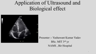
  • 1. Application of Ultrasound and Biological effect Presenter :- Yashawant Kumar Yadav BSc. MIT 3rd yr NAMS , Bir Hospital 1
  • 2. OUTLINE • Introduction of Ultrasound • Modes of Ultrasound with application • Doppler US and types with application • Different kinds of probes with medical application • Biological effect • References 2
  • 3. Intro….. • Any sound with a frequency above 20,000 Hz (or 20 kHz)that is, above the highest audible frequency—is defined to be ultrasound. • 1Hz = 1 cycle / vibration • 1kHz = 1000 cycle per second • 1MHz = 1 million vibrations per second. 3
  • 4. Contd…. • A sound wave can be described as a mechanical, longitudinal wave composed of cyclic compressions and rarefactions of molecules in a medium. 4
  • 5. Interaction • Reflection • Refraction • Absorption • Scattering • Acoustic impedance • Z = ρv, The units for Z are therefore kg/(m2 · s). • Matching layer ??? • Z of PZC is 15 times higher than skin 5
  • 6. Parameters • Amplitude: • Power :- (joules /s) (A2) • Intensity :- The energy per unit cross-sectional area in a sound beam, • Intensity is expressed in joules/second/square centimeter. • Joule/second = watt • (w/cm2) 6
  • 7. Contd… Relative scale intensity is defined in units of the decibel (dB) In common practice, the lowest-intensity audible sound (10-12 W /M2) is assigned the value of 0 dB. 10dB = 102 Times of 0dB So 120 dB = 1012 times i.e 1 trillion times of 0 dB 7
  • 8. Production of US in Medicine Piezoelectric US generation. Natural -- - (quartz, and lead titanate) Piezoelectric crystals I. Barium titanate,(BaTiO3) (1st discovered ) II. Rochelle salts, (KNaC4H4O6·4H2O) III. Potassium niobate (KNbO3) IV. Ammonium dihydrogen phosphate (ADP) V. lead zirconate titanate . (Pb[ZRxTi]O3) VI. Sodium tungstate (N2WO3) VII. Sodium potassium niobate (NKN) lead free 8
  • 9. Pulse acoustic phenomenon • Continuous waves are not useful for structural imaging. • One pulse typically consists of three to five cycles. Pulse US parameters • Pulse duration (0.5 to 3 µs). • Pulse repetition period: ( 0.1 to 1 ms.) • Spatial pulse length: (0.1 to 1 mm). • Duty factor (0.1 % to 1 %. ) • Pulse repetition frequency (PRF): (1 OOO to 10,000 Hz ) PRF is inversely proportional to imaging depth. 9
  • 10. Tissues attenuation • Bone is represented as a very bright structure and appears ‘hyperechoic’. • Muscle presents as hypoechoic, with some internal signals as a result of collagen fibers. • Tendon show tightly packed hyperechoic lines representing the fibrils of the tendon. 10
  • 11. Contd…. • Nerves as ‘honeycomb’ like structures composed of hypoechoic spots embedded in a hyperechoic background. • Fluid presents has an anechoic appearance on ultrasound. • Air bubbles reflect much of the US that engages them and appear very echo dense (bright). • Ligament 11
  • 12. Medical application • Ultrasound is used in medicine for therapy and diagnosis. In diagnosis : • Ultrasound have 4 basic mode :- 1. A – amplitude mode 2. B – brightness mode 3. M- motion mode 4. Doppler 12
  • 13. A-amplitude mode (10–20 MHz) • Mode A is a one-dimensional process, and simplest type of US . • Basically used to determine depth of abnormality . (function of depth ) • Therapeutic ultrasound aimed at a specific tumor or calculus is also A-mode, to allow for pinpoint accurate focus of the destructive wave energy. • As an example on how Mode A can be applied, • US encephalography, and US ophthalmology 13
  • 14. 14
  • 15. Contd… In a case where the middle brain structures have been displaced (for example by the presence of an odd mass in a lobe that pushes them into the other lobe) 15
  • 17. B – Brightness mode • The B-mode is a two-dimensional (2D) image of the area that is simultaneously scanned by a linear array of 100–300 piezoelectric elements rather than a single one as in A-mode • B-Mode composed of bright dots representing the ultrasound echoes. • The brightness of each dot is determined by the amplitude of the returned echo signal. 17
  • 18. 18
  • 19. M- Motion Mode • A continuous B-mode display. Displays one scan line versus time. • Allows for a high frame rate, accuracy of linear measurements, and tracking of motion of reflectors (x-axis: time; y-axis: reflector depth).. • M-mode is used extensively in cardiac and fetal cardiac imaging. • Its an application of B - mode imaging 19
  • 20. Doppler mode • The Doppler effect is defined as the change in the frequency of sound emitted or reflected by a moving object. The amount of change is termed the Doppler shift. 20
  • 21. Th is angle is known as the angle of insonation. 21
  • 22. Contd… • The highest Doppler frequency shift from any specific vessel occurs when the vessel and the beam are aligned; that is, when the angle of insonation is zero and the cosine function has the value of 1.0. • The least desirable situation occurs when the angle approaches 90° as then the Doppler frequency shift will be minimum. • In clinical practice it is often not possible to align the beam and the vessel. • As a rule, provided the angle is less than about 60°, good-quality spectral waveforms can be obtained. 22
  • 23. Application of doppler 1. Peripheral vascular system - Arteries and veins of the extremities 2. Central arteries / veins - Abdominal aorta, IVC, mesenteric vessels 3. Cerebrovascular system - 4. Cardiology 5. Obstetrics and Gynecology - IUGR, Uterine arteries, Ovarian torsion 6. Renovascular hypertension 7. Male infertility / erectile dysfunction, testicular torsion 8. Tumors, AV malformations. 23
  • 24. Contd… There are five methods of imaging by employing Doppler principle. a) Continuous wave doppler b) Pulsed wave Doppler c) Color Doppler d) Power Doppler e) Spectral Doppler or Duplex scanning 24
  • 25. CONTINIOUS WAVE DOPPLER • This employs two piezoelectric crystals, both contained in a single head. One crystal transmits a continuous sonic signal at a known frequency (3-8 MHz). • The other crystal receives the returning echoes and records their frequency. • It is employed in detection of blood flow but does not give information of depth, direction and velocity of flow. • From a practical standpoint, continuous wave Doppler should be used when measuring velocities > 1.2 m/s. (e.g., regurgitant jets, stenotic valves). • No depth resolution 25
  • 26. 26
  • 27. Contd… Pulsed wave Doppler (PW):- Provide depth information , variable depth can be acquired by changing beam frequency (low for high penetration and high for low penetration)  Three factors affects PWD To evaluate mobile structure I. High pulse repetition frequency II. Optimum transducer frequency III. Insonation angle (less than 600) It is customarily combined with a B mode / 2-Dimensional mode imaging, which is then called duplex scanning. measuring relatively low-flow velocities ( < ~ 1.2 m/s) in specific areas of interest (e.g., pulmonary vein flow, mitral valve inflow). 27
  • 28. Contd.. • Because sampling is intermittent, the pulse repetition frequency limits the maximum Doppler shift (and thus maximum velocity) that can be measured accurately. • Velocities higher than this maximum velocity will appear to wrap around on the display, a phenomenon known as aliasing. • The Doppler frequency shift at which aliasing occurs, equal to PRF divided by 2, is termed the Nyquist limit. 28
  • 29. Contd.. • Doppler shift (expressed in Hz) = (2 · v · Fi cosine ϴ)/c (if ϴ= 0/180 =1/-1) Doppler shift= (v· Fi)/770 v = 770· (Doppler shift/Fi) • if a 5-MHz transducer can only send out about 15,000 pulses per second, the Nyquist limit is 7500 Hz (15,000/2). • Using the velocity equation shown earlier, the maximum velocity that can be measured without aliasing is about 1.15m/s (770 X (7500/5,000,000)). 29
  • 30. 30
  • 31. Color doppler(Also called color flow imaging (CFI) or color velocity imaging (CVI) ) • Ten or more pulse packets are used to produce an estimate of mean velocity of all reflectors. • This data is assigned color by the machine and is superimposed on B mode data from stationary structures. 31
  • 32. Contd… • Flow that travels away from the transducer (negative Doppler shift) is depicted in blue, • Flow that is traveling toward the transducer (positive Doppler shift) is depicted in red, with lighter shades of each color denoting higher velocities. • A third color, usually green or yellow, is often used to denote areas of high flow turbulence. Clinical use :- • overall blood flow to a region of interest. Depiction of the general velocity and direction of blood flow within the heart and blood vessels. • It also allows the. generation of unique phenomena such as the fluid color sign 32
  • 33. Contd … • The fluid color sign is a diagnostic sign to differentiate a pleural effusion from pleural thickening by means of color Doppler ultrasound. • In the case of pleural effusion a color signal is seen in the pleural fluid during respiratory and cardiac movement, whereas this color signal is not seen in the case of pleural thickening. 33
  • 34. Power Doppler or Energy mode imaging • An alternative processing method. which ignores the velocity and simply estimates the strength/amplitude of the Doppler signal detected from each location. And encoded as a color . • The Energy mode image is continuous and hence not sensitive to relative flow direction. It does not give the absolute values of velocity but only gives the strength of the Doppler signal. 34
  • 35. Contd…. • The higher sensitivity to slow flow. • Power Doppler is particularly useful when examining superficial structures, like thyroid, testis, renal grafts and subcutaneous lesions. It may be used to look for tumor vessels, to evaluate tiny low-flow vessels and detect subtle ischemic areas. 35
  • 36. Contd… Advantages of Power Doppler over Color flow imaging I. More sensitive to flow states (3-5% more sensitive than CFD) II. Angle effects are ignored unless the angle is close to 90° III. Aliasing is not applicable. There are disadvantages also (i) Values of velocity and the direction of blood flow cannot be assessed. (ii) Because of more averaging of information at slower frame rates. slow moving soft tissue signals appear as flash artifacts 36
  • 37. Spectral doppler • Spectral Doppler displays the blood flow measurements graphically, displaying flow velocities recorded over time. • For spectral Doppler , the machine represents the movement of blood via an audible flow sound, and a graph depicting flow velocities in relation to time. • This mode is ideal for measuring lower velocities at chosen anatomic locations, 37
  • 38. ULTRASOUND PROBES • Linear array • Curvilinear array • Phased array • linear array, “L” • electronic linear scanning, “E” in the xz plane • Curve scan “C” • “E<xz” electronically focus 38
  • 39. Acoustic window’s An acoustic window is the location from which an ultrasound probe makes it's scan. 39
  • 40. Linear (5-15MHz) • Designed to make contact with flat surfaces, with the footprint decreasing in size with increasing frequency. • Here rectangular and trapezoidal formats provide appropriate viewing areas. With abdominal imaging, to increase the viewing area with minimal increases of the contact area • Use for high-resolution imaging of superficial structures, 40
  • 41. Contd.. • This probe is ideal for ultrasound-guided needle entry (particularly vascular access), • evaluation of deep venous thrombosis, • chest imaging for pleural lines (pneumothorax), • skin and soft tissue imaging for abscesses. • Nerve blocks • Ophthalmologic • Joint /tendon • Thyroid/breast • Evaluation of superficial structure 41
  • 42. Curve linear array (1-8MHz) • convex arrays designed to make surface contact in deformable soft areas of the body. • The curvilinear (convex) array probe is used for scanning deeper structures, • The crystal alignment along a curved surface generates a beam that fans outward. The result is a field of view wider than the probe’s footprint, • These probes are most often used in abdominal and pelvic diagnostic applications. 42
  • 43. Contd…. • These probes are most often used in abdominal and pelvic diagnostic applications. • Hepatobiliary • Renal • Appendix (pediatric ) • Aorta • Focus assessment in case of trauma • Fetal heart tones and obstetric complication (pregnancy ) • Emergency lung examination • Paracentesis and thoracentesis 43
  • 44. Phase array (2-7.5MHz) • Consisting of 64-128 elements. It is a smaller assembly . • The phased array probe is a lower frequency probe. • Beam can be steer electronically , variable field shape can be acquired . • The smaller footprint allows for image acquisition between the ribs, and the slightly convex surface produces a fanning beam with a sector-shaped image. 44
  • 45. Contd….. • This probe is used for echocardiography and thoracic imaging but can be applied to abdominal and pelvic imaging as well. • Procedure guide • Alternative to curvilinear for abdominal structure evaluation • Pericardiocentesis • Thoracentesis 45
  • 46. 46
  • 47. Biological effect • Energy deposition in biological tissue. • With CW ultrasound, the tissues are irradiated throughout the duration of the exposure, and the total energy deposited in tissue will be much more in comparison to PW irradiation over the same total exposure duration, • Consequences of high intensity, prolonged exposures 1. Elevation of tissue temperature (thermal effect ) 2. Cavitation in liquids (non thermal effect ) Interlinked 47
  • 48. Elevation of temperature • The mechanical energy of ultrasound can be transformed into heat through absorption in tissue, resulting in the elevation of tissue temperature. • Temperatures exceeding 4°C above normal body temperature, maintained for long periods of time, may be harmful to the embryo and fetus, leading to a variety of biological changes which can result in abortion, fetal death, or developmental abnormalities,(teratogenic effect ) • These effects have been seen following whole body heating of both the mother and the offspring, (approx. 0.5°C min−1 ) 48
  • 49. Contd.. The World Federation for Ultrasound in Medicine and Biology (WFUMB) recommends that diagnostic exposures that produce a maximum temperature rise of 1.50 c above the normal physiological level of 37°C • Ultrasound heating is very rapid . In addition, ultrasound exposures in pregnancy only subject a small fraction of the maternal volume to a potential temperature rise, and thus any heating of the embryo is likely to be rapidly dissipated. 49
  • 50. Cavitation in liquids( non thermal effect ) • Cavitation is a phenomenon which takes place in liquid cavities exposed to very high intensities of ultrasound • The presence of gas within the volume of the liquid may lead to the formation of micro bubbles during the low pressure phase (rarefaction) of the ultrasound wave cycle. With changes in pressure, the bubbles may grow in size. During the high pressure phase (compression), • The micro bubbles may collapse a with the release of large quantities of mechanical energy which can cause biological damage. 50
  • 51. Contd.. • cavitation can also be the cause of free radical formation. • Free radicals are highly reactive chemical species which can attack bio molecules through a chain of chemical reactions, producing toxic bio products. • This leads to damage of biological matter not only locally, but also remotely through diffusion of toxic products. • cavitation is unlikely to occur with PW ultrasound 51
  • 52. Drug delivery (Sonoporation ) • Refers to a technique capable of controlling drug delivery efficiency by maximizing the drug permeability of surrounding tissues or cells using a reaction mechanism between microbubbles and ultrasonic waves. • Injecting microbubbles intravenously can minimize drug side effects that may cause nonspecific delivery of drugs into healthy organs because the microbubbles only collapse in specific diseased areas due to focused ultrasound irradiation. 52
  • 53. 53
  • 54. Contd … • ultrasound-mediated microbubble destruction (UMMD) • ultrasound-targeted microbubble destruction (UTMD). 54
  • 56. References • https://theultrasoundsite.co.uk/ultrasound-case-studies/starting-article-2- different-tissues-ultrasound-look-like/ • https://www.nysora.com/foundations-of-regional- anesthesia/equipment/physics-of-ultrasound/ • https://radiologykey.com/doppler-imaging-2/ • https://radiopaedia.org/articles/phased-array • https://www.ncbi.nlm.nih.gov/pmc/articles/PMC6208473/ • Martinoli, C., Pretolesi, F., Crespi, G., Bianchi, S., Gandolfo, N., Valle, M., & Derchi, L. E. (1998). Power Doppler sonography: clinical applications. European journal of radiology, 27 Suppl 2, S133–S140. https://doi.org/10.1016/s0720-048x(98)00054-0 • https://echocardiographer.org/Echo%20Physics/spectral%20doppler.html 56
  • 57. 1. Cavitation effect in microbubble contrast agent ?? 57

Editor's Notes

  1. Specular reflection = greater spacing of surface than US wavelength ( mirror effect ) Diffused / Scattering reflection = small or equal spacing of surface than US wavelength Rayleigh reflection = very small spacing than US
  2. The rate of energy transfer, expressed in watts (joules/second). Power is proportional to the square of the amplitude. This is the parameter used most frequently when describing the biological safety of US.
  3. We can never observe details significantly smaller than the wavelength of our probe.
  4. It is important to note that the piezoelectric properties of an artificial crystal can be destroyed if the crystal is heated to high temperatures US generation by magnetostriction. Electrodynamic US generation. Electrostatic US generation. The general principle involved in generating ultrasonic waves is to cause some dense material to vibrate very rapidly
  5. Gradient time , slew rate and duty time – mri Interrogation time , duty cycle and extinction time - fluoroscopy
  6.  fluid within the superficial infrapatellar bursa of the knee.
  7. The major application is ophthalmic corneal pachymetry, which is a noninvasive technique for measuring corneal thickness.
  8. Echoencephalography detects brain anomalies like tumors, hematoma, injuries, displacement of the brain ventricles, etc
  9. , the actual Doppler shift is in the audible range (20 to 20,000 Hz).
  10. motion of the blood, which now acts as a moving source
  11. Intrauterine Growth Restriction, Movement , direction and velocity
  12. PW ultrasound does not provide the information about the structure / site of origin of an echo,
  13. 5-7.5Mhz
  14. Doppler sample volume gate , doppler sampling volume and insonation angle
  15. Thus, one might expect a 6-cm penetration from a 10-MHz center frequency transducer
  16. A procedure in which a thin needle or tube is put into the abdomen to remove fluid from the peritoneal cavity  Thoracentesis is a procedure to remove fluid from the space between the lining of the outside of the lungs (pleura) and the wall of the chest.
  17. Particularly sensitive to temperature raises is the embryo and fetus in utero. It requires ultrasonic beam intensities of several hundred mill watts per square centimeter (mW/cm2) to elevate body temperature by just 1°C,
  18.  (a temperature rise of 4°C for 5 min). reduced the threshold to lie at a temperature rise of 4°C maintained for 30 s. The temperature rise may be limited by the cooling effects of blood flow in the exposed volume. Highly vascular organs, such as the liver and kidney, are thus more difficult to heat than bone,
  19. often termed breathing oscillations
  20. Ex. for microbubbles composed of lipids, hydrophobic anticancer drugs can be very efficiently loaded onto the hydrophobic tail of the lipid found between the gas core and the shell . Thus, the loaded drug can be safely delivered to the target tissue without being attacked by various enzymes present in the blood and can then be released in response to ultrasonic waves as an external stimulus-triggered drug release strategy.