SlideShare a Scribd company logo
1 of 59
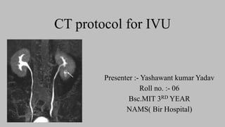
CT protocol for IVU
Presenter :- Yashawant kumar Yadav
Roll no. :- 06
Bsc.MIT 3RD YEAR
NAMS( Bir Hospital)
Outline
• Anatomy
• Indications
• Preparation for procedure IVU
• Technique and modification
• Radiation dose on procedure
• Findings in IVU
• Advantage and disadvantage
• References
Introduction
• A computerized tomography (CT) urogram is an imaging exam used to evaluate
your urinary tract, including kidneys, bladder and the tubes (ureters) that carry
urine from your kidneys to your bladder.
• MDCT urography is today recognized as state-of-the-art imaging modality in the
evaluation of hematuria and choice for renal evaluation
• Unenhanced CT is generally reserved to demonstrate calcifications and calculi that
may be obscured by contrast agent or it is used as a baseline for attenuation
measurements when enhancement is calculated as a feature of renal mass
characterization. (KUB)
Anatomy of Urinary System
Organs:
• Kidneys – clean and filter blood
• Ureters – tubes that take urine to bladder ‘
• Bladder – stores urine until eliminated
• Urethra – removes urine from body
Kidney
• The kidneys are situated against the posterior abdominal wall and are
retro peritoneal organ .
 however, that the two kidneys differ slightly in position—
1.the left kidney extends from about T12 to L3, whereas
2.the right kidney sits slightly lower on the abdominal wall because
of the position of the liver.
• The superior portion of both kidneys are partially protected by the 11th and
12th pairs of ribs.
External Anatomy of the
Kidneys
• The kidneys are held in place on the posterior body wall and protected by
three external layers of connective tissue.
1.Renal fascia.
2.Adipose capsule. The middle and thickest layer, called the adipose
capsule, consists of adipose tissue that wedges each kidney in place
and shields it from physical shock.
3. Renal capsule. The renal capsule is an extremely thin layer of dense
irregular connective tissue that covers the exterior of each kidney like
plastic wrap. It protects the kidney from infection and physical trauma.
Internal Anatomy of the Kidneys
• A frontal section
of the kidney
reveals the
three distinct
regions of this
organ: the
outermost renal
cortex, the
middle renal
medulla, and
the inner renal
pelvis
Contd….
• Nephrons are the functional units of the kidney
• The renal cortex and renal medulla of each
kidney contain over one million microscopic
filtering structures called nephrons.
• which consists of two main components: the
globe-shaped renal corpuscle, and a long,
snaking tube of epithelium called the renal tubule.
• The renal corpuscle and the majority of the renal
tubule reside in the renal cortex, whereas varying
amounts of the renal tubule dip into the renal
medulla.
Contd …
• These two types of nephrons labeled
as cortical and juxtamedullary
• About 80% of nephrons are cortical
nephrons, so named because they are
located primarily in the renal cortex and
have very short nephron loops
• less numerous, type of nephron in the
kidney is the juxtamedullary nephron.
• It has a long nephron loop that
burrows deeply into the renal medulla.
• The loop is surrounded by a ladder-like
network of capillaries called the vasa
Contd..
• The tip of each renal pyramid tapers into a
slender papilla , which borders on the first
urine draining structure, a cup-shaped tube
called a minor calyx
• Urine from three to four minor calyces
drains into a larger major calyx
• Two to three major calyces, in turn, drain
urine into the large collecting chamber that
is the renal pelvis, which leads into the
ureter Smooth muscle tissue in the walls of
the calyces and renal pelvis contracts to
help propel urine toward the ureter.
Blood supply
• The kidneys receive approximately one-fourth of the total cardiac
output—about 1200 ml per minute—from the right and left renal arteries,
which branch from the abdominal aorta.
•
Contd..
•1 Renal artery → 2 Segmental artery → 3 Interlobar
artery → 4 Arcuate artery → 5 Interlobular (cortical
radiate) artery → 6 Afferent arteriole → 7 Glomerulus →
8 Efferent arteriole → 9 Peritubular capillaries. → 10
Interlobular vein → 11 Arcuate vein → 12 Interlobar vein
→ 13 Renal vein.
Ureters• The ureters transport urine from the kidneys to the urinary bladder. The
ureters are generally about 25–30 cm long and 3–4 mm in diameter in an
adult.
• They begin at roughly the level of the second lumbar vertebra, travel
behind the peritoneum, and empty into the urinary bladder.
• The ureters drain posteriorly into the inferior urinary bladder. In this region
a mechanism prevents urine from flowing backward through the ureter.
• As each ureter passes along the posterior urinary bladder, it travels
obliquely through a “tunnel” in the bladder wall. As urine collects in the
bladder, the pressure rises and compresses this tunnel, pinching the
ureter closed and preventing backflow of urine.
Urinary Bladder
• The urinary bladder is a hollow, distensible organ that sits on
the floor of the pelvic cavity, suspended by a fold of parietal
peritoneum.
Indications of IVU
• Hematuria
a) Micro
b) Macro
• Urinary bladder tumors/stone
• Detection, staging and surveillance of
urothelial tumors
• Obstruction,
• trauma, congenital abnormalities,
• percutaneous nephrolithotomy &
complex infections
• Kidney stones/ tumor or cysts
 Relative or absolute
contraindications
• renal insufficiency,
• contrast allergy,
• pregnancy,
• young age and frequent
examinations
Pt. preparation
• Have any allergies, particularly to iodine or had a previous severe reaction to CM
• Are pregnant or think you might be pregnant ( risk vs benefit )
• Are taking any medications, such as metformin (Fortamet, Glucophage, Glumetza), nonsteroidal
anti-inflammatory drugs (NSAIDS), anti-rejection drugs or antibiotics
• Have had a recent illness
• Have a medical condition, including heart disease, asthma, diabetes, kidney disease or a prior
organ transplantation
• Written and verbal consents are followed
• In order to expand (distend) your bladder, you may be asked to drink water before a CT urogram
and not to urinate until after the procedure.
• However, depending on condition, guidelines about eating and drinking before your CT
urogram may vary .
• Urea & creatinine should be in normal range (15-50mg/dl---0.45-1.4mg/dl)
Contrast media
• The volume of the contrast material bolus ranges from 100 to 150 ml,
administered at a rate of 2–4 ml/sec
• Nonionic contrast agent of 300 mg I/mL is preferred .
Technique
 For CT Urography there are different phase are followed
1. Unenhanced or non- contrast phase
2. Enhance corticomedullary phase
3. Nephrographic phase
4. Execratory phase &
5. Delayed phase
-tailored according to the specific clinical problem involved
Unenhanced or non- contrast phase
• A digital scout radiograph is obtained to ensure coverage from the diaphragm to
the iliac crests.
• A non-contrast CT scan is obtained scanning from the top of the kidneys through
to the pubic symphysis.
• Malignant urologic tumors, such as renal cell carcinoma and transitional cell
carcinoma, are potentially detectable during unenhanced imaging examinations.
• Renal cell carcinoma and transitional cell carcinoma typically appear solid on
unenhanced images and have higher attenuation (5–30 HU) than urine
Contd…
• A renal lesion and also ensure that renal parenchymal calcifications,
renal calculi,
• renal and perinephric hemorrhage and fat, and calcification in a renal
mass
Non enhanced scans also help differentiate hyperdense cyst from renal
solid tumor
Corticomedullary
• This phase is acquired 25-70 sec after contrast
injection
• contrast material enters the cortical capillaries
and peritubular spaces and filters into the
proximal cortical tubules.
Renal cortex can be differentiated from renal
medulla at this stage because
• the vascularity of the cortex is greater than that
of the medulla, &
• contrast material has not yet reached the distal
aspect of the renal tubules
• Renal vasculature or Detected renal mass --represent an aneurysm or an AV malformation
or fistula.
Maximal opacification of the renal vein and arteries, allowing confident diagnosis of tumor
extension to the vein.
left renal artery (black arrow) and
vein (white arrow).
Nephrographic phase
• Nephrographic phase images are
acquired 90–100 seconds after
administration of a nonionic contrast
agent .
• Imaging (2.5- to 5-mm slice thickness) is
typically confined to the kidneys during
this phase.
• Nephrographic phase imaging has the
highest sensitivity in the detection of
renal masses, and correlation with
unenhanced images is required to show
unequivocal enhancement.
• Nephrographic phase axial CT at level of renal hilum.
Excretory phase
• Begins approx 5-8 min after the start of contrast injection.
• The contrast excretes into the collecting system decreasing the attenuation of the
nephrogram
• helpful to better delineate the relationship of a centrally located mass with the
collecting system.
• also useful for evaluating urothelial masses.
• McNicholas et al (28) showed that excretory phase CT scans obtained with
patients in a prone position also improved opacification of the distal ureters
compared to CT scans obtained in supine patients without abdominal compression.
Contd…
• Alternative techniques for achieving optimal visualization of the collecting
systems include supplemental use of normal saline infusion and diuretic injection.
• McTavish et al . reported that supplemental infusion of 250 mL of physiologic
saline immediately after injecting intravenous contrast material significantly
improved opacification of the distal ureters.
• Nolte-Ernsting et al . reported that intravenous injection of low-dose diuretics (10
mg of furosemide) before intravenous contrast material injection also permitted
less dense, homogeneous opacification of the collecting systems compared to
supplemental infusion of 300 mL of normal saline.
• Excretory phase axial CT at level of renal hilum.
• CT urogram: Excretory phase obtained 7 minutes following
contrast administration. This phase is used to look for filling
defects in the urinary collecting system.
Contd…
• Classic renal vascular anatomy in a
36-year-old potential renal donor.
Coronal volume-rendering images in
the (a) arterial phase
Contd…
• Venous phase
Protocol of BIR
Phase Timing Range Slice thickness What to detect
Nonenhanced Pre contrast Lung bases to pubic
symphysis
5 mm Calculi,
hemorrhage/hemorrha
gic cysts
Nephrogram 90–100 seconds Lung bases to pubic
symphysis
3mm Renal tumors, renal
vein thrombosis
Excretory 5-8 min Lung bases to pubic
symphysis
2-3mm Papillary necrosis,
urothelial carcinoma
Sacn :- craniocaudal
Kvp:- 100-120
mA :-80-500
Pitch :- 1-1.375
Unenhanced
Delay
Split-bolus technique
• generally done in DECT
• Radiation doses can be reduced with use of a split-bolus (two-phase) technique
• In which an unenhanced acquisition is followed by IV administration of 30–50
mL of contrast material, and a second bolus of 80–100 mL of IV contrast material
is given after an 8- to 10-minute delay, during which the acquisition is performed.
• Thus in a single nephropyelographic phase acquisition, the renal parenchyma
(nephrographic phase) and the collecting system, ureters, and bladder
(pyelographic phase) are assessed in a reduced the number of phases at a reduced
radiation dose.
Contd…
• excretory-parenchymal
• arterial –portal venous
• vascular –parenchymal
• Provides vascular and parenchymal information or arterial and venous information
in a single acquisition
• A possible disadvantage of the split-bolus technique is that the presence of
contrast material within the ureter at imaging can obscure the subtle isoattenuating
tumors that are not seen in the low-dose unenhanced phase
• CT urogram utilizing a split dose technique: Axial image through
the kidneys and collecting systems demonstrates both
nephrographic and excretory phases of enhancement in the
• CT urogram utilizing a split dose technique: Axial images
displayed with wider window settings are suitable for display of
the opacified collecting systems and urinary tract calculi.
Radiation dose
• Many variations of the standard CT urographic protocol have been investigated
with the goal of reducing radiation exposure and optimizing imaging of the
urothelium.
• Caoili et al. [12] reported radiation doses of 25– 35 mSv for four-phase CT
urography compared with a mean effective dose of 3.6 mSv for excretory
urography
• Radiation doses in CT urography can be reduced by limiting the number of
imaging phases through the use of dual-energy CT or split-bolus
technique .
• Dual-energy CT obviates an unenhanced phase of imaging because
virtual unenhanced CT scans can be postprocessed from a contrast-
enhanced study acquired with two tube potentials operating
simultaneously .
Contd…
• In the early days of CTU, a three-phase CTU protocol could be associated with
effective dose levels of 25–35 mSv .
• A phantom analysis of single-bolus three-phase and split-bolus two-phase CTU
protocols showed mean effective doses of 20–40 mSv, with individual effective
doses as high as 66 mSv .
• Proper stepwise protocol optimization toward use of a three-phase CTU protocol
with unenhanced, corticomedullary, and excretory phases resulted in a reduction in
CTU effective doses from 29.9 to 11.7 mSv for women and from 22.5 to 8.8 mSv
for men.
• A recent CT dose survey performed in The Netherlands showed that, for (split-
bolus two-phase) CTU, comparable median effective doses would be 3.6 mSv for
the unenhanced phase and 6.6 mSv for the concurrent nephrographic and
excretory phases
Advantages
• Multidetector CT technology allows faster data acquisition times.
• Multidetector CT improved z-axis spatial resolution.
• Thinner collimation improves the quality of three-dimensional (3D) data sets and allows
generation of exquisite 3D images of the renal arteries , veins, ureter and kidney
• Motion Artifacts reduction due to the faster acquisition
• Radiation dose minimized
• Example of 3D postprocessing work performed by the
radiologist after the acquisition of the imaging data.
• The image has been obliqued to
optimally visualize the left distal ureter
and left ureterovesicular junction.
• The image has been obliqued in order
to optimally visualize the right distal
ureter and right ureterovesicular
DECT
• DECT is currently not widely available in our clinical practice.
• Compared with single-energy CT, it can enhance tissue and calculi
characterization .
• Two synchronous CT acquisitions at various tube voltage levels allow tissue
discrimination (e.g., bone) from iodine because of their diverse x-ray
absorptions at low and high peak kV levels .
• Omitting iodine from contrast-enhanced DECT images ------virtual unenhanced
images -----for detecting calculi within an iodine-filled urinary collecting
system.
• This process can obviate the requirement for unenhanced CT images and
thereby reduce radiation exposure.
Advantages of DECT urography
• Easy and reliable subtraction of bony structures, 3D visualization of the
collecting systems without concealment by bony structures
• Visualization of renal parenchyma, vascular structures, urinary tracts, calculi,
and tumour's
• Radiation dose reduction by split bolus combined in DECT eliminating the
requirement for an unenhanced scan.
Cons. (consensus) PCNL, (percutaneous nephro-lithotomy)
Findings
Contd..
Contd…
Contd…
• Evidence of right renal infarct at prior
examination. Coronal volume-rendered
image nicely demonstrates an 80% stenosis
in the proximal right renal artery caused by a
partially calcified plaque (white arrow).
• There is a large infarct involving the upper
pole of the right kidney (black arrows).
References
• https://www.ajronline.org/doi/pdf/10.2214/AJR.10.4198
• https://radiologykey.com/ct-urography/
• https://pubs.rsna.org/doi/10.1148/rg.24si045513#TABLE1
• https://emedicine.medscape.com/article/1890669-overview#a3
• https://pubs.rsna.org/doi/full/10.1148/rg.e20
• https://www.ajronline.org/doi/10.2214/AJR.14.13862
Ct protocol for ivu

More Related Content

What's hot

magnetic resonance angiography
magnetic resonance angiographymagnetic resonance angiography
magnetic resonance angiographyqavi786
 
Mrcp radiology
Mrcp radiologyMrcp radiology
Mrcp radiologyVidya TK
 
CT perfusion physics and its application in Neuroimaging
CT perfusion physics and its application in NeuroimagingCT perfusion physics and its application in Neuroimaging
CT perfusion physics and its application in NeuroimagingDr.Suhas Basavaiah
 
Ct mri urography
Ct mri urographyCt mri urography
Ct mri urographyDev Lakhera
 
Artifacts usg, ct, mri radiology ppt,pdf pk
Artifacts usg, ct, mri radiology  ppt,pdf pkArtifacts usg, ct, mri radiology  ppt,pdf pk
Artifacts usg, ct, mri radiology ppt,pdf pkDr pradeep Kumar
 
Portable ct mobile ct
Portable ct mobile ctPortable ct mobile ct
Portable ct mobile ctanilayyakutty
 
Physics of Multidetector CT Scan
Physics of Multidetector CT ScanPhysics of Multidetector CT Scan
Physics of Multidetector CT ScanDr Varun Bansal
 
TRIPLE PHASE CT AND ITS TECHNIQUE BY VANI PUSHPA (2).pptx
TRIPLE PHASE CT AND ITS TECHNIQUE BY VANI PUSHPA (2).pptxTRIPLE PHASE CT AND ITS TECHNIQUE BY VANI PUSHPA (2).pptx
TRIPLE PHASE CT AND ITS TECHNIQUE BY VANI PUSHPA (2).pptxVANI PUSHPA MUDAVATH
 
Gb nd biliary tree imaging
Gb nd biliary tree imagingGb nd biliary tree imaging
Gb nd biliary tree imagingPaul Joy
 
Perfusion imaging: CT, MRI, Nuclear Medicine- Avinesh Shrestha
Perfusion imaging: CT, MRI, Nuclear Medicine- Avinesh ShresthaPerfusion imaging: CT, MRI, Nuclear Medicine- Avinesh Shrestha
Perfusion imaging: CT, MRI, Nuclear Medicine- Avinesh ShresthaAvinesh Shrestha
 
CT Angiography presentation
CT Angiography presentation CT Angiography presentation
CT Angiography presentation Shatha M
 
CT ITS BASIC PHYSICS
CT ITS BASIC PHYSICSCT ITS BASIC PHYSICS
CT ITS BASIC PHYSICSDEEPAK
 
Ct & mr enterography
Ct & mr enterographyCt & mr enterography
Ct & mr enterographyRakesh Ca
 

What's hot (20)

CT RADIATION DOSE REDUCTION
CT RADIATION DOSE REDUCTION CT RADIATION DOSE REDUCTION
CT RADIATION DOSE REDUCTION
 
magnetic resonance angiography
magnetic resonance angiographymagnetic resonance angiography
magnetic resonance angiography
 
Basics in ct
Basics in ctBasics in ct
Basics in ct
 
Mrcp radiology
Mrcp radiologyMrcp radiology
Mrcp radiology
 
CT perfusion physics and its application in Neuroimaging
CT perfusion physics and its application in NeuroimagingCT perfusion physics and its application in Neuroimaging
CT perfusion physics and its application in Neuroimaging
 
Ct mri urography
Ct mri urographyCt mri urography
Ct mri urography
 
CT perfusion
CT perfusionCT perfusion
CT perfusion
 
CT Angiography Lower Limb
CT Angiography Lower LimbCT Angiography Lower Limb
CT Angiography Lower Limb
 
Artifacts usg, ct, mri radiology ppt,pdf pk
Artifacts usg, ct, mri radiology  ppt,pdf pkArtifacts usg, ct, mri radiology  ppt,pdf pk
Artifacts usg, ct, mri radiology ppt,pdf pk
 
Portable ct mobile ct
Portable ct mobile ctPortable ct mobile ct
Portable ct mobile ct
 
Physics of Multidetector CT Scan
Physics of Multidetector CT ScanPhysics of Multidetector CT Scan
Physics of Multidetector CT Scan
 
TRIPLE PHASE CT AND ITS TECHNIQUE BY VANI PUSHPA (2).pptx
TRIPLE PHASE CT AND ITS TECHNIQUE BY VANI PUSHPA (2).pptxTRIPLE PHASE CT AND ITS TECHNIQUE BY VANI PUSHPA (2).pptx
TRIPLE PHASE CT AND ITS TECHNIQUE BY VANI PUSHPA (2).pptx
 
Gb nd biliary tree imaging
Gb nd biliary tree imagingGb nd biliary tree imaging
Gb nd biliary tree imaging
 
Perfusion imaging: CT, MRI, Nuclear Medicine- Avinesh Shrestha
Perfusion imaging: CT, MRI, Nuclear Medicine- Avinesh ShresthaPerfusion imaging: CT, MRI, Nuclear Medicine- Avinesh Shrestha
Perfusion imaging: CT, MRI, Nuclear Medicine- Avinesh Shrestha
 
CT Angiography presentation
CT Angiography presentation CT Angiography presentation
CT Angiography presentation
 
CT ITS BASIC PHYSICS
CT ITS BASIC PHYSICSCT ITS BASIC PHYSICS
CT ITS BASIC PHYSICS
 
Phase pdf
Phase pdfPhase pdf
Phase pdf
 
Ct head protocols
Ct head protocolsCt head protocols
Ct head protocols
 
Ct & mr enterography
Ct & mr enterographyCt & mr enterography
Ct & mr enterography
 
MR imaging of liver
MR imaging of liverMR imaging of liver
MR imaging of liver
 

Similar to Ct protocol for ivu

Kidney anatomy - Almaskhan .khorfakhan hospital
Kidney anatomy - Almaskhan  .khorfakhan hospitalKidney anatomy - Almaskhan  .khorfakhan hospital
Kidney anatomy - Almaskhan .khorfakhan hospitalalmasmkm
 
medicine.Kidney 2.(dr.ala)
medicine.Kidney 2.(dr.ala)medicine.Kidney 2.(dr.ala)
medicine.Kidney 2.(dr.ala)student
 
Intravenous urography and its modifications.pptx 01
Intravenous urography and its modifications.pptx 01Intravenous urography and its modifications.pptx 01
Intravenous urography and its modifications.pptx 01SUJAN KARKI
 
Kidney by ismail Surchi
Kidney by ismail SurchiKidney by ismail Surchi
Kidney by ismail SurchiIsmail Surchi
 
Intravenous urography
Intravenous urographyIntravenous urography
Intravenous urographyrajen ray
 
Excretion Urography / Intravenous Urography (IVU)
Excretion Urography / Intravenous Urography (IVU)Excretion Urography / Intravenous Urography (IVU)
Excretion Urography / Intravenous Urography (IVU)Sudil Paudyal
 
Azhar kappil tumer bla and kid
Azhar kappil tumer bla and kidAzhar kappil tumer bla and kid
Azhar kappil tumer bla and kidazharkappil
 
URINARY SYSTEM - Copy.pptx
URINARY SYSTEM - Copy.pptxURINARY SYSTEM - Copy.pptx
URINARY SYSTEM - Copy.pptxGokulnathMbbs
 
Radiology 5th year, 1st & 2nd lectures (Dr. Nasrin Alatrushi)
Radiology 5th year, 1st & 2nd lectures (Dr. Nasrin Alatrushi)Radiology 5th year, 1st & 2nd lectures (Dr. Nasrin Alatrushi)
Radiology 5th year, 1st & 2nd lectures (Dr. Nasrin Alatrushi)College of Medicine, Sulaymaniyah
 
Management of penetrating renal injury
Management of penetrating renal injury Management of penetrating renal injury
Management of penetrating renal injury George Owusu
 
An Approach to Gastrointestinal Bleeding
An Approach to Gastrointestinal BleedingAn Approach to Gastrointestinal Bleeding
An Approach to Gastrointestinal Bleedingmohamedrafi112
 
Urinary tract
Urinary tractUrinary tract
Urinary tractmr_koky
 
Partial nephrectomy
Partial nephrectomy Partial nephrectomy
Partial nephrectomy Bright Singh
 
imaging in urology copy.pptx
imaging in urology copy.pptximaging in urology copy.pptx
imaging in urology copy.pptxPirfa Jo
 
IVU- Intravenous urography
IVU- Intravenous urographyIVU- Intravenous urography
IVU- Intravenous urographyAshim Budhathoki
 

Similar to Ct protocol for ivu (20)

Kidney anatomy - Almaskhan .khorfakhan hospital
Kidney anatomy - Almaskhan  .khorfakhan hospitalKidney anatomy - Almaskhan  .khorfakhan hospital
Kidney anatomy - Almaskhan .khorfakhan hospital
 
medicine.Kidney 2.(dr.ala)
medicine.Kidney 2.(dr.ala)medicine.Kidney 2.(dr.ala)
medicine.Kidney 2.(dr.ala)
 
Gut
GutGut
Gut
 
Intravenous urography and its modifications.pptx 01
Intravenous urography and its modifications.pptx 01Intravenous urography and its modifications.pptx 01
Intravenous urography and its modifications.pptx 01
 
Kidney by ismail Surchi
Kidney by ismail SurchiKidney by ismail Surchi
Kidney by ismail Surchi
 
Endoüroloji 2011 ankara
Endoüroloji 2011 ankaraEndoüroloji 2011 ankara
Endoüroloji 2011 ankara
 
Intravenous urography
Intravenous urographyIntravenous urography
Intravenous urography
 
Excretion Urography / Intravenous Urography (IVU)
Excretion Urography / Intravenous Urography (IVU)Excretion Urography / Intravenous Urography (IVU)
Excretion Urography / Intravenous Urography (IVU)
 
Excretionurography
Excretionurography Excretionurography
Excretionurography
 
Azhar kappil tumer bla and kid
Azhar kappil tumer bla and kidAzhar kappil tumer bla and kid
Azhar kappil tumer bla and kid
 
Urological investigations
Urological investigationsUrological investigations
Urological investigations
 
URINARY SYSTEM - Copy.pptx
URINARY SYSTEM - Copy.pptxURINARY SYSTEM - Copy.pptx
URINARY SYSTEM - Copy.pptx
 
Radiology 5th year, 1st & 2nd lectures (Dr. Nasrin Alatrushi)
Radiology 5th year, 1st & 2nd lectures (Dr. Nasrin Alatrushi)Radiology 5th year, 1st & 2nd lectures (Dr. Nasrin Alatrushi)
Radiology 5th year, 1st & 2nd lectures (Dr. Nasrin Alatrushi)
 
Surgeical interventions on urinary system in horses
Surgeical interventions on urinary system in horsesSurgeical interventions on urinary system in horses
Surgeical interventions on urinary system in horses
 
Management of penetrating renal injury
Management of penetrating renal injury Management of penetrating renal injury
Management of penetrating renal injury
 
An Approach to Gastrointestinal Bleeding
An Approach to Gastrointestinal BleedingAn Approach to Gastrointestinal Bleeding
An Approach to Gastrointestinal Bleeding
 
Urinary tract
Urinary tractUrinary tract
Urinary tract
 
Partial nephrectomy
Partial nephrectomy Partial nephrectomy
Partial nephrectomy
 
imaging in urology copy.pptx
imaging in urology copy.pptximaging in urology copy.pptx
imaging in urology copy.pptx
 
IVU- Intravenous urography
IVU- Intravenous urographyIVU- Intravenous urography
IVU- Intravenous urography
 

More from Yashawant Yadav

Mr procedure of pelvis and hip joint
Mr procedure of pelvis and hip jointMr procedure of pelvis and hip joint
Mr procedure of pelvis and hip jointYashawant Yadav
 
Positron emission tomography pet scan and its applications
Positron emission tomography pet scan and its applicationsPositron emission tomography pet scan and its applications
Positron emission tomography pet scan and its applicationsYashawant Yadav
 
Application of ultrasound
Application of ultrasoundApplication of ultrasound
Application of ultrasoundYashawant Yadav
 
Ptc )percutaneous transhepatic cholangiography
Ptc )percutaneous transhepatic cholangiographyPtc )percutaneous transhepatic cholangiography
Ptc )percutaneous transhepatic cholangiographyYashawant Yadav
 
Ct coronary angiography edited 1st
Ct   coronary  angiography edited 1stCt   coronary  angiography edited 1st
Ct coronary angiography edited 1stYashawant Yadav
 
Mri spin echo pulse sequences its variations and
Mri spin echo pulse sequences its variations andMri spin echo pulse sequences its variations and
Mri spin echo pulse sequences its variations andYashawant Yadav
 
Technique of thoracic and lumber spine radiography
Technique  of thoracic and lumber spine radiographyTechnique  of thoracic and lumber spine radiography
Technique of thoracic and lumber spine radiographyYashawant Yadav
 
Digital breast tomosynthesis
Digital breast tomosynthesisDigital breast tomosynthesis
Digital breast tomosynthesisYashawant Yadav
 
Ptc )percutaneous transhepatic cholangiography
Ptc )percutaneous transhepatic cholangiographyPtc )percutaneous transhepatic cholangiography
Ptc )percutaneous transhepatic cholangiographyYashawant Yadav
 
High voltage transformer and high frequency generator
High voltage transformer and high frequency generatorHigh voltage transformer and high frequency generator
High voltage transformer and high frequency generatorYashawant Yadav
 
Radiological contrast media
Radiological contrast mediaRadiological contrast media
Radiological contrast mediaYashawant Yadav
 
Procedure of ercp and t tube cholangiography
Procedure of ercp and t tube cholangiographyProcedure of ercp and t tube cholangiography
Procedure of ercp and t tube cholangiographyYashawant Yadav
 
Presentation1.pptx thoraccic and lumber spine
Presentation1.pptx thoraccic and lumber spinePresentation1.pptx thoraccic and lumber spine
Presentation1.pptx thoraccic and lumber spineYashawant Yadav
 
Technique of dental radiographic
Technique of dental radiographicTechnique of dental radiographic
Technique of dental radiographicYashawant Yadav
 
Presentation2.pptx technique chest 2
Presentation2.pptx technique chest 2Presentation2.pptx technique chest 2
Presentation2.pptx technique chest 2Yashawant Yadav
 
Image characteristics of x ray film
Image characteristics of x ray filmImage characteristics of x ray film
Image characteristics of x ray filmYashawant Yadav
 
Mammographic equipment and its advancement
Mammographic equipment and its advancementMammographic equipment and its advancement
Mammographic equipment and its advancementYashawant Yadav
 
Dacrocystography and sialography
Dacrocystography and sialographyDacrocystography and sialography
Dacrocystography and sialographyYashawant Yadav
 
Ct image quality artifacts and it remedy
Ct image quality artifacts and it remedyCt image quality artifacts and it remedy
Ct image quality artifacts and it remedyYashawant Yadav
 

More from Yashawant Yadav (20)

Mr procedure of pelvis and hip joint
Mr procedure of pelvis and hip jointMr procedure of pelvis and hip joint
Mr procedure of pelvis and hip joint
 
Positron emission tomography pet scan and its applications
Positron emission tomography pet scan and its applicationsPositron emission tomography pet scan and its applications
Positron emission tomography pet scan and its applications
 
Application of ultrasound
Application of ultrasoundApplication of ultrasound
Application of ultrasound
 
Ptc )percutaneous transhepatic cholangiography
Ptc )percutaneous transhepatic cholangiographyPtc )percutaneous transhepatic cholangiography
Ptc )percutaneous transhepatic cholangiography
 
Ct coronary angiography edited 1st
Ct   coronary  angiography edited 1stCt   coronary  angiography edited 1st
Ct coronary angiography edited 1st
 
Mri spin echo pulse sequences its variations and
Mri spin echo pulse sequences its variations andMri spin echo pulse sequences its variations and
Mri spin echo pulse sequences its variations and
 
Technique of thoracic and lumber spine radiography
Technique  of thoracic and lumber spine radiographyTechnique  of thoracic and lumber spine radiography
Technique of thoracic and lumber spine radiography
 
Digital breast tomosynthesis
Digital breast tomosynthesisDigital breast tomosynthesis
Digital breast tomosynthesis
 
Ptc )percutaneous transhepatic cholangiography
Ptc )percutaneous transhepatic cholangiographyPtc )percutaneous transhepatic cholangiography
Ptc )percutaneous transhepatic cholangiography
 
High voltage transformer and high frequency generator
High voltage transformer and high frequency generatorHigh voltage transformer and high frequency generator
High voltage transformer and high frequency generator
 
Radiological contrast media
Radiological contrast mediaRadiological contrast media
Radiological contrast media
 
Procedure of ercp and t tube cholangiography
Procedure of ercp and t tube cholangiographyProcedure of ercp and t tube cholangiography
Procedure of ercp and t tube cholangiography
 
Presentation1.pptx thoraccic and lumber spine
Presentation1.pptx thoraccic and lumber spinePresentation1.pptx thoraccic and lumber spine
Presentation1.pptx thoraccic and lumber spine
 
Technique of dental radiographic
Technique of dental radiographicTechnique of dental radiographic
Technique of dental radiographic
 
Presentation2.pptx technique chest 2
Presentation2.pptx technique chest 2Presentation2.pptx technique chest 2
Presentation2.pptx technique chest 2
 
Image characteristics of x ray film
Image characteristics of x ray filmImage characteristics of x ray film
Image characteristics of x ray film
 
Procedure of upper gi
Procedure of upper giProcedure of upper gi
Procedure of upper gi
 
Mammographic equipment and its advancement
Mammographic equipment and its advancementMammographic equipment and its advancement
Mammographic equipment and its advancement
 
Dacrocystography and sialography
Dacrocystography and sialographyDacrocystography and sialography
Dacrocystography and sialography
 
Ct image quality artifacts and it remedy
Ct image quality artifacts and it remedyCt image quality artifacts and it remedy
Ct image quality artifacts and it remedy
 

Recently uploaded

Historical philosophical, theoretical, and legal foundations of special and i...
Historical philosophical, theoretical, and legal foundations of special and i...Historical philosophical, theoretical, and legal foundations of special and i...
Historical philosophical, theoretical, and legal foundations of special and i...jaredbarbolino94
 
DATA STRUCTURE AND ALGORITHM for beginners
DATA STRUCTURE AND ALGORITHM for beginnersDATA STRUCTURE AND ALGORITHM for beginners
DATA STRUCTURE AND ALGORITHM for beginnersSabitha Banu
 
Presiding Officer Training module 2024 lok sabha elections
Presiding Officer Training module 2024 lok sabha electionsPresiding Officer Training module 2024 lok sabha elections
Presiding Officer Training module 2024 lok sabha electionsanshu789521
 
call girls in Kamla Market (DELHI) 🔝 >༒9953330565🔝 genuine Escort Service 🔝✔️✔️
call girls in Kamla Market (DELHI) 🔝 >༒9953330565🔝 genuine Escort Service 🔝✔️✔️call girls in Kamla Market (DELHI) 🔝 >༒9953330565🔝 genuine Escort Service 🔝✔️✔️
call girls in Kamla Market (DELHI) 🔝 >༒9953330565🔝 genuine Escort Service 🔝✔️✔️9953056974 Low Rate Call Girls In Saket, Delhi NCR
 
Difference Between Search & Browse Methods in Odoo 17
Difference Between Search & Browse Methods in Odoo 17Difference Between Search & Browse Methods in Odoo 17
Difference Between Search & Browse Methods in Odoo 17Celine George
 
ECONOMIC CONTEXT - PAPER 1 Q3: NEWSPAPERS.pptx
ECONOMIC CONTEXT - PAPER 1 Q3: NEWSPAPERS.pptxECONOMIC CONTEXT - PAPER 1 Q3: NEWSPAPERS.pptx
ECONOMIC CONTEXT - PAPER 1 Q3: NEWSPAPERS.pptxiammrhaywood
 
Hierarchy of management that covers different levels of management
Hierarchy of management that covers different levels of managementHierarchy of management that covers different levels of management
Hierarchy of management that covers different levels of managementmkooblal
 
Pharmacognosy Flower 3. Compositae 2023.pdf
Pharmacognosy Flower 3. Compositae 2023.pdfPharmacognosy Flower 3. Compositae 2023.pdf
Pharmacognosy Flower 3. Compositae 2023.pdfMahmoud M. Sallam
 
Earth Day Presentation wow hello nice great
Earth Day Presentation wow hello nice greatEarth Day Presentation wow hello nice great
Earth Day Presentation wow hello nice greatYousafMalik24
 
Introduction to AI in Higher Education_draft.pptx
Introduction to AI in Higher Education_draft.pptxIntroduction to AI in Higher Education_draft.pptx
Introduction to AI in Higher Education_draft.pptxpboyjonauth
 
ECONOMIC CONTEXT - LONG FORM TV DRAMA - PPT
ECONOMIC CONTEXT - LONG FORM TV DRAMA - PPTECONOMIC CONTEXT - LONG FORM TV DRAMA - PPT
ECONOMIC CONTEXT - LONG FORM TV DRAMA - PPTiammrhaywood
 
MICROBIOLOGY biochemical test detailed.pptx
MICROBIOLOGY biochemical test detailed.pptxMICROBIOLOGY biochemical test detailed.pptx
MICROBIOLOGY biochemical test detailed.pptxabhijeetpadhi001
 
MARGINALIZATION (Different learners in Marginalized Group
MARGINALIZATION (Different learners in Marginalized GroupMARGINALIZATION (Different learners in Marginalized Group
MARGINALIZATION (Different learners in Marginalized GroupJonathanParaisoCruz
 
Crayon Activity Handout For the Crayon A
Crayon Activity Handout For the Crayon ACrayon Activity Handout For the Crayon A
Crayon Activity Handout For the Crayon AUnboundStockton
 
Organic Name Reactions for the students and aspirants of Chemistry12th.pptx
Organic Name Reactions  for the students and aspirants of Chemistry12th.pptxOrganic Name Reactions  for the students and aspirants of Chemistry12th.pptx
Organic Name Reactions for the students and aspirants of Chemistry12th.pptxVS Mahajan Coaching Centre
 
Proudly South Africa powerpoint Thorisha.pptx
Proudly South Africa powerpoint Thorisha.pptxProudly South Africa powerpoint Thorisha.pptx
Proudly South Africa powerpoint Thorisha.pptxthorishapillay1
 
Capitol Tech U Doctoral Presentation - April 2024.pptx
Capitol Tech U Doctoral Presentation - April 2024.pptxCapitol Tech U Doctoral Presentation - April 2024.pptx
Capitol Tech U Doctoral Presentation - April 2024.pptxCapitolTechU
 
Enzyme, Pharmaceutical Aids, Miscellaneous Last Part of Chapter no 5th.pdf
Enzyme, Pharmaceutical Aids, Miscellaneous Last Part of Chapter no 5th.pdfEnzyme, Pharmaceutical Aids, Miscellaneous Last Part of Chapter no 5th.pdf
Enzyme, Pharmaceutical Aids, Miscellaneous Last Part of Chapter no 5th.pdfSumit Tiwari
 
“Oh GOSH! Reflecting on Hackteria's Collaborative Practices in a Global Do-It...
“Oh GOSH! Reflecting on Hackteria's Collaborative Practices in a Global Do-It...“Oh GOSH! Reflecting on Hackteria's Collaborative Practices in a Global Do-It...
“Oh GOSH! Reflecting on Hackteria's Collaborative Practices in a Global Do-It...Marc Dusseiller Dusjagr
 

Recently uploaded (20)

Historical philosophical, theoretical, and legal foundations of special and i...
Historical philosophical, theoretical, and legal foundations of special and i...Historical philosophical, theoretical, and legal foundations of special and i...
Historical philosophical, theoretical, and legal foundations of special and i...
 
DATA STRUCTURE AND ALGORITHM for beginners
DATA STRUCTURE AND ALGORITHM for beginnersDATA STRUCTURE AND ALGORITHM for beginners
DATA STRUCTURE AND ALGORITHM for beginners
 
Presiding Officer Training module 2024 lok sabha elections
Presiding Officer Training module 2024 lok sabha electionsPresiding Officer Training module 2024 lok sabha elections
Presiding Officer Training module 2024 lok sabha elections
 
call girls in Kamla Market (DELHI) 🔝 >༒9953330565🔝 genuine Escort Service 🔝✔️✔️
call girls in Kamla Market (DELHI) 🔝 >༒9953330565🔝 genuine Escort Service 🔝✔️✔️call girls in Kamla Market (DELHI) 🔝 >༒9953330565🔝 genuine Escort Service 🔝✔️✔️
call girls in Kamla Market (DELHI) 🔝 >༒9953330565🔝 genuine Escort Service 🔝✔️✔️
 
Difference Between Search & Browse Methods in Odoo 17
Difference Between Search & Browse Methods in Odoo 17Difference Between Search & Browse Methods in Odoo 17
Difference Between Search & Browse Methods in Odoo 17
 
ECONOMIC CONTEXT - PAPER 1 Q3: NEWSPAPERS.pptx
ECONOMIC CONTEXT - PAPER 1 Q3: NEWSPAPERS.pptxECONOMIC CONTEXT - PAPER 1 Q3: NEWSPAPERS.pptx
ECONOMIC CONTEXT - PAPER 1 Q3: NEWSPAPERS.pptx
 
Hierarchy of management that covers different levels of management
Hierarchy of management that covers different levels of managementHierarchy of management that covers different levels of management
Hierarchy of management that covers different levels of management
 
Pharmacognosy Flower 3. Compositae 2023.pdf
Pharmacognosy Flower 3. Compositae 2023.pdfPharmacognosy Flower 3. Compositae 2023.pdf
Pharmacognosy Flower 3. Compositae 2023.pdf
 
Earth Day Presentation wow hello nice great
Earth Day Presentation wow hello nice greatEarth Day Presentation wow hello nice great
Earth Day Presentation wow hello nice great
 
Introduction to AI in Higher Education_draft.pptx
Introduction to AI in Higher Education_draft.pptxIntroduction to AI in Higher Education_draft.pptx
Introduction to AI in Higher Education_draft.pptx
 
ECONOMIC CONTEXT - LONG FORM TV DRAMA - PPT
ECONOMIC CONTEXT - LONG FORM TV DRAMA - PPTECONOMIC CONTEXT - LONG FORM TV DRAMA - PPT
ECONOMIC CONTEXT - LONG FORM TV DRAMA - PPT
 
MICROBIOLOGY biochemical test detailed.pptx
MICROBIOLOGY biochemical test detailed.pptxMICROBIOLOGY biochemical test detailed.pptx
MICROBIOLOGY biochemical test detailed.pptx
 
MARGINALIZATION (Different learners in Marginalized Group
MARGINALIZATION (Different learners in Marginalized GroupMARGINALIZATION (Different learners in Marginalized Group
MARGINALIZATION (Different learners in Marginalized Group
 
OS-operating systems- ch04 (Threads) ...
OS-operating systems- ch04 (Threads) ...OS-operating systems- ch04 (Threads) ...
OS-operating systems- ch04 (Threads) ...
 
Crayon Activity Handout For the Crayon A
Crayon Activity Handout For the Crayon ACrayon Activity Handout For the Crayon A
Crayon Activity Handout For the Crayon A
 
Organic Name Reactions for the students and aspirants of Chemistry12th.pptx
Organic Name Reactions  for the students and aspirants of Chemistry12th.pptxOrganic Name Reactions  for the students and aspirants of Chemistry12th.pptx
Organic Name Reactions for the students and aspirants of Chemistry12th.pptx
 
Proudly South Africa powerpoint Thorisha.pptx
Proudly South Africa powerpoint Thorisha.pptxProudly South Africa powerpoint Thorisha.pptx
Proudly South Africa powerpoint Thorisha.pptx
 
Capitol Tech U Doctoral Presentation - April 2024.pptx
Capitol Tech U Doctoral Presentation - April 2024.pptxCapitol Tech U Doctoral Presentation - April 2024.pptx
Capitol Tech U Doctoral Presentation - April 2024.pptx
 
Enzyme, Pharmaceutical Aids, Miscellaneous Last Part of Chapter no 5th.pdf
Enzyme, Pharmaceutical Aids, Miscellaneous Last Part of Chapter no 5th.pdfEnzyme, Pharmaceutical Aids, Miscellaneous Last Part of Chapter no 5th.pdf
Enzyme, Pharmaceutical Aids, Miscellaneous Last Part of Chapter no 5th.pdf
 
“Oh GOSH! Reflecting on Hackteria's Collaborative Practices in a Global Do-It...
“Oh GOSH! Reflecting on Hackteria's Collaborative Practices in a Global Do-It...“Oh GOSH! Reflecting on Hackteria's Collaborative Practices in a Global Do-It...
“Oh GOSH! Reflecting on Hackteria's Collaborative Practices in a Global Do-It...
 

Ct protocol for ivu

  • 1. CT protocol for IVU Presenter :- Yashawant kumar Yadav Roll no. :- 06 Bsc.MIT 3RD YEAR NAMS( Bir Hospital)
  • 2. Outline • Anatomy • Indications • Preparation for procedure IVU • Technique and modification • Radiation dose on procedure • Findings in IVU • Advantage and disadvantage • References
  • 3. Introduction • A computerized tomography (CT) urogram is an imaging exam used to evaluate your urinary tract, including kidneys, bladder and the tubes (ureters) that carry urine from your kidneys to your bladder. • MDCT urography is today recognized as state-of-the-art imaging modality in the evaluation of hematuria and choice for renal evaluation • Unenhanced CT is generally reserved to demonstrate calcifications and calculi that may be obscured by contrast agent or it is used as a baseline for attenuation measurements when enhancement is calculated as a feature of renal mass characterization. (KUB)
  • 4. Anatomy of Urinary System Organs: • Kidneys – clean and filter blood • Ureters – tubes that take urine to bladder ‘ • Bladder – stores urine until eliminated • Urethra – removes urine from body
  • 5. Kidney • The kidneys are situated against the posterior abdominal wall and are retro peritoneal organ .  however, that the two kidneys differ slightly in position— 1.the left kidney extends from about T12 to L3, whereas 2.the right kidney sits slightly lower on the abdominal wall because of the position of the liver. • The superior portion of both kidneys are partially protected by the 11th and 12th pairs of ribs.
  • 6.
  • 7. External Anatomy of the Kidneys • The kidneys are held in place on the posterior body wall and protected by three external layers of connective tissue. 1.Renal fascia. 2.Adipose capsule. The middle and thickest layer, called the adipose capsule, consists of adipose tissue that wedges each kidney in place and shields it from physical shock. 3. Renal capsule. The renal capsule is an extremely thin layer of dense irregular connective tissue that covers the exterior of each kidney like plastic wrap. It protects the kidney from infection and physical trauma.
  • 8.
  • 9. Internal Anatomy of the Kidneys • A frontal section of the kidney reveals the three distinct regions of this organ: the outermost renal cortex, the middle renal medulla, and the inner renal pelvis
  • 10. Contd…. • Nephrons are the functional units of the kidney • The renal cortex and renal medulla of each kidney contain over one million microscopic filtering structures called nephrons. • which consists of two main components: the globe-shaped renal corpuscle, and a long, snaking tube of epithelium called the renal tubule. • The renal corpuscle and the majority of the renal tubule reside in the renal cortex, whereas varying amounts of the renal tubule dip into the renal medulla.
  • 11. Contd … • These two types of nephrons labeled as cortical and juxtamedullary • About 80% of nephrons are cortical nephrons, so named because they are located primarily in the renal cortex and have very short nephron loops • less numerous, type of nephron in the kidney is the juxtamedullary nephron. • It has a long nephron loop that burrows deeply into the renal medulla. • The loop is surrounded by a ladder-like network of capillaries called the vasa
  • 12. Contd.. • The tip of each renal pyramid tapers into a slender papilla , which borders on the first urine draining structure, a cup-shaped tube called a minor calyx • Urine from three to four minor calyces drains into a larger major calyx • Two to three major calyces, in turn, drain urine into the large collecting chamber that is the renal pelvis, which leads into the ureter Smooth muscle tissue in the walls of the calyces and renal pelvis contracts to help propel urine toward the ureter.
  • 13. Blood supply • The kidneys receive approximately one-fourth of the total cardiac output—about 1200 ml per minute—from the right and left renal arteries, which branch from the abdominal aorta. •
  • 14. Contd.. •1 Renal artery → 2 Segmental artery → 3 Interlobar artery → 4 Arcuate artery → 5 Interlobular (cortical radiate) artery → 6 Afferent arteriole → 7 Glomerulus → 8 Efferent arteriole → 9 Peritubular capillaries. → 10 Interlobular vein → 11 Arcuate vein → 12 Interlobar vein → 13 Renal vein.
  • 15. Ureters• The ureters transport urine from the kidneys to the urinary bladder. The ureters are generally about 25–30 cm long and 3–4 mm in diameter in an adult. • They begin at roughly the level of the second lumbar vertebra, travel behind the peritoneum, and empty into the urinary bladder. • The ureters drain posteriorly into the inferior urinary bladder. In this region a mechanism prevents urine from flowing backward through the ureter. • As each ureter passes along the posterior urinary bladder, it travels obliquely through a “tunnel” in the bladder wall. As urine collects in the bladder, the pressure rises and compresses this tunnel, pinching the ureter closed and preventing backflow of urine.
  • 16.
  • 17. Urinary Bladder • The urinary bladder is a hollow, distensible organ that sits on the floor of the pelvic cavity, suspended by a fold of parietal peritoneum.
  • 18. Indications of IVU • Hematuria a) Micro b) Macro • Urinary bladder tumors/stone • Detection, staging and surveillance of urothelial tumors • Obstruction, • trauma, congenital abnormalities, • percutaneous nephrolithotomy & complex infections • Kidney stones/ tumor or cysts  Relative or absolute contraindications • renal insufficiency, • contrast allergy, • pregnancy, • young age and frequent examinations
  • 19. Pt. preparation • Have any allergies, particularly to iodine or had a previous severe reaction to CM • Are pregnant or think you might be pregnant ( risk vs benefit ) • Are taking any medications, such as metformin (Fortamet, Glucophage, Glumetza), nonsteroidal anti-inflammatory drugs (NSAIDS), anti-rejection drugs or antibiotics • Have had a recent illness • Have a medical condition, including heart disease, asthma, diabetes, kidney disease or a prior organ transplantation • Written and verbal consents are followed • In order to expand (distend) your bladder, you may be asked to drink water before a CT urogram and not to urinate until after the procedure. • However, depending on condition, guidelines about eating and drinking before your CT urogram may vary . • Urea & creatinine should be in normal range (15-50mg/dl---0.45-1.4mg/dl)
  • 20. Contrast media • The volume of the contrast material bolus ranges from 100 to 150 ml, administered at a rate of 2–4 ml/sec • Nonionic contrast agent of 300 mg I/mL is preferred .
  • 21. Technique  For CT Urography there are different phase are followed 1. Unenhanced or non- contrast phase 2. Enhance corticomedullary phase 3. Nephrographic phase 4. Execratory phase & 5. Delayed phase -tailored according to the specific clinical problem involved
  • 22. Unenhanced or non- contrast phase • A digital scout radiograph is obtained to ensure coverage from the diaphragm to the iliac crests. • A non-contrast CT scan is obtained scanning from the top of the kidneys through to the pubic symphysis. • Malignant urologic tumors, such as renal cell carcinoma and transitional cell carcinoma, are potentially detectable during unenhanced imaging examinations. • Renal cell carcinoma and transitional cell carcinoma typically appear solid on unenhanced images and have higher attenuation (5–30 HU) than urine
  • 23. Contd… • A renal lesion and also ensure that renal parenchymal calcifications, renal calculi, • renal and perinephric hemorrhage and fat, and calcification in a renal mass Non enhanced scans also help differentiate hyperdense cyst from renal solid tumor
  • 24.
  • 25. Corticomedullary • This phase is acquired 25-70 sec after contrast injection • contrast material enters the cortical capillaries and peritubular spaces and filters into the proximal cortical tubules. Renal cortex can be differentiated from renal medulla at this stage because • the vascularity of the cortex is greater than that of the medulla, & • contrast material has not yet reached the distal aspect of the renal tubules
  • 26. • Renal vasculature or Detected renal mass --represent an aneurysm or an AV malformation or fistula. Maximal opacification of the renal vein and arteries, allowing confident diagnosis of tumor extension to the vein. left renal artery (black arrow) and vein (white arrow).
  • 27. Nephrographic phase • Nephrographic phase images are acquired 90–100 seconds after administration of a nonionic contrast agent . • Imaging (2.5- to 5-mm slice thickness) is typically confined to the kidneys during this phase. • Nephrographic phase imaging has the highest sensitivity in the detection of renal masses, and correlation with unenhanced images is required to show unequivocal enhancement.
  • 28. • Nephrographic phase axial CT at level of renal hilum.
  • 29. Excretory phase • Begins approx 5-8 min after the start of contrast injection. • The contrast excretes into the collecting system decreasing the attenuation of the nephrogram • helpful to better delineate the relationship of a centrally located mass with the collecting system. • also useful for evaluating urothelial masses. • McNicholas et al (28) showed that excretory phase CT scans obtained with patients in a prone position also improved opacification of the distal ureters compared to CT scans obtained in supine patients without abdominal compression.
  • 30. Contd… • Alternative techniques for achieving optimal visualization of the collecting systems include supplemental use of normal saline infusion and diuretic injection. • McTavish et al . reported that supplemental infusion of 250 mL of physiologic saline immediately after injecting intravenous contrast material significantly improved opacification of the distal ureters. • Nolte-Ernsting et al . reported that intravenous injection of low-dose diuretics (10 mg of furosemide) before intravenous contrast material injection also permitted less dense, homogeneous opacification of the collecting systems compared to supplemental infusion of 300 mL of normal saline.
  • 31. • Excretory phase axial CT at level of renal hilum.
  • 32. • CT urogram: Excretory phase obtained 7 minutes following contrast administration. This phase is used to look for filling defects in the urinary collecting system.
  • 33. Contd… • Classic renal vascular anatomy in a 36-year-old potential renal donor. Coronal volume-rendering images in the (a) arterial phase
  • 35. Protocol of BIR Phase Timing Range Slice thickness What to detect Nonenhanced Pre contrast Lung bases to pubic symphysis 5 mm Calculi, hemorrhage/hemorrha gic cysts Nephrogram 90–100 seconds Lung bases to pubic symphysis 3mm Renal tumors, renal vein thrombosis Excretory 5-8 min Lung bases to pubic symphysis 2-3mm Papillary necrosis, urothelial carcinoma Sacn :- craniocaudal Kvp:- 100-120 mA :-80-500 Pitch :- 1-1.375
  • 37.
  • 38. Delay
  • 39. Split-bolus technique • generally done in DECT • Radiation doses can be reduced with use of a split-bolus (two-phase) technique • In which an unenhanced acquisition is followed by IV administration of 30–50 mL of contrast material, and a second bolus of 80–100 mL of IV contrast material is given after an 8- to 10-minute delay, during which the acquisition is performed. • Thus in a single nephropyelographic phase acquisition, the renal parenchyma (nephrographic phase) and the collecting system, ureters, and bladder (pyelographic phase) are assessed in a reduced the number of phases at a reduced radiation dose.
  • 40. Contd… • excretory-parenchymal • arterial –portal venous • vascular –parenchymal • Provides vascular and parenchymal information or arterial and venous information in a single acquisition • A possible disadvantage of the split-bolus technique is that the presence of contrast material within the ureter at imaging can obscure the subtle isoattenuating tumors that are not seen in the low-dose unenhanced phase
  • 41. • CT urogram utilizing a split dose technique: Axial image through the kidneys and collecting systems demonstrates both nephrographic and excretory phases of enhancement in the
  • 42. • CT urogram utilizing a split dose technique: Axial images displayed with wider window settings are suitable for display of the opacified collecting systems and urinary tract calculi.
  • 43.
  • 44. Radiation dose • Many variations of the standard CT urographic protocol have been investigated with the goal of reducing radiation exposure and optimizing imaging of the urothelium. • Caoili et al. [12] reported radiation doses of 25– 35 mSv for four-phase CT urography compared with a mean effective dose of 3.6 mSv for excretory urography • Radiation doses in CT urography can be reduced by limiting the number of imaging phases through the use of dual-energy CT or split-bolus technique . • Dual-energy CT obviates an unenhanced phase of imaging because virtual unenhanced CT scans can be postprocessed from a contrast- enhanced study acquired with two tube potentials operating simultaneously .
  • 45. Contd… • In the early days of CTU, a three-phase CTU protocol could be associated with effective dose levels of 25–35 mSv . • A phantom analysis of single-bolus three-phase and split-bolus two-phase CTU protocols showed mean effective doses of 20–40 mSv, with individual effective doses as high as 66 mSv . • Proper stepwise protocol optimization toward use of a three-phase CTU protocol with unenhanced, corticomedullary, and excretory phases resulted in a reduction in CTU effective doses from 29.9 to 11.7 mSv for women and from 22.5 to 8.8 mSv for men. • A recent CT dose survey performed in The Netherlands showed that, for (split- bolus two-phase) CTU, comparable median effective doses would be 3.6 mSv for the unenhanced phase and 6.6 mSv for the concurrent nephrographic and excretory phases
  • 46. Advantages • Multidetector CT technology allows faster data acquisition times. • Multidetector CT improved z-axis spatial resolution. • Thinner collimation improves the quality of three-dimensional (3D) data sets and allows generation of exquisite 3D images of the renal arteries , veins, ureter and kidney • Motion Artifacts reduction due to the faster acquisition • Radiation dose minimized
  • 47. • Example of 3D postprocessing work performed by the radiologist after the acquisition of the imaging data.
  • 48. • The image has been obliqued to optimally visualize the left distal ureter and left ureterovesicular junction. • The image has been obliqued in order to optimally visualize the right distal ureter and right ureterovesicular
  • 49. DECT • DECT is currently not widely available in our clinical practice. • Compared with single-energy CT, it can enhance tissue and calculi characterization . • Two synchronous CT acquisitions at various tube voltage levels allow tissue discrimination (e.g., bone) from iodine because of their diverse x-ray absorptions at low and high peak kV levels . • Omitting iodine from contrast-enhanced DECT images ------virtual unenhanced images -----for detecting calculi within an iodine-filled urinary collecting system. • This process can obviate the requirement for unenhanced CT images and thereby reduce radiation exposure.
  • 50. Advantages of DECT urography • Easy and reliable subtraction of bony structures, 3D visualization of the collecting systems without concealment by bony structures • Visualization of renal parenchyma, vascular structures, urinary tracts, calculi, and tumour's • Radiation dose reduction by split bolus combined in DECT eliminating the requirement for an unenhanced scan.
  • 51. Cons. (consensus) PCNL, (percutaneous nephro-lithotomy)
  • 55.
  • 56.
  • 57. Contd… • Evidence of right renal infarct at prior examination. Coronal volume-rendered image nicely demonstrates an 80% stenosis in the proximal right renal artery caused by a partially calcified plaque (white arrow). • There is a large infarct involving the upper pole of the right kidney (black arrows).
  • 58. References • https://www.ajronline.org/doi/pdf/10.2214/AJR.10.4198 • https://radiologykey.com/ct-urography/ • https://pubs.rsna.org/doi/10.1148/rg.24si045513#TABLE1 • https://emedicine.medscape.com/article/1890669-overview#a3 • https://pubs.rsna.org/doi/full/10.1148/rg.e20 • https://www.ajronline.org/doi/10.2214/AJR.14.13862

Editor's Notes

  1. organs, meaning they are located posterior to the peritoneal membranes
  2. During prolonged starvation, the body uses the fatty acids in the adipose capsule of the kidney for fuel. This causes the kidney to droop, a condition called nephroptosis.
  3. a typical adult kidney is about the size of a large bar of soap (11 cm long, 6 cm wide, 3cm thick ) & 150gm wt
  4. Notice in Figure 24.3a that the renal cortex is reddish-brown. This is due to its rich blood supply—it houses 90–95% of the kidney’s blood vessels.
  5. The renal arteries fan out into ever-smaller vessels
  6. Note :- that no segmental veins are present in the kidneys; the interlobar veins merge in the renal sinus to form the large renal vein.
  7. It collapses when empty, but when distended, it becomes pear shaped, and can hold up to about 700–800 ml of urine in males (slightly less in females because of the position of the uterus).
  8. Area of scan should be clean of anything that May cause artefact
  9. Nephrographic phase. The renal parenchyma enhances uniformly, and corticomedullary differentiation is lost.
  10. exquisite = very beautiful
  11. Xanthogranulomatous pyelonephritis (XGP), first described by Schlagenhaufer in 1916, is a rare, serious, chronic inflammatory disorder of the kidney characterized by a destructive mass that invades the renal parenchyma