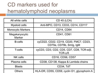
FLOW
CYTOMETRY
MODERATOR: DR. R.M. JAISWAL
By: Dr. Megha Gupta & Dr. Tashi Agarwal
FLOW CYTOMETRY
 Definition:
Measuring properties of cell as they flow in a
fluid suspension across an illuminated light
path.
Basic mechanism
Biological sample
Label it with a fluorescent marker
Cells move in a linear stream through a focused light
source (laser beam)
Fluorescent molecule gets activated and emits light
that is filtered and detected by sensitive light detectors
(usually a photomultiplier tube)
Conversion of analog fluorescent signals to digital
signals
Flow Cytometry
 This method allows the quantitative and
qualitative analysis of several properties of cell
populations from virtually any type of fresh
unfixed tissue or body fluid.
 The properties measured include a particle’s
related size, relative granularity or internal
complexity, and relative fluorescence intensity
Most commonly analyzed materials are:
 blood,
 bone marrow aspirate and
 lymph node suspensions.
Principle of Flow Cytometry
 Flow cytometer is composed of three main
components:
 The Flow system (fluidics)
Cells in suspension are brought in single file past
 The Optical system (light sensing)
a focused laser which scatter light and emit
fluorescence that is filtered and collected
 The Electronic system (signal processing)
emitted light is converted to digitized values that
are stored in a file for analysis
The Flow System
 One of the fundamentals of flow cytometry is the ability
to measure the properties of individual particles, which
is managed by the fluidics system.
 When a sample is injected into a flow cytometer, it is
ordered into a stream of single particles.
 The fluidic system consists of a FLOW CELL (Quartz
Chamber):
 Central channel/ core - through which the sample is
injected.
 Outer sheath - contains faster flowing fluid k/a
Sheath fluid (0.9% Saline / PBS) , enclosing the
Hydrodynamic Focusing
Once the sample is injected
into a stream of sheath fluid
within the flow chamber, they
are forced into the center of
the stream forming a single
file by the PRINCIPLE OF
HYDRODYNAMIC
FOCUSING.
'Only one cell or particle can
pass through the laser beam
at a given moment.'
• The sample pressure is always higher than the
sheath fluid pressure, ensuring a high flow rate
allowing more cells to enter the stream at a given
moment.
• High Flow Rate - Immunophenotyping analysis of
cells
• Low Flow Rate - DNA Analysis
Sheath
Tank
Waste
Tank
Line PressureVacuum
Sample
Pressure
(Variable)
Sheath
Pressure
(Constant)
Sample
Tube
OPTICS
 After the cell delivery system, the need is to excite the
cells using a light source.
 The light source used in a flow cytometer:
 Laser (more commonly)
 Arc lamp
 Why Lasers are more common?
 They are highly coherent and uniform. They can be easily
focused on a very small area (like a sample stream).
 They are monochromatic, emitting single wavelengths of light.
 ARGON Lasers - 488nm wavelength (blue to blue
green)
When a light intersects a laser beam at the so called
'interogation point' two events occur:
a) light scattering
b) emission of light (fluorescence )
Fluorescence is light emitted during decay of excited
electron to its basal state.
OPTICS
a) LIGHT SCATTER
 When light from a laser interrogates a cell, that cell
scatters light in all directions.
 The scattered light can travel from the interrogation point
down a path to a detector.
OPTICS - FORWARD SCATTER
(FSC)
• Light that is scattered in the forward direction
(along the same axis the laser is traveling) is
detected in the Forward Scatter Channel.
• The intensity of this signal has been attributed to
cell size, refractive index (membrane
permeability).
OPTICS - SIDE SCATTER
(SSC)
 Laser light that is scattered at 90 degrees to the axis of
the laser path is detected in the Side Scatter Channel.
 The intensity of this signal is proportional to the amount
of cytosolic structure in the cell (eg. granules, cell
inclusions, etc.) Side scatter detector
Measuring cell granularity
FSC
Detector
Collection
Lens
SSC
Detector
Laser Beam
FSC
SSC
Lymphocytes
Monocytes
Granulocytes
RBCs, Debris,
Dead Cells
Study of FSC and SSC allows us to know the
differentiation of different types of cells.
Why FSC & SSC?
 The light scattered in the forward direction is
proportional to the square of the radius of a sphere, and
so to the size of the cell or particle.
 The cells are labelled with fluorochrome-linked
antibodies or stained with fluorescent membrane,
cytoplasmic or nuclear dye.
Commonly used Fluorochromes
FLUOROCHROMES EMISSION
MAXIMUM
Fluorescein Isothiocynate (FITC) 530nm
Phycoerythrin (PE) 576nm
Peridin-chlorophyll alpha complex
(PerCP)
680nm
Allophycocyanin (APC) 660nm
Texas red 620nm
ECD( PE - Texas Red Tandem) 615nm
PC5 (PE - cyanin 5 dye tandem) 667nm
Optics
B) EMISSION OF FLUORESCENT LIGHT
(FLUORESCENCE)
 As the fluorescent molecule present in or on the particle
is interrogated by the laser light, it will absorb energy
from the laser light and release the absorbed energy at
longer wave length.
 Emitted photons pass through the collection lens and are
split and steered down specific channels with the use of
filters.
 Emitted fluorescence intensity is proportional to the
Optics- Filters
 Different wavelengths of light are scattered
simultaneously from a cell
 Need to split the light into its specific wavelengths in
order to measure and quantify them independently.
This is done with filters.
 The system of filters ensures that each photodetector
receives light bands of various wavelengths.
 Optical filters are designed such that they absorb or
reflect some wavelengths of light, while transmitting
others.
 Types of filters
1. Long Pass 2. Short Pass
3. Band Pass 4. Dichroic
Optics- Long Pass Filters
 Transmit all wavelengths greater than specified
wavelength
 Example: 500LP will transmit all wavelengths greater
than 500nm
400nm 500nm 600nm 700nm
Transmittance
Original from Cytomation Training Manual
Optics- Short Pass Filter
 Transmits all wavelengths less than specified
wavelength
 Example: 600SP will transmit all wavelengths less
than 600nm.
400nm 500nm 600nm 700nm
Transmittance
Original from Cytomation Training Manual
Optics- Band Pass Filter
 Transmits a specific band of wavelengths
 Example: 550/20BP Filter will transmit wavelengths
of light between 540nm and 560nm (550/20 = 550+/-
10, not 550+/-20)
400nm 500nm 600nm 700nm
Transmittance
Original from Cytomation Training Manual
Optics- Dichroic Filters
 Long pass or short pass filters
 Placed at a 45º angle of incidence
 Part of the light is reflected at 90º , and part of the light is
transmitted and continues.
Dichroic Filter
Detector 1
Detector 2
OPTICS - DETECTORS
 The photodetectors convert the photons to electrical
impulses.
 Two common types of detectors used in flow cytometry:
 Photodiode
used for strong signals, when saturation is a potential
problem (eg, forward scatter detector).
 Photomultiplier tube (PMT)
more sensitive than photodiode but can be destroyed
by exposure to too much light.
used for side scatter and fluorescent signols.
ELECTRONICS
 The electronic subsystem converts photons to
photoelectrons.
 Measures amplitude, area and width of photoelectron
pulse.
 It amplifies pulse either linearly or logarithmically and
then digitalizing the amplified pulse.
Time
Electronics- Creation of a Voltage Pulse
Data Analysis- Plot Types
 There are several plot choices:
 Single Color Histogram
 Fluorescence intensity (FI) versus the number of cells
counted.
 Two Color Dot Plot
 FI of parameter 1 versus FI of Parameter 2
 Two Color Contour Plot
 Concentric rings form around populations. The more
dense the population, the closer the rings are to each
other
 Two Color Density Plot
 Areas of higher density will have a different color than
Plot Types
Contour Plot Density Plot
Greyscale Density Dot Plot
www.treestar.com
Histogram
DATA ANALYSIS - GATING
 Gating is in essence electronic window that sets
upper and lower limits on the type and amount
of material that passes through.
 Selection of only a certain population of cells
for analysis on a plot.
 Allows the ability to look at parameters specific
to only that subset.
Interpretation of Graphs
 An important tool for evaluating data is the dot
plot.
 The instrument detects each cell as a point on
an X-Y graph. This form of data presentation
looks at two parameters of the sample at the
same time.
Three common modes for dot plots
are:
 Forward scatter (FSC) vs. side scatter (SSC)
To look at the distribution of cells based upon size &
granularity
 Single color vs. side scatter
To visualize the expression of the fluorescence of the
cells
 Two-color fluorescence plot.
To differentiate between those cells that express only one
of the particular fluorescent markers, those that express
neither, and those that express both.
used to discriminate dead cells from the live ones that
are expressing the desired fluorescence.
When to say an antigen is positive
or negative?
 A sample that has some
cells single positives for
CD8 along the x-axis
(green arrow)
 some single positives for
CD4 along the y-axis (red
arrow).
 Upper right quadrant of the
plot - cells positive for both
fluorescent markers
(purple arrow).
 Lower left quadrant - cells
negative for both markers
(orange arrow).
How to differentiate dim & bright
expression of an antigen?
 Dim : cells are
present more towards
the origin(0) on x(red)
- y axis (pink)
 Bright : cells are
present away from
the origin(0) on
x(green) & y(yellow)
axis.
DIM
BRIGHT
Y-axis
CD4
X-axis
CD8
WHAT IS UNIQUE IN
FLOWCYTOMETRY
 MULTIPARAMETRIC
 RAPID ANALYSIS OF LARGE NUMBER OF
CELLS
 INFORMATION AT A SINGLE CELL LEVEL
 DETECTION OF RARE CELL POPULATIONS
 ALLOWS PHYSICAL ISOLATION OF CELLS
OF INTEREST
USES OF
FLOWCYTOMETRY
APPLICATIONS
 ANALYSIS
 Immunophenotyping
 Dyes that bind to nucleic acids (DNA, RNA)
 Functional assays
 CELL COUNTING
 CELL SORTING
CLINICAL APPLICATIONS
• Absolute CD4 counts
HIV/AIDS
• HLA B27 assay
Joint Pain
• Diagnosis and Classification
• Detection of MRD
Hematological
Malignancies
• DNA Ploidy
• S Phase fractionSolid Tumours
• TBNK
• Phagocytic function defect
Primary
Immunodeficiency
disorders
Cont..
• Reticulocyte count
• PNH
• Osmotic fragility assay
Hemolytic anaemia
• Feto- maternal Hemorrhage
• treatment response in Sickle Cell AnemiaFetal Hb detection
• Platelet receptor assays (Platelet count, GT,
BSS)
• Platelet function assay (CD62P, PAC-1)
Bleeding Disorders
• CD34 STEM CELL COUNTS
• Residual WBC count in leukodepleted blood
packs
• Flow cytometry Crossmatch
Transfusion and
Transplant
• Surface markers in PMN, Monocytes
• Cytokine response
Host Immune
response in Sepsis
CLPD ON
FLOWCYTOMETRY
Objectives
 Diagnosis of lymphoma
 Classification of lymphoma
 Ploidy analysis
Flow cytometric approach to the diagnosis
and classification of B- cell lymphoid
neoplasms.
B Cell Lymphoma
B CELL DIFFERENTIATION
How to differentiate between
Normal and Neoplastic B
cells
1) Imunoglobulin light
chain class restriction.
2) Aberrant antigen
expression.
MONOCLONALITY CD 13, CD 33, CD 5 ON B
CELLS
Normal, polyclonal B-cells are a
mixture of kappa-B-cells and
lambda-B-cells.
A B-cell carries either kappa- or
lambda-light chain on its surface. And
normal polyclonal B-cells are a mixture
of kappa-B-cells and lambda B-cells as
can be seen in the left-hand figure.
Monoclonal mature B-cells are either
kappa or lambda.
If a malignant B-cell clone proliferates
this will result in a B-cell population
consisting of either only kappa- or only
lambda-B-cells. The latter case (i.e.
lambda-monoclonal B-cells) is
symbolized in the left-hand figure.
Expression of CD5
The arrow in the right panel
points to the abnormal,
strong expression of CD5 by
B-cells. CD5 expression as
strong as this can usually
only be found on T-cells.
Normal B-cells show no or
only a weak expression of
CD5 (left-hand panel)
Weak expression of CD20
The B-cells in the right panel
show only a weak
expression of CD20 (arrow).
For comparison: normal
CD20 expression in the left-
hand panel.
APPROACH TO B CELL
LYMPHOMA
CD5
POSITIVE NEGATIVE
CD23 -
FMC7 +
CD23 + FMC7 -
CD23 + FMC7 +
MCL MARG: CD38-,CD23-,
FMC7 +
DLBCL: CD38+
LPL: CD38+
CLL
PLL
CD10 POSITIVE
NEGATIVE
1. FOLLICULAR: FMC7 +
2. DLBCL
3. BURKITT: CD23 - FMC7 +
4. B-ALL: CD23 - FMC7 -
CD103,CD25, CD123
POSITIVE-HCL
NEGATIVE
Chronic lymphocytic leukemia
 Typical phenotype: CD20 (d), CD22 (d), sIg
(d), CD23+
 FMC-7-
 Characteristic morphology
 Testing for the prognostic markers CD38 and
ZAP-70 can be considered
Mantle cell lymphoma
 Variable phenotype not typical for CLL;
 often CD20 (i), sIg (i), CD23-, FMC-7 +
 IHC : Cyclin-D1
 FISH : t(11;14)/CCND1 rearrangement
Hairy cell lekaemia
 Typical pheotype: CD20 (b), CD22 (b), CD11c
(b),
 CD25+, CD103+, sIg (i)
 Confirm characteristic morphology of a hairy
cell and TRAP +
 A small subset of HCL are CD10+ but are
morphologically similar to CD10- HCL.
Follicular lymphoma
 Usually bcl-2, CD43.
 Some follicular growth.
 t(14;18)/BCL-2 rearrangement.
 D/D
1. DLBCL : diffuse growth pattern against the
nodular growth pattern in FL
2. BL : morphologial (vacuoles), High S phase
fraction.
 80/F
 c/o cervical adenopathy
 On CBC : an absolute
lymphocytosis ≥5 ×
109/L;
 PBF is flooded with
small mature
lymphocytes with
condensed chromatin
and scant cytoplasm
along with numerous
smudge cells.
CASE
On flow
A diagnosis of CLL can be made. CD5+ CD23+
Not that simple
 A certain immunophenotype may be typical but
is by no means obligatory.
 The significance of one marker depends on
the expression of other markers.
 The strength of antigen expression is
important.
Flow cytometric approach to the diagnosis
and classification of T- cell lymphoid
neoplasms.
T CELL LYMPHOMA
Finding abnormal T/ NK cells.
1) Search for
monoclonal T cells
2) T cells with aberrant
antigen expression.
CD4/CD8 Ratio
Loss of CD3
Overexpression of CD5
Normally, the CD4/CD8-T-
Cell ratio in peripheral
blood is about 2:1.
In a T-lymphocytic leukemia
this ratio can shift
dramatically. Unfortunately,
this ratio may also be altered
by many non-malignant
diseases. eg viral infections.
Therefore, only extreme
alterations of this ratio can
be regarded as a sign for T-
lymphocytic malignancy.
CD4/CD8 coexpression
In the right-hand dot-plot you can
see cells that express both the CD4
and the CD8-antigen (arrow) which
is highly irregular. In addition both
antigens are expressed weakly
(compared to normal T-cells). Left-
hand panel shows a normal
situation.
Loss of CD3
Overexpression of CD5
In the right-hand dot-plot you can
see T-cells which overexpress CD5
while they lack CD3 (arrow). Only a
few normal T-cells are present.
(blue oval). Left-hand panel shows
a normal situation.
PROBLEMS IN DIAGNOSIS OF T-
CLPD
 Relatively low incidence.
 5-25% of all lympoid neoplasms.
 Clinico-biological heterogeneity.
 Lack of distinctive genetic markers.
T CELL DIFFERENTIATION
T- CLPD BY
FLOWCYTOMETRY
CTCL/Sézary syndrome
 Often CD7-, CD26-, CD4+, CD25+/- (with
heterogeneous staining intensity).
 Confirm characteristic morphology and clinical
presentation.
 HTLV-1-
• DNA PLOIDY
• S PHASE FRACTION
PLOIDY ANALYSIS
Cell cycle analysis
 The percentage of the cells in each region is
analyzed. In normal tissues –
 95% cells - G0/G1 phase
 2.5% cells - S phase
 2.5% cells - G2/M phase
 In neoplasm, percentage of cells in S and G2/M
phase increases which is expressed as S phase
fraction or the proliferation index
S Phase, synthesis phase.
It is the part of cell cycle in which DNA is
replicated occurring between G1phase and G2
phase.
S phase has strong correlation with
grading.
DNA ploidy has no correlation with
grading.
ADVANTAGES OVER IHC
Highest proliferative
activity: mean SPF,
35.3%
6.6%
6.5%
20.4%
DIAGNOSIS OF ACUTE
LEUKAEMIAS ON
FLOWCYTOMETRY
STEPS
 Finding the blast population
 Defining the immunophenotype
 Diagnosis
A malignant blast population
may be detected because of
Increase of
immature cells
Abnormal marker
expression of
immature cells
CD38 / CD45 AGAINST
SSC
CD19, 7 on non lymphoid
cell
Finding immature
cells using CD45-
CD34 dot-plots
The arrows points at the
blast populations, which
is very conspicuous in
case AL 1 (upper right)
and AL 3 (lower right).
In case AL 2 (lower left),
the difference between
the normal picture is
more subtle and the
blasts may be missed
because in this case the
blasts are CD34
negative.
Intermediate
CD45 and
low side
scatter
BLAST
WINDOW
NEUTROPHI
LS
LYMPHOCYT
ES
MONOCYTE
S
RBC’S AND
DEBRIS
B CELLS
CD45/SSC gating strategy is more sensitive than FSC/SSC
gating and it dilineates the blasts well.
Finding immature
cells using CD45-
Side Scatter dot-plots
The three cases of
acute leukemia: The
arrows point to the
blast populations which
are clearly visible in all
three cases.
Even the blasts of case
AL 2 can easily be
spotted.
BLAST
WINDOW
B CELLS
MONOCYTES
RBC’S AND
DEBRIS
LYMPHOCYT
ES
NEUTROPHIL
S
CD45/SSC gating strategy is more sensitive than FSC/SSC
gating and it dilineates the blasts well.
Example of an abnormal antigen
expression on myeloid blasts
Compare the normal blasts (upper
dot-plot, blue oval) with those of an
acute myeloid leukemia (lower dot-
plot, red oval)): the malignant
blasts abnormally express CD15
and they show an increased
expression of CD34.
Note: CD34-negative cells have
been removed for reason of clarity.
DIAGNOSIS
 WHICH ONES TO IMMUNOPHENOTYPE?
1. Equivocal morphology
2. Cytochemistry is noncontributory
3. Specific subtypes
 LEUKAEMIA VS NON LEUKAEMIA
1. Overlapping morphology. Eg: hematogones, viral
infections.
2. Partially treated acute leukaemia
PROGNOSTIFICATION
 CYTOGENIC AND MOLECULAR
ABNORMALITIES
• Association with specific cytogenic
abnormalities
• DNA ploidy
 RESIDUAL DISEASE MONITORING
CLASSIFICATION
Acute leukaemia is classified on the basis of
immunological markers into
 B lineage ALL
 T lineage ALL
 Acute myeloid leukaemia
 Acute leukaemia of ambiguous lineage
How to define the lineage of
leukaemia
THE FLOW CYTOMETRIC EVALUATION OF HEMATOPOIETIC
NEOPLASIA Brent L. Wood, Michael J. Borowitz. Henry’s, 22nd edition,
Chapter 34
Flow cytometric approach to the diagnosis
and classification of ALL.
ACUTE LYMPHOID
LEUKAEMIA
How to diff ALL from NHL
 CD 34
 TdT
 Bcl2
 CD99
 NHL cases with spillover demonstrate bright
CD45 expression while it is moderate in B
ALL.
Subtypes of ALL
 Flow cytometric immunophenotyping does not
provide a suitable surrogate tool for detection
of these subtypes of ALL.
Subtype
ALL
HLA-
DR
TdT CD 10 CD 19 SmIg CyCD79
a
Pro- B ALL +/- + - + - +
Common
ALL
+ + + + - +
Pre B ALL + - - + - +
Mature B
ALL
- - - + +K/L +
Precursor B cell lymphoblastic
leukemia/lymphoma
Flow cytometric approach to the diagnosis
and classification of AML.
ACUTE MYELOID
LEUKAEMIA
Diagnosis of AML
Morphology Auer rod
Cytochemistry >3% MPO positive
Immunophenotyping CD33, CD13, CD117, anti-
MPO
Cytogenetics t(8;21), t(15;17), inv16,
MLL, t(9;11), t(6;9), t(3;3),
t(1;22)
CD markers used for
hematolymphoid neoplasms
All white cells CD 45 (LCA)
Myeloid cells Anti-MPO, CD13, CD33, CD14, CD117
Monocytic Markers CD14, CD64
Megakaryocytic
Marker
CD41, CD61
B-cells cyCD22, CD22, CD19, CD20, FMC7, CD23,
CD79a, CD79b, SmIg, IgM
T-cells cyCD3, CD3, CD2, CD5, CD7, CD8, TCR-α/β,
TCR-γ/δ
NK cells CD16, CD56, CD57
Plasma cells CD38, CD138, Kappa & Lambda chains
Blasts CD34, TdT
Others HLA-DR, CD55, CD59, cyclin D1, glycophorin A
Myeloblast characterization
13+, 15+, 33+,
anti-MPO+
Clinical, Genetic, Morphologic
Erythroi
d
Megakaryocyti
c
Myeloid Monocytic
41+
61+
71++
GlyA+
36+, 64+,
14+, 33++
36+
Classification - FAB
 M0 : AML-minimal differentiation
 M1 : AML-without maturation
 M2 : AML-with maturation (blast<80%)
 M3 : AML-promyelocytic
 M4 : AML-myelomonocytic (>20% monocytes)
 M5 : AML-monocytic
 M6 : AML-erythroid
 M7 : AML-megakaryocytic
AML- minimal differentiation
(M0)
 Myeloblasts - < 3% positivity with SBB, MPO &
PAS-, NSE-
 Myeloid antigens - CD13+, CD33+, CD117+,
and/or MPO+
 CD34, CD38, HLA-DR, and TdT - often
expressed
AML- Promyelocytic Leukemia
(M3)
 Phenotype - CD13h+, CD33++, CD34-, HLA-
DR-
 Diagnostic molecular alteration - PML/RARA
t(15;17) translocation
 Strongly positive - MPO, SBB, PAS
cytoplasmic positivity.
 Characteristic morphology
 D/D : AML-monocytic leukemia (M5) -
 HLA-DR+, CD11c+, CD14+ & CD64+
AML - Myelomonocytic Leukemia
(M4)
 Phenotype:
 myeloid antigens - CD13+ & CD33+, HLA-DR+
 monocytic markers: CD14+, CD4+, CD11b+,
CD11c+, CD64+, CD36+, CD68+
 Blasts >20% of marrow NEC
 Monocytic component >20% of NEC &
monocytes in blood >5 x 10⁹/L
AML - Monocytic Leukemia
(M5)
 Phenotype: CD33 (b), CD13+, HLA-DR+
 Characteristic CD14+, CD11b+, CD11c+,
CD64+, CD68+
 Cytochemistry : NSE +
 M5a : Acute monoblastic leukemia
 M5b : Acute monocytic leukemia
CD34 APC CD15 FITC CD56 A488
CD45
CD4
AML - Megakaryocytic leukemia
(M7)
 CD41+, CD61+, CD13+, CD33+
 CD34, HLA-DR - Negative
CASE
 29yrs/ F
 O/E : Fever, Pallor, Gum
hyperplasia,
Hepatosplenomegaly.
 CBC : Hb-5.2 g%, Plt-
19,000/cu.mm
 PBF : shows blasts and dual
differentiation to
granulocytes and
monocytoid cells (large cells,
abundant pale blue
cytoplasm, lobulated or
indented nucleus with
variable nucleoli).
DLC
Blasts40 P8 L10 Monocytoid41
E1
red - dim CD45and low
side scatter.
Positive for - CD13,
cyMPO, CD34, HLA-
DR, CD33, CD11c
Negative for - CD10,
CD19, CD3, CD79a.
Blue - bright CD45 &
moderate side scatter.
Positive for - CD14,
CD11c, CD13.
Negative for - CD34,
cyMPO, CD3, CD10,
CD19
ACUTE MYELOMONOCYTIC LEUKEM
Hematogones
 Physiologic precursors of maturing B-cells.
 Confused with neoplastic immature lymphoid
cells of B lymphoblastic leukemia/ lymphoma
or B-ALL.
 Increased in:
 Autoimmune or congenital cytopenias
 Solid organ tumors e.g. neuroblastoma
 AIDS
 NHL
 Post-chemotherapy and after BMT
 Copper deficiency
 Morphologically, hematogones resemble
lymphoblasts.
 Hematogones can be differentiated from
lymphoblasts by
 Unique Immunophenotypic pattern :
 CD34 < TdT < CD20 < PAX5
 Variable CD10 & CD20
 "J shaped trail pattern" : on CD10/20 Dot plot
Lymphocytes
Hematogone
Immunophenotypic analysis of hematogones in 662 consecutive bone marrow
specimens by 4-color flow cytometry. Mckenna et al, BLOOD, 15 OCTOBER 2001
Acute leukaemia of ambiguous
lineage
 Mixed phenotype acute leukaemia
 Acute undifferentiated leukaemia
 NK/plasmacytoid dendritic cell leukaemia
MPAL – WHO 2008
Myeloid
lineage
MPO
FC, IHC,
Cytochemistry
Monocytic
diferentiation
Atleast 2: NSE,
CD11c, CD14,
CD64, lyzozyme
T lineage
Cytoplasmic CD3
Surface CD3
B lineage
Strong CD19 with
atleast 1 : CD79a,
cytoplasmic
CD22, CD10
Weak CD19 with
atleast 2 strongly
expressed
CD79a,
cytoplasmic
CD22, CD10
Or Or Or
EGIL scoring system
The European group for the Immunological Classification of Leukaemias (EGIL)
scoring system
Flowcytometry analysis in MRD detection
Minimal Residual Disease
detection
 At diagnosis the tumour burden is
approximately 10^12 leukaemic cells.
 Induction chemothereapy achieves a 3 log cell
kill bringing it down to 10^9 leukaemic cells.
 Light microscopy of BMA can detect leukaemia
only when there are more than 5 blasts/ 100
nucleated cells. Anything less than that is
termed remission.
Introduction
Introduction
 What is Minimal residual disease or MRD?
It is that submicroscopic disease that cannot be
detected by conventional light microscopic
examination of the BMA.
 It could be as high as 1 billion leukaemic cells.
 It can be performed by two techniques: FCM &
PCR.
Used mainly in -
1. Acute leukaemia for guiding thereapy as well
as prognostic purposes.
2. Patients with low grade B cell malignancies
undergoing high dose chemotherapy.
3. Post stem cell transplant and
immunothereapy.
4. Lymphoma spillover.
REFERENCES
 THE FLOW CYTOMETRIC EVALUATION OF
HEMATOPOIETIC NEOPLASIA Brent L. Wood, Michael
J. Borowitz. Henry’s, 22nd edition, Chapter 34
 ATLAS AND TEXT OF HEMATOLOGY. Dr Tejinder
singh
 Manual: 6th Advanced TCS Flowcytometry workshop on
hematological malignancies.
 Flow Cytometry in Hematopathology. A visual approach
to data analysis and interpretation. Doyen, Lawrence
and Raul.
 Flow Cytometric Analysis of Leukemia and Lymphoma -
The Basics Univ.Doz.Dr.med. Wolfgang Hübl

Flow cytometry

  • 1.
    FLOW CYTOMETRY MODERATOR: DR. R.M.JAISWAL By: Dr. Megha Gupta & Dr. Tashi Agarwal
  • 2.
    FLOW CYTOMETRY  Definition: Measuringproperties of cell as they flow in a fluid suspension across an illuminated light path.
  • 3.
    Basic mechanism Biological sample Labelit with a fluorescent marker Cells move in a linear stream through a focused light source (laser beam) Fluorescent molecule gets activated and emits light that is filtered and detected by sensitive light detectors (usually a photomultiplier tube) Conversion of analog fluorescent signals to digital signals
  • 4.
    Flow Cytometry  Thismethod allows the quantitative and qualitative analysis of several properties of cell populations from virtually any type of fresh unfixed tissue or body fluid.  The properties measured include a particle’s related size, relative granularity or internal complexity, and relative fluorescence intensity Most commonly analyzed materials are:  blood,  bone marrow aspirate and  lymph node suspensions.
  • 5.
    Principle of FlowCytometry  Flow cytometer is composed of three main components:  The Flow system (fluidics) Cells in suspension are brought in single file past  The Optical system (light sensing) a focused laser which scatter light and emit fluorescence that is filtered and collected  The Electronic system (signal processing) emitted light is converted to digitized values that are stored in a file for analysis
  • 6.
    The Flow System One of the fundamentals of flow cytometry is the ability to measure the properties of individual particles, which is managed by the fluidics system.  When a sample is injected into a flow cytometer, it is ordered into a stream of single particles.  The fluidic system consists of a FLOW CELL (Quartz Chamber):  Central channel/ core - through which the sample is injected.  Outer sheath - contains faster flowing fluid k/a Sheath fluid (0.9% Saline / PBS) , enclosing the
  • 7.
    Hydrodynamic Focusing Once thesample is injected into a stream of sheath fluid within the flow chamber, they are forced into the center of the stream forming a single file by the PRINCIPLE OF HYDRODYNAMIC FOCUSING. 'Only one cell or particle can pass through the laser beam at a given moment.'
  • 8.
    • The samplepressure is always higher than the sheath fluid pressure, ensuring a high flow rate allowing more cells to enter the stream at a given moment. • High Flow Rate - Immunophenotyping analysis of cells • Low Flow Rate - DNA Analysis Sheath Tank Waste Tank Line PressureVacuum Sample Pressure (Variable) Sheath Pressure (Constant) Sample Tube
  • 9.
    OPTICS  After thecell delivery system, the need is to excite the cells using a light source.  The light source used in a flow cytometer:  Laser (more commonly)  Arc lamp  Why Lasers are more common?  They are highly coherent and uniform. They can be easily focused on a very small area (like a sample stream).  They are monochromatic, emitting single wavelengths of light.  ARGON Lasers - 488nm wavelength (blue to blue green)
  • 10.
    When a lightintersects a laser beam at the so called 'interogation point' two events occur: a) light scattering b) emission of light (fluorescence ) Fluorescence is light emitted during decay of excited electron to its basal state.
  • 11.
    OPTICS a) LIGHT SCATTER When light from a laser interrogates a cell, that cell scatters light in all directions.  The scattered light can travel from the interrogation point down a path to a detector.
  • 12.
    OPTICS - FORWARDSCATTER (FSC) • Light that is scattered in the forward direction (along the same axis the laser is traveling) is detected in the Forward Scatter Channel. • The intensity of this signal has been attributed to cell size, refractive index (membrane permeability).
  • 13.
    OPTICS - SIDESCATTER (SSC)  Laser light that is scattered at 90 degrees to the axis of the laser path is detected in the Side Scatter Channel.  The intensity of this signal is proportional to the amount of cytosolic structure in the cell (eg. granules, cell inclusions, etc.) Side scatter detector Measuring cell granularity
  • 14.
  • 15.
    FSC SSC Lymphocytes Monocytes Granulocytes RBCs, Debris, Dead Cells Studyof FSC and SSC allows us to know the differentiation of different types of cells. Why FSC & SSC?
  • 16.
     The lightscattered in the forward direction is proportional to the square of the radius of a sphere, and so to the size of the cell or particle.  The cells are labelled with fluorochrome-linked antibodies or stained with fluorescent membrane, cytoplasmic or nuclear dye.
  • 17.
    Commonly used Fluorochromes FLUOROCHROMESEMISSION MAXIMUM Fluorescein Isothiocynate (FITC) 530nm Phycoerythrin (PE) 576nm Peridin-chlorophyll alpha complex (PerCP) 680nm Allophycocyanin (APC) 660nm Texas red 620nm ECD( PE - Texas Red Tandem) 615nm PC5 (PE - cyanin 5 dye tandem) 667nm
  • 18.
    Optics B) EMISSION OFFLUORESCENT LIGHT (FLUORESCENCE)  As the fluorescent molecule present in or on the particle is interrogated by the laser light, it will absorb energy from the laser light and release the absorbed energy at longer wave length.  Emitted photons pass through the collection lens and are split and steered down specific channels with the use of filters.  Emitted fluorescence intensity is proportional to the
  • 19.
    Optics- Filters  Differentwavelengths of light are scattered simultaneously from a cell  Need to split the light into its specific wavelengths in order to measure and quantify them independently. This is done with filters.  The system of filters ensures that each photodetector receives light bands of various wavelengths.  Optical filters are designed such that they absorb or reflect some wavelengths of light, while transmitting others.  Types of filters 1. Long Pass 2. Short Pass 3. Band Pass 4. Dichroic
  • 20.
    Optics- Long PassFilters  Transmit all wavelengths greater than specified wavelength  Example: 500LP will transmit all wavelengths greater than 500nm 400nm 500nm 600nm 700nm Transmittance Original from Cytomation Training Manual
  • 21.
    Optics- Short PassFilter  Transmits all wavelengths less than specified wavelength  Example: 600SP will transmit all wavelengths less than 600nm. 400nm 500nm 600nm 700nm Transmittance Original from Cytomation Training Manual
  • 22.
    Optics- Band PassFilter  Transmits a specific band of wavelengths  Example: 550/20BP Filter will transmit wavelengths of light between 540nm and 560nm (550/20 = 550+/- 10, not 550+/-20) 400nm 500nm 600nm 700nm Transmittance Original from Cytomation Training Manual
  • 23.
    Optics- Dichroic Filters Long pass or short pass filters  Placed at a 45º angle of incidence  Part of the light is reflected at 90º , and part of the light is transmitted and continues. Dichroic Filter Detector 1 Detector 2
  • 24.
    OPTICS - DETECTORS The photodetectors convert the photons to electrical impulses.  Two common types of detectors used in flow cytometry:  Photodiode used for strong signals, when saturation is a potential problem (eg, forward scatter detector).  Photomultiplier tube (PMT) more sensitive than photodiode but can be destroyed by exposure to too much light. used for side scatter and fluorescent signols.
  • 25.
    ELECTRONICS  The electronicsubsystem converts photons to photoelectrons.  Measures amplitude, area and width of photoelectron pulse.  It amplifies pulse either linearly or logarithmically and then digitalizing the amplified pulse.
  • 26.
  • 27.
    Data Analysis- PlotTypes  There are several plot choices:  Single Color Histogram  Fluorescence intensity (FI) versus the number of cells counted.  Two Color Dot Plot  FI of parameter 1 versus FI of Parameter 2  Two Color Contour Plot  Concentric rings form around populations. The more dense the population, the closer the rings are to each other  Two Color Density Plot  Areas of higher density will have a different color than
  • 28.
    Plot Types Contour PlotDensity Plot Greyscale Density Dot Plot www.treestar.com Histogram
  • 29.
    DATA ANALYSIS -GATING  Gating is in essence electronic window that sets upper and lower limits on the type and amount of material that passes through.  Selection of only a certain population of cells for analysis on a plot.  Allows the ability to look at parameters specific to only that subset.
  • 30.
    Interpretation of Graphs An important tool for evaluating data is the dot plot.  The instrument detects each cell as a point on an X-Y graph. This form of data presentation looks at two parameters of the sample at the same time.
  • 31.
    Three common modesfor dot plots are:  Forward scatter (FSC) vs. side scatter (SSC) To look at the distribution of cells based upon size & granularity  Single color vs. side scatter To visualize the expression of the fluorescence of the cells  Two-color fluorescence plot. To differentiate between those cells that express only one of the particular fluorescent markers, those that express neither, and those that express both. used to discriminate dead cells from the live ones that are expressing the desired fluorescence.
  • 32.
    When to sayan antigen is positive or negative?  A sample that has some cells single positives for CD8 along the x-axis (green arrow)  some single positives for CD4 along the y-axis (red arrow).  Upper right quadrant of the plot - cells positive for both fluorescent markers (purple arrow).  Lower left quadrant - cells negative for both markers (orange arrow).
  • 33.
    How to differentiatedim & bright expression of an antigen?  Dim : cells are present more towards the origin(0) on x(red) - y axis (pink)  Bright : cells are present away from the origin(0) on x(green) & y(yellow) axis. DIM BRIGHT Y-axis CD4 X-axis CD8
  • 34.
    WHAT IS UNIQUEIN FLOWCYTOMETRY  MULTIPARAMETRIC  RAPID ANALYSIS OF LARGE NUMBER OF CELLS  INFORMATION AT A SINGLE CELL LEVEL  DETECTION OF RARE CELL POPULATIONS  ALLOWS PHYSICAL ISOLATION OF CELLS OF INTEREST
  • 35.
  • 36.
    APPLICATIONS  ANALYSIS  Immunophenotyping Dyes that bind to nucleic acids (DNA, RNA)  Functional assays  CELL COUNTING  CELL SORTING
  • 37.
    CLINICAL APPLICATIONS • AbsoluteCD4 counts HIV/AIDS • HLA B27 assay Joint Pain • Diagnosis and Classification • Detection of MRD Hematological Malignancies • DNA Ploidy • S Phase fractionSolid Tumours • TBNK • Phagocytic function defect Primary Immunodeficiency disorders
  • 38.
    Cont.. • Reticulocyte count •PNH • Osmotic fragility assay Hemolytic anaemia • Feto- maternal Hemorrhage • treatment response in Sickle Cell AnemiaFetal Hb detection • Platelet receptor assays (Platelet count, GT, BSS) • Platelet function assay (CD62P, PAC-1) Bleeding Disorders • CD34 STEM CELL COUNTS • Residual WBC count in leukodepleted blood packs • Flow cytometry Crossmatch Transfusion and Transplant • Surface markers in PMN, Monocytes • Cytokine response Host Immune response in Sepsis
  • 39.
  • 40.
    Objectives  Diagnosis oflymphoma  Classification of lymphoma  Ploidy analysis
  • 41.
    Flow cytometric approachto the diagnosis and classification of B- cell lymphoid neoplasms. B Cell Lymphoma
  • 42.
  • 43.
    How to differentiatebetween Normal and Neoplastic B cells 1) Imunoglobulin light chain class restriction. 2) Aberrant antigen expression. MONOCLONALITY CD 13, CD 33, CD 5 ON B CELLS
  • 44.
    Normal, polyclonal B-cellsare a mixture of kappa-B-cells and lambda-B-cells. A B-cell carries either kappa- or lambda-light chain on its surface. And normal polyclonal B-cells are a mixture of kappa-B-cells and lambda B-cells as can be seen in the left-hand figure. Monoclonal mature B-cells are either kappa or lambda. If a malignant B-cell clone proliferates this will result in a B-cell population consisting of either only kappa- or only lambda-B-cells. The latter case (i.e. lambda-monoclonal B-cells) is symbolized in the left-hand figure.
  • 45.
    Expression of CD5 Thearrow in the right panel points to the abnormal, strong expression of CD5 by B-cells. CD5 expression as strong as this can usually only be found on T-cells. Normal B-cells show no or only a weak expression of CD5 (left-hand panel) Weak expression of CD20 The B-cells in the right panel show only a weak expression of CD20 (arrow). For comparison: normal CD20 expression in the left- hand panel.
  • 46.
    APPROACH TO BCELL LYMPHOMA CD5 POSITIVE NEGATIVE CD23 - FMC7 + CD23 + FMC7 - CD23 + FMC7 + MCL MARG: CD38-,CD23-, FMC7 + DLBCL: CD38+ LPL: CD38+ CLL PLL CD10 POSITIVE NEGATIVE 1. FOLLICULAR: FMC7 + 2. DLBCL 3. BURKITT: CD23 - FMC7 + 4. B-ALL: CD23 - FMC7 - CD103,CD25, CD123 POSITIVE-HCL NEGATIVE
  • 47.
    Chronic lymphocytic leukemia Typical phenotype: CD20 (d), CD22 (d), sIg (d), CD23+  FMC-7-  Characteristic morphology  Testing for the prognostic markers CD38 and ZAP-70 can be considered
  • 49.
    Mantle cell lymphoma Variable phenotype not typical for CLL;  often CD20 (i), sIg (i), CD23-, FMC-7 +  IHC : Cyclin-D1  FISH : t(11;14)/CCND1 rearrangement
  • 51.
    Hairy cell lekaemia Typical pheotype: CD20 (b), CD22 (b), CD11c (b),  CD25+, CD103+, sIg (i)  Confirm characteristic morphology of a hairy cell and TRAP +  A small subset of HCL are CD10+ but are morphologically similar to CD10- HCL.
  • 53.
    Follicular lymphoma  Usuallybcl-2, CD43.  Some follicular growth.  t(14;18)/BCL-2 rearrangement.  D/D 1. DLBCL : diffuse growth pattern against the nodular growth pattern in FL 2. BL : morphologial (vacuoles), High S phase fraction.
  • 55.
     80/F  c/ocervical adenopathy  On CBC : an absolute lymphocytosis ≥5 × 10⁹/L;  PBF is flooded with small mature lymphocytes with condensed chromatin and scant cytoplasm along with numerous smudge cells. CASE
  • 56.
    On flow A diagnosisof CLL can be made. CD5+ CD23+
  • 57.
    Not that simple A certain immunophenotype may be typical but is by no means obligatory.  The significance of one marker depends on the expression of other markers.  The strength of antigen expression is important.
  • 58.
    Flow cytometric approachto the diagnosis and classification of T- cell lymphoid neoplasms. T CELL LYMPHOMA
  • 59.
    Finding abnormal T/NK cells. 1) Search for monoclonal T cells 2) T cells with aberrant antigen expression. CD4/CD8 Ratio Loss of CD3 Overexpression of CD5
  • 60.
    Normally, the CD4/CD8-T- Cellratio in peripheral blood is about 2:1. In a T-lymphocytic leukemia this ratio can shift dramatically. Unfortunately, this ratio may also be altered by many non-malignant diseases. eg viral infections. Therefore, only extreme alterations of this ratio can be regarded as a sign for T- lymphocytic malignancy.
  • 61.
    CD4/CD8 coexpression In theright-hand dot-plot you can see cells that express both the CD4 and the CD8-antigen (arrow) which is highly irregular. In addition both antigens are expressed weakly (compared to normal T-cells). Left- hand panel shows a normal situation. Loss of CD3 Overexpression of CD5 In the right-hand dot-plot you can see T-cells which overexpress CD5 while they lack CD3 (arrow). Only a few normal T-cells are present. (blue oval). Left-hand panel shows a normal situation.
  • 62.
    PROBLEMS IN DIAGNOSISOF T- CLPD  Relatively low incidence.  5-25% of all lympoid neoplasms.  Clinico-biological heterogeneity.  Lack of distinctive genetic markers.
  • 63.
  • 64.
  • 65.
    CTCL/Sézary syndrome  OftenCD7-, CD26-, CD4+, CD25+/- (with heterogeneous staining intensity).  Confirm characteristic morphology and clinical presentation.  HTLV-1-
  • 67.
    • DNA PLOIDY •S PHASE FRACTION PLOIDY ANALYSIS
  • 68.
    Cell cycle analysis The percentage of the cells in each region is analyzed. In normal tissues –  95% cells - G0/G1 phase  2.5% cells - S phase  2.5% cells - G2/M phase  In neoplasm, percentage of cells in S and G2/M phase increases which is expressed as S phase fraction or the proliferation index
  • 69.
    S Phase, synthesisphase. It is the part of cell cycle in which DNA is replicated occurring between G1phase and G2 phase.
  • 70.
    S phase hasstrong correlation with grading. DNA ploidy has no correlation with grading. ADVANTAGES OVER IHC
  • 71.
    Highest proliferative activity: meanSPF, 35.3% 6.6% 6.5% 20.4%
  • 72.
  • 73.
    STEPS  Finding theblast population  Defining the immunophenotype  Diagnosis
  • 74.
    A malignant blastpopulation may be detected because of Increase of immature cells Abnormal marker expression of immature cells CD38 / CD45 AGAINST SSC CD19, 7 on non lymphoid cell
  • 75.
    Finding immature cells usingCD45- CD34 dot-plots The arrows points at the blast populations, which is very conspicuous in case AL 1 (upper right) and AL 3 (lower right). In case AL 2 (lower left), the difference between the normal picture is more subtle and the blasts may be missed because in this case the blasts are CD34 negative.
  • 76.
    Intermediate CD45 and low side scatter BLAST WINDOW NEUTROPHI LS LYMPHOCYT ES MONOCYTE S RBC’SAND DEBRIS B CELLS CD45/SSC gating strategy is more sensitive than FSC/SSC gating and it dilineates the blasts well.
  • 77.
    Finding immature cells usingCD45- Side Scatter dot-plots The three cases of acute leukemia: The arrows point to the blast populations which are clearly visible in all three cases. Even the blasts of case AL 2 can easily be spotted.
  • 78.
    BLAST WINDOW B CELLS MONOCYTES RBC’S AND DEBRIS LYMPHOCYT ES NEUTROPHIL S CD45/SSCgating strategy is more sensitive than FSC/SSC gating and it dilineates the blasts well.
  • 79.
    Example of anabnormal antigen expression on myeloid blasts Compare the normal blasts (upper dot-plot, blue oval) with those of an acute myeloid leukemia (lower dot- plot, red oval)): the malignant blasts abnormally express CD15 and they show an increased expression of CD34. Note: CD34-negative cells have been removed for reason of clarity.
  • 80.
    DIAGNOSIS  WHICH ONESTO IMMUNOPHENOTYPE? 1. Equivocal morphology 2. Cytochemistry is noncontributory 3. Specific subtypes  LEUKAEMIA VS NON LEUKAEMIA 1. Overlapping morphology. Eg: hematogones, viral infections. 2. Partially treated acute leukaemia
  • 81.
    PROGNOSTIFICATION  CYTOGENIC ANDMOLECULAR ABNORMALITIES • Association with specific cytogenic abnormalities • DNA ploidy  RESIDUAL DISEASE MONITORING
  • 82.
    CLASSIFICATION Acute leukaemia isclassified on the basis of immunological markers into  B lineage ALL  T lineage ALL  Acute myeloid leukaemia  Acute leukaemia of ambiguous lineage
  • 83.
    How to definethe lineage of leukaemia THE FLOW CYTOMETRIC EVALUATION OF HEMATOPOIETIC NEOPLASIA Brent L. Wood, Michael J. Borowitz. Henry’s, 22nd edition, Chapter 34
  • 84.
    Flow cytometric approachto the diagnosis and classification of ALL. ACUTE LYMPHOID LEUKAEMIA
  • 85.
    How to diffALL from NHL  CD 34  TdT  Bcl2  CD99  NHL cases with spillover demonstrate bright CD45 expression while it is moderate in B ALL.
  • 86.
    Subtypes of ALL Flow cytometric immunophenotyping does not provide a suitable surrogate tool for detection of these subtypes of ALL. Subtype ALL HLA- DR TdT CD 10 CD 19 SmIg CyCD79 a Pro- B ALL +/- + - + - + Common ALL + + + + - + Pre B ALL + - - + - + Mature B ALL - - - + +K/L +
  • 87.
    Precursor B celllymphoblastic leukemia/lymphoma
  • 88.
    Flow cytometric approachto the diagnosis and classification of AML. ACUTE MYELOID LEUKAEMIA
  • 89.
    Diagnosis of AML MorphologyAuer rod Cytochemistry >3% MPO positive Immunophenotyping CD33, CD13, CD117, anti- MPO Cytogenetics t(8;21), t(15;17), inv16, MLL, t(9;11), t(6;9), t(3;3), t(1;22)
  • 90.
    CD markers usedfor hematolymphoid neoplasms All white cells CD 45 (LCA) Myeloid cells Anti-MPO, CD13, CD33, CD14, CD117 Monocytic Markers CD14, CD64 Megakaryocytic Marker CD41, CD61 B-cells cyCD22, CD22, CD19, CD20, FMC7, CD23, CD79a, CD79b, SmIg, IgM T-cells cyCD3, CD3, CD2, CD5, CD7, CD8, TCR-α/β, TCR-γ/δ NK cells CD16, CD56, CD57 Plasma cells CD38, CD138, Kappa & Lambda chains Blasts CD34, TdT Others HLA-DR, CD55, CD59, cyclin D1, glycophorin A
  • 91.
    Myeloblast characterization 13+, 15+,33+, anti-MPO+ Clinical, Genetic, Morphologic Erythroi d Megakaryocyti c Myeloid Monocytic 41+ 61+ 71++ GlyA+ 36+, 64+, 14+, 33++ 36+
  • 92.
    Classification - FAB M0 : AML-minimal differentiation  M1 : AML-without maturation  M2 : AML-with maturation (blast<80%)  M3 : AML-promyelocytic  M4 : AML-myelomonocytic (>20% monocytes)  M5 : AML-monocytic  M6 : AML-erythroid  M7 : AML-megakaryocytic
  • 93.
    AML- minimal differentiation (M0) Myeloblasts - < 3% positivity with SBB, MPO & PAS-, NSE-  Myeloid antigens - CD13+, CD33+, CD117+, and/or MPO+  CD34, CD38, HLA-DR, and TdT - often expressed
  • 95.
    AML- Promyelocytic Leukemia (M3) Phenotype - CD13h+, CD33++, CD34-, HLA- DR-  Diagnostic molecular alteration - PML/RARA t(15;17) translocation  Strongly positive - MPO, SBB, PAS cytoplasmic positivity.  Characteristic morphology  D/D : AML-monocytic leukemia (M5) -  HLA-DR+, CD11c+, CD14+ & CD64+
  • 97.
    AML - MyelomonocyticLeukemia (M4)  Phenotype:  myeloid antigens - CD13+ & CD33+, HLA-DR+  monocytic markers: CD14+, CD4+, CD11b+, CD11c+, CD64+, CD36+, CD68+  Blasts >20% of marrow NEC  Monocytic component >20% of NEC & monocytes in blood >5 x 10⁹/L
  • 99.
    AML - MonocyticLeukemia (M5)  Phenotype: CD33 (b), CD13+, HLA-DR+  Characteristic CD14+, CD11b+, CD11c+, CD64+, CD68+  Cytochemistry : NSE +  M5a : Acute monoblastic leukemia  M5b : Acute monocytic leukemia
  • 100.
    CD34 APC CD15FITC CD56 A488 CD45 CD4
  • 101.
    AML - Megakaryocyticleukemia (M7)  CD41+, CD61+, CD13+, CD33+  CD34, HLA-DR - Negative
  • 103.
    CASE  29yrs/ F O/E : Fever, Pallor, Gum hyperplasia, Hepatosplenomegaly.  CBC : Hb-5.2 g%, Plt- 19,000/cu.mm  PBF : shows blasts and dual differentiation to granulocytes and monocytoid cells (large cells, abundant pale blue cytoplasm, lobulated or indented nucleus with variable nucleoli). DLC Blasts40 P8 L10 Monocytoid41 E1
  • 104.
    red - dimCD45and low side scatter. Positive for - CD13, cyMPO, CD34, HLA- DR, CD33, CD11c Negative for - CD10, CD19, CD3, CD79a. Blue - bright CD45 & moderate side scatter. Positive for - CD14, CD11c, CD13. Negative for - CD34, cyMPO, CD3, CD10, CD19 ACUTE MYELOMONOCYTIC LEUKEM
  • 105.
    Hematogones  Physiologic precursorsof maturing B-cells.  Confused with neoplastic immature lymphoid cells of B lymphoblastic leukemia/ lymphoma or B-ALL.  Increased in:  Autoimmune or congenital cytopenias  Solid organ tumors e.g. neuroblastoma  AIDS  NHL  Post-chemotherapy and after BMT  Copper deficiency
  • 106.
     Morphologically, hematogonesresemble lymphoblasts.  Hematogones can be differentiated from lymphoblasts by  Unique Immunophenotypic pattern :  CD34 < TdT < CD20 < PAX5  Variable CD10 & CD20  "J shaped trail pattern" : on CD10/20 Dot plot
  • 107.
    Lymphocytes Hematogone Immunophenotypic analysis ofhematogones in 662 consecutive bone marrow specimens by 4-color flow cytometry. Mckenna et al, BLOOD, 15 OCTOBER 2001
  • 108.
    Acute leukaemia ofambiguous lineage  Mixed phenotype acute leukaemia  Acute undifferentiated leukaemia  NK/plasmacytoid dendritic cell leukaemia
  • 109.
    MPAL – WHO2008 Myeloid lineage MPO FC, IHC, Cytochemistry Monocytic diferentiation Atleast 2: NSE, CD11c, CD14, CD64, lyzozyme T lineage Cytoplasmic CD3 Surface CD3 B lineage Strong CD19 with atleast 1 : CD79a, cytoplasmic CD22, CD10 Weak CD19 with atleast 2 strongly expressed CD79a, cytoplasmic CD22, CD10 Or Or Or
  • 110.
    EGIL scoring system TheEuropean group for the Immunological Classification of Leukaemias (EGIL) scoring system
  • 111.
    Flowcytometry analysis inMRD detection Minimal Residual Disease detection
  • 112.
     At diagnosisthe tumour burden is approximately 10^12 leukaemic cells.  Induction chemothereapy achieves a 3 log cell kill bringing it down to 10^9 leukaemic cells.  Light microscopy of BMA can detect leukaemia only when there are more than 5 blasts/ 100 nucleated cells. Anything less than that is termed remission. Introduction
  • 113.
    Introduction  What isMinimal residual disease or MRD? It is that submicroscopic disease that cannot be detected by conventional light microscopic examination of the BMA.  It could be as high as 1 billion leukaemic cells.  It can be performed by two techniques: FCM & PCR.
  • 114.
    Used mainly in- 1. Acute leukaemia for guiding thereapy as well as prognostic purposes. 2. Patients with low grade B cell malignancies undergoing high dose chemotherapy. 3. Post stem cell transplant and immunothereapy. 4. Lymphoma spillover.
  • 116.
    REFERENCES  THE FLOWCYTOMETRIC EVALUATION OF HEMATOPOIETIC NEOPLASIA Brent L. Wood, Michael J. Borowitz. Henry’s, 22nd edition, Chapter 34  ATLAS AND TEXT OF HEMATOLOGY. Dr Tejinder singh  Manual: 6th Advanced TCS Flowcytometry workshop on hematological malignancies.  Flow Cytometry in Hematopathology. A visual approach to data analysis and interpretation. Doyen, Lawrence and Raul.  Flow Cytometric Analysis of Leukemia and Lymphoma - The Basics Univ.Doz.Dr.med. Wolfgang Hübl