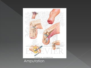
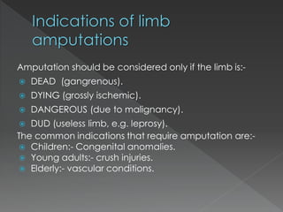
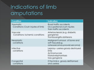
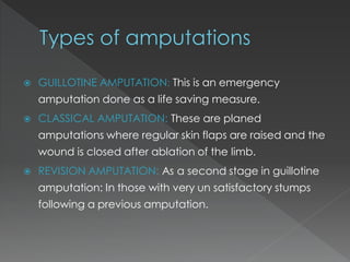
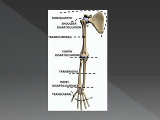
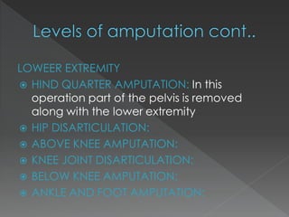
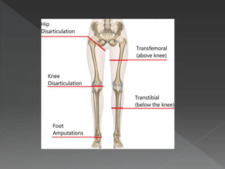
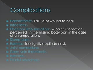
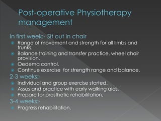
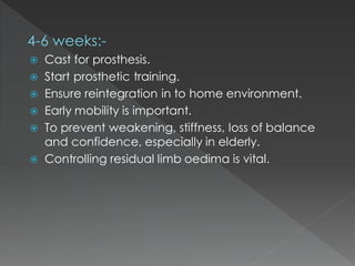
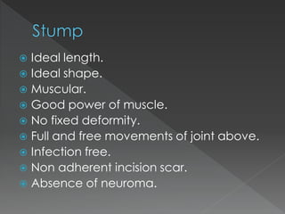
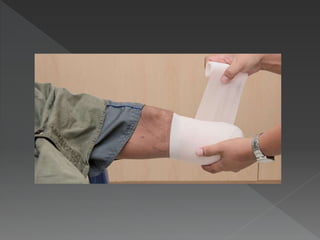
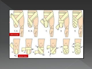
Amputation Is defined as the surgical removal of
a part or whole of a limb.
Disarticulation means removal of the limb
through joint.
Amputation
Hip Disarticulation
Amputation should be considered only if the limb is:-
 DEAD (gangrenous).
 DYING (grossly ischemic).
 DANGEROUS (due to malignancy).
 DUD (useless limb, e.g. leprosy).
The common indications that require amputation are:-
 Children:- Congenital anomalies.
 Young adults:- crush injuries.
 Elderly:- vascular conditions.
 GUILLOTINE AMPUTATION: This is an emergency
amputation done as a life saving measure.
 CLASSICAL AMPUTATION: These are planed
amputations where regular skin flaps are raised and the
wound is closed after ablation of the limb.
 REVISION AMPUTATION: As a second stage in guillotine
amputation: In those with very un satisfactory stumps
following a previous amputation.
Right thigh stump
after guillotine
amputation
Classical amputation
UPPER EXTRIMITY
 FORQUARTER AMPUTATION: Is carried out proximal to
the shoulder joint in this part of the scapula and clavicle
are removed along with the shoulder gurdle muscle
 SHOULDER DISARTICULATION: Not so popular
 ABOVE ELBOW AMPUTATION:
 ELBOW DSARTICULATION:
 BELOW ELBOW AMPUTATION: KRUKENBERG
AMPUTATION(fork like forearm bones)
 WRIST DISARTICULATION:
LOWEER EXTREMITY
 HIND QUARTER AMPUTATION: In this
operation part of the pelvis is removed
along with the lower extremity
 HIP DISARTICULATION:
 ABOVE KNEE AMPUTATION:
 KNEE JOINT DISARTICULATION:
 BELOW KNEE AMPUTATION:
 ANKLE AND FOOT AMPUTATION:
 Haematoma:- Failure of wound to heal.
 Infections:-
 Phantom limb sensation:- A painful sensation
perceived in the missing body part in the case
of an amputation.
 Stump pain:-
 Edema:- Too tightly appliede cast.
 Joint contracture:-
 Deep vain thrombosis:-
 Necrosis:-
 Painful neuromas:-
 Syme’s amputation:- In this operation the tibia and
fibula are divided jest above the ankle joint the
intact skin over the heel is attached back to the
end of the stump with or without a part of the
calcaneum.
 Lisfranc amputation:- Through intertarsal joint.
 Chopart’s amputation:- Through talonavicular joint.
 Pigroff’s amputation:- Anterior part of calcaneum is
cut across and row bone is fixed to the row under
surface of tibia.
 Boyd’s amputation:- Talus is excised, calcaneum is
advanced and attached to the row under surface
of tibia.
Syme’s
amputation:-
 The greatest skin length possible should be
maintained for muscle coverage and a tension
free closure.
 Muscle is placed over the cut end of bones via
a myodesis.
 Nerves are transected under tension, proximal
to the cut end of bones in a tension free
environment.
 The larger arteries and veins are dissected and
separately ligated, this prevents the
development of arteriovenous fistulas and
aneurysms.
 Bony prominence around disarticulation
are removed with a saw and tied
smooth, Maintaining the maximal
extremity length possible is desirable.
 Make a limb 2.5cm long for every 30cm
of the body height.
 Disarticulation when possible ,
disarticulation completely eliminates the
problem of terminal over growth and
subsequent revision surgery.
 Mental preparation of the patient is vital to accept
the loss.
 A thorough evaluation of the skin, ROM, muscle
strength, circulation sensory system of the affected
and un affected limbs.
 The status of vision and hearing analyzed as, a new
proprioceptive system needs to be developed and
understood by the patient after amputation.
 Treat the underlying disease like TAO, Diabetes, etc
 Study his mental makeup, his family , his work and
his society and offer suitable guidance.
 Chest exercises.
 ROM exercise to all joints to prevent thrombosis,
improve mobility, strength etc.
 Mobility training for trunk, shoulder, pelvis , etc.
 Walking training with special emphasis.
 Resistive exercise practiced on the sound limb.
 Training for weight transfer, use of crutches ,
wheelchairs etc.
 Training for single limb standing and balancing.
 Training for lying, turning and limb positioning
on bed.
In first week:- Sit out in chair
 Range of movement and strength for all limbs and
trunks.
 Balance training and transfer practice, wheel chair
provision.
 Oedema control.
 Continue exercise for strength range and balance.
2-3 weeks:-
 Individual and group exercise started.
 Asses and practice with early walking aids.
 Prepare for prosthetic rehabilitation.
3-4 weeks:-
 Progress rehabilitation.
4-6 weeks:-
 Cast for prosthesis.
 Start prosthetic training.
 Ensure reintegration in to home environment.
 Early mobility is important.
 To prevent weakening, stiffness, loss of balance
and confidence, especially in elderly.
 Controlling residual limb oedima is vital.
 Ideal length.
 Ideal shape.
 Muscular.
 Good power of muscle.
 No fixed deformity.
 Full and free movements of joint above.
 Infection free.
 Non adherent incision scar.
 Absence of neuroma.
 Stump drainage.
 Stump splinting.
 Stump bandaging.
 Stump exercise early.
 Stump hygiene.
 Elastic stump compression socks methods
reduce edema and conditioning the
stump.
 Bandaging is controversial method of
controlling stump edema particularly in
vascular patient.
 Pressure should be firm, even and
decreasing pressure proximally.
 Diagonal oblique and spiral turn should be
used rather than circular turns prevent
tourniquet effect.
Bandage size:
 Upper limb - 4”
 Lower limb - 6/8”
 Above knee - 6”
 Below knee -4”
Above knee bandaging:-
 It should extend up to groin to prevent
follicle infection due to friction with
socket of prosthesis.
 It should bandage with hip in extension
and adduction.
Below knee bandaging:-
 Stump should bandage with knee in
slight flexion.
 Encourage prone lying to prevent hip flexion
contracture.
 Avoid pillows beneath the knee.
 Avoid long periods of sitting to prevent deformities
of the knee.
 Avoid dragging on the bed, rather push up the for
arm to get up.
 Advise vigorous ROM and strengthening to all
joints of the body.
 Strong isometrics to the stump after 3-4 days.
 Introduce transfer activity ones the patient is able
to sit up and push his body in sitting.
 Do prosthetic preparation: compression and
shaping of the residual limb using elastic bandages
or stocking, elevation, exercise and early walking
aids.
 Improve cardiovascular fitness.
 Provide education and information for patient and
cares.
 Refer appropriate agencies for prosthetic provision.
 Carry out physical, psychological and functional
assessment.
 Exercise for strengthening, stretching, stability and
balance.
 Gait re-education
 Pain control measures
 Progress walking aids and skills with prosthesis.
 Practice activities of daily living.
 Provide information regarding care of the residual
and remaining limb and prosthesis.
 Prescribe maintenance exercise.
 Provide further intervention when the
situation changes.
 Organize review appointment to re assess.
 Facilitate reintegration to normal living.
 Act as long term resource for patients,
family.
Thank you.

AMPUTATION.pdf

  • 2.
    Amputation Is definedas the surgical removal of a part or whole of a limb. Disarticulation means removal of the limb through joint.
  • 3.
  • 4.
  • 5.
    Amputation should beconsidered only if the limb is:-  DEAD (gangrenous).  DYING (grossly ischemic).  DANGEROUS (due to malignancy).  DUD (useless limb, e.g. leprosy). The common indications that require amputation are:-  Children:- Congenital anomalies.  Young adults:- crush injuries.  Elderly:- vascular conditions.
  • 7.
     GUILLOTINE AMPUTATION:This is an emergency amputation done as a life saving measure.  CLASSICAL AMPUTATION: These are planed amputations where regular skin flaps are raised and the wound is closed after ablation of the limb.  REVISION AMPUTATION: As a second stage in guillotine amputation: In those with very un satisfactory stumps following a previous amputation.
  • 8.
    Right thigh stump afterguillotine amputation Classical amputation
  • 9.
    UPPER EXTRIMITY  FORQUARTERAMPUTATION: Is carried out proximal to the shoulder joint in this part of the scapula and clavicle are removed along with the shoulder gurdle muscle  SHOULDER DISARTICULATION: Not so popular  ABOVE ELBOW AMPUTATION:  ELBOW DSARTICULATION:  BELOW ELBOW AMPUTATION: KRUKENBERG AMPUTATION(fork like forearm bones)  WRIST DISARTICULATION:
  • 11.
    LOWEER EXTREMITY  HINDQUARTER AMPUTATION: In this operation part of the pelvis is removed along with the lower extremity  HIP DISARTICULATION:  ABOVE KNEE AMPUTATION:  KNEE JOINT DISARTICULATION:  BELOW KNEE AMPUTATION:  ANKLE AND FOOT AMPUTATION:
  • 13.
     Haematoma:- Failureof wound to heal.  Infections:-  Phantom limb sensation:- A painful sensation perceived in the missing body part in the case of an amputation.  Stump pain:-  Edema:- Too tightly appliede cast.  Joint contracture:-  Deep vain thrombosis:-  Necrosis:-  Painful neuromas:-
  • 14.
     Syme’s amputation:-In this operation the tibia and fibula are divided jest above the ankle joint the intact skin over the heel is attached back to the end of the stump with or without a part of the calcaneum.  Lisfranc amputation:- Through intertarsal joint.  Chopart’s amputation:- Through talonavicular joint.  Pigroff’s amputation:- Anterior part of calcaneum is cut across and row bone is fixed to the row under surface of tibia.  Boyd’s amputation:- Talus is excised, calcaneum is advanced and attached to the row under surface of tibia.
  • 15.
  • 18.
     The greatestskin length possible should be maintained for muscle coverage and a tension free closure.  Muscle is placed over the cut end of bones via a myodesis.  Nerves are transected under tension, proximal to the cut end of bones in a tension free environment.  The larger arteries and veins are dissected and separately ligated, this prevents the development of arteriovenous fistulas and aneurysms.
  • 19.
     Bony prominencearound disarticulation are removed with a saw and tied smooth, Maintaining the maximal extremity length possible is desirable.  Make a limb 2.5cm long for every 30cm of the body height.  Disarticulation when possible , disarticulation completely eliminates the problem of terminal over growth and subsequent revision surgery.
  • 20.
     Mental preparationof the patient is vital to accept the loss.  A thorough evaluation of the skin, ROM, muscle strength, circulation sensory system of the affected and un affected limbs.  The status of vision and hearing analyzed as, a new proprioceptive system needs to be developed and understood by the patient after amputation.  Treat the underlying disease like TAO, Diabetes, etc  Study his mental makeup, his family , his work and his society and offer suitable guidance.
  • 21.
     Chest exercises. ROM exercise to all joints to prevent thrombosis, improve mobility, strength etc.  Mobility training for trunk, shoulder, pelvis , etc.  Walking training with special emphasis.  Resistive exercise practiced on the sound limb.  Training for weight transfer, use of crutches , wheelchairs etc.  Training for single limb standing and balancing.  Training for lying, turning and limb positioning on bed.
  • 22.
    In first week:-Sit out in chair  Range of movement and strength for all limbs and trunks.  Balance training and transfer practice, wheel chair provision.  Oedema control.  Continue exercise for strength range and balance. 2-3 weeks:-  Individual and group exercise started.  Asses and practice with early walking aids.  Prepare for prosthetic rehabilitation. 3-4 weeks:-  Progress rehabilitation.
  • 23.
    4-6 weeks:-  Castfor prosthesis.  Start prosthetic training.  Ensure reintegration in to home environment.  Early mobility is important.  To prevent weakening, stiffness, loss of balance and confidence, especially in elderly.  Controlling residual limb oedima is vital.
  • 24.
     Ideal length. Ideal shape.  Muscular.  Good power of muscle.  No fixed deformity.  Full and free movements of joint above.  Infection free.  Non adherent incision scar.  Absence of neuroma.
  • 26.
     Stump drainage. Stump splinting.  Stump bandaging.  Stump exercise early.  Stump hygiene.
  • 27.
     Elastic stumpcompression socks methods reduce edema and conditioning the stump.  Bandaging is controversial method of controlling stump edema particularly in vascular patient.  Pressure should be firm, even and decreasing pressure proximally.  Diagonal oblique and spiral turn should be used rather than circular turns prevent tourniquet effect.
  • 28.
    Bandage size:  Upperlimb - 4”  Lower limb - 6/8”  Above knee - 6”  Below knee -4”
  • 29.
    Above knee bandaging:- It should extend up to groin to prevent follicle infection due to friction with socket of prosthesis.  It should bandage with hip in extension and adduction. Below knee bandaging:-  Stump should bandage with knee in slight flexion.
  • 31.
     Encourage pronelying to prevent hip flexion contracture.  Avoid pillows beneath the knee.  Avoid long periods of sitting to prevent deformities of the knee.  Avoid dragging on the bed, rather push up the for arm to get up.  Advise vigorous ROM and strengthening to all joints of the body.  Strong isometrics to the stump after 3-4 days.  Introduce transfer activity ones the patient is able to sit up and push his body in sitting.
  • 32.
     Do prostheticpreparation: compression and shaping of the residual limb using elastic bandages or stocking, elevation, exercise and early walking aids.  Improve cardiovascular fitness.  Provide education and information for patient and cares.  Refer appropriate agencies for prosthetic provision.
  • 33.
     Carry outphysical, psychological and functional assessment.  Exercise for strengthening, stretching, stability and balance.  Gait re-education  Pain control measures  Progress walking aids and skills with prosthesis.  Practice activities of daily living.  Provide information regarding care of the residual and remaining limb and prosthesis.
  • 34.
     Prescribe maintenanceexercise.  Provide further intervention when the situation changes.  Organize review appointment to re assess.  Facilitate reintegration to normal living.  Act as long term resource for patients, family.
  • 35.