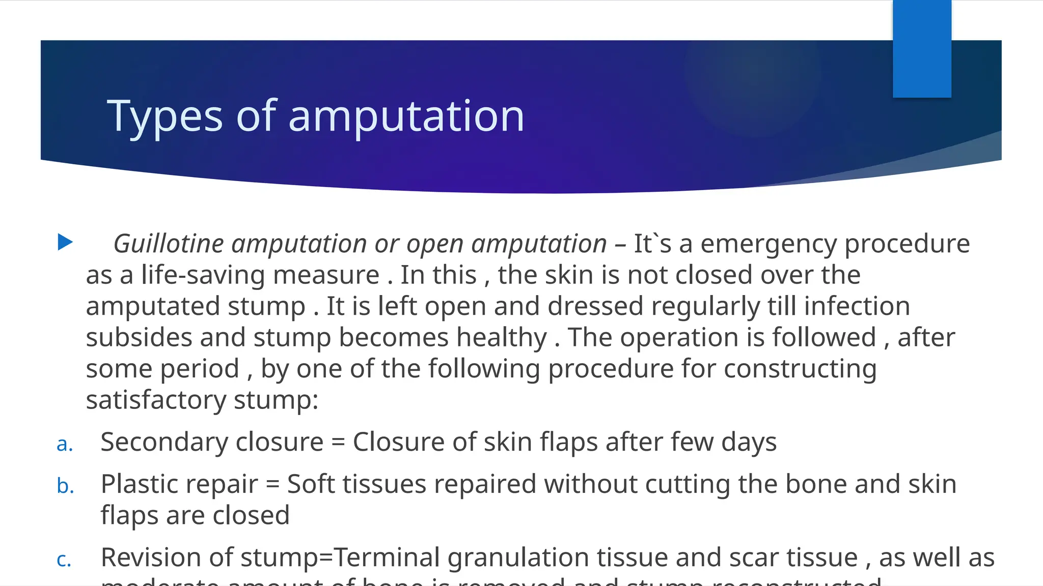
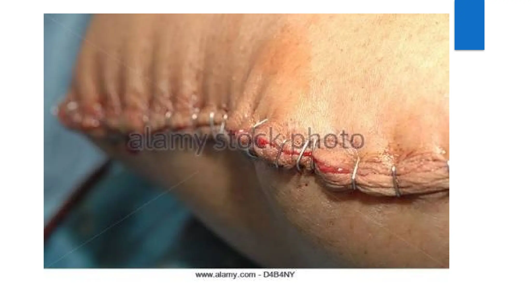
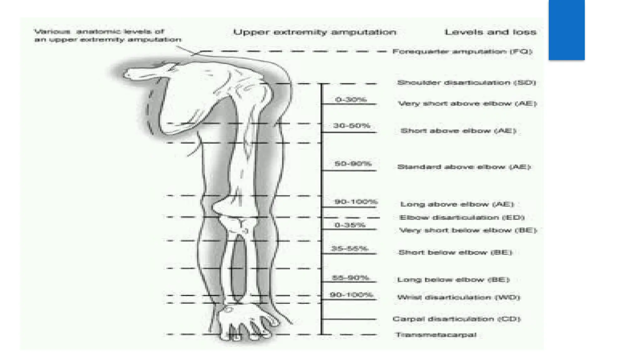
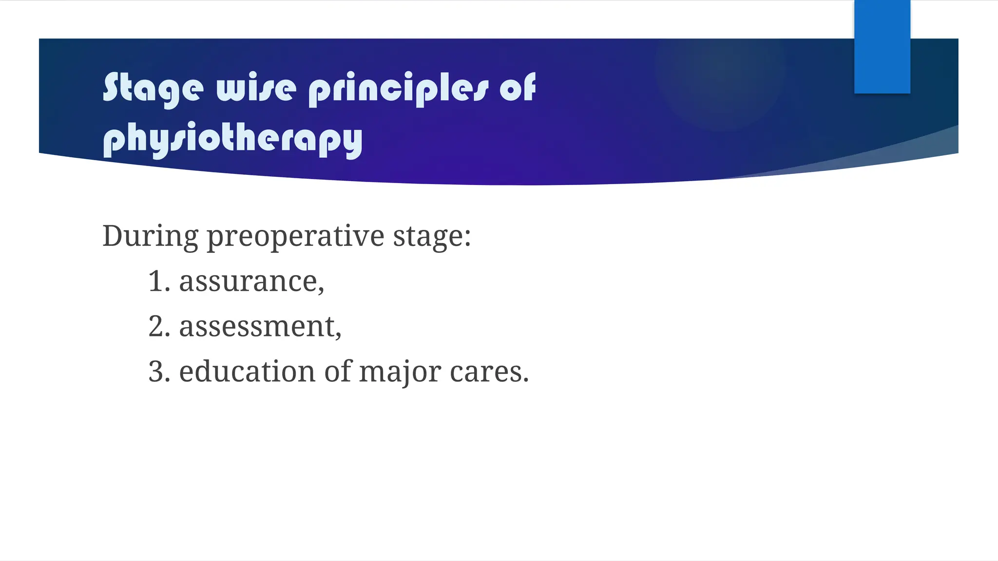
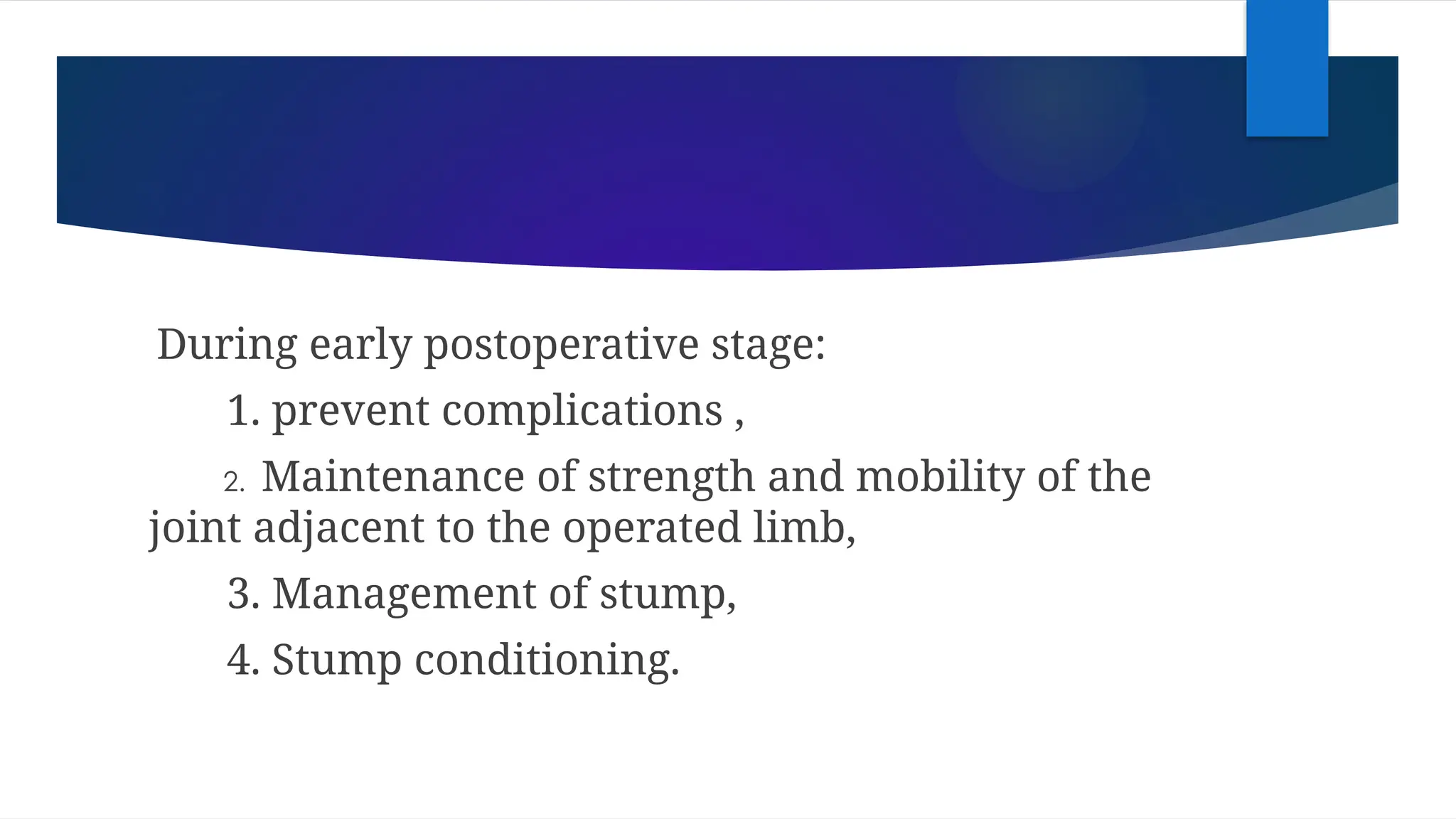
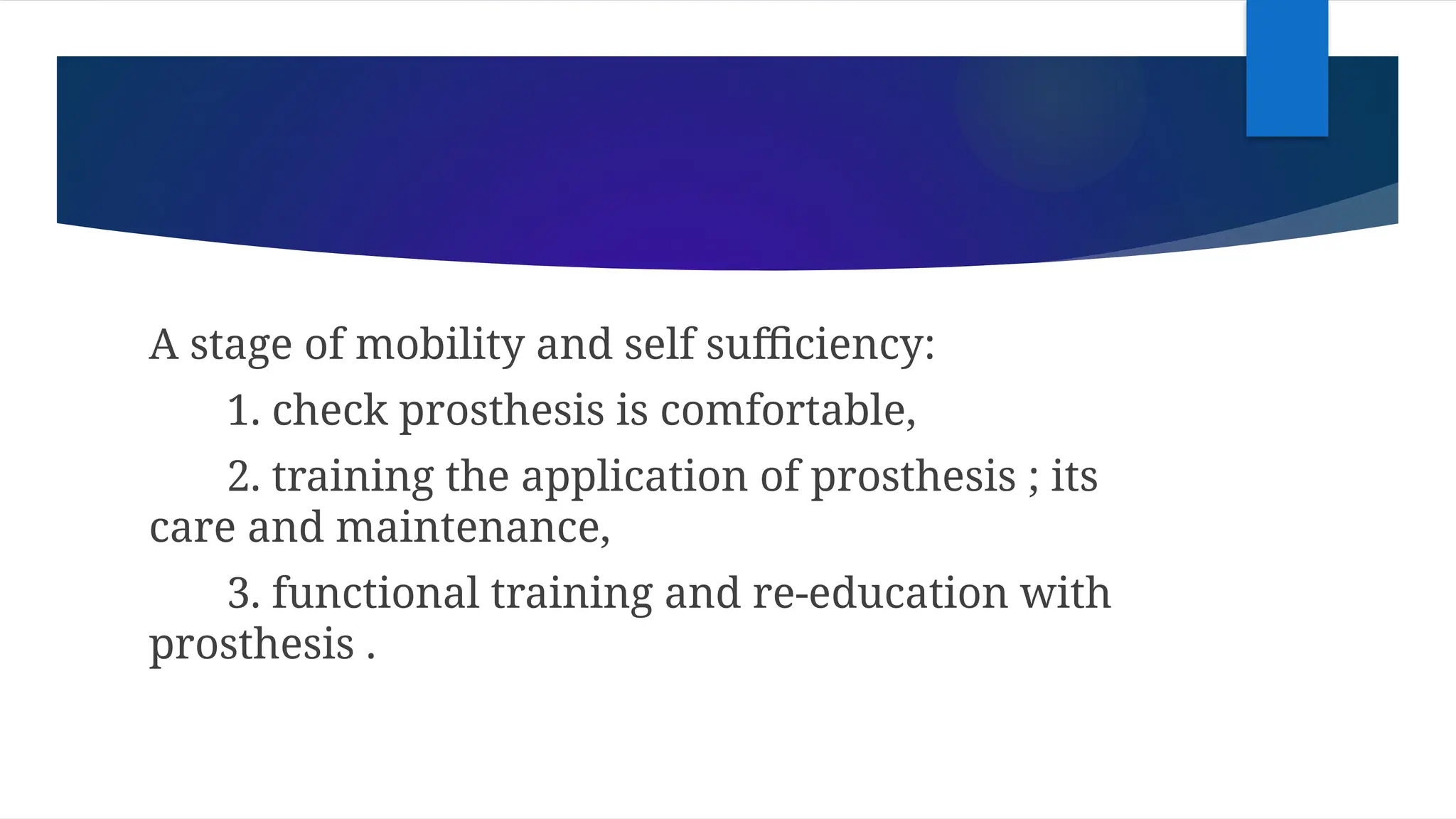
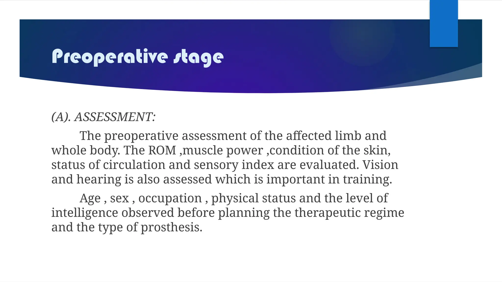
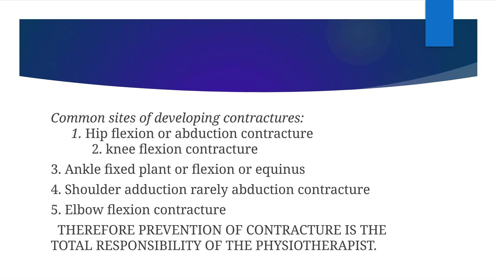
AMPUTATION
DEFINITION
 Amputation is defined as the surgical
removal of the limb, partly or totally
through one or more bones.
 Disarticulation means removal of the
limb through the joints.
 Amputation are common in men and often in
the lower limb.
 Amputation is performed for life threatening
disease, pain or unsalvageable injury, where all
other treatment have failed.
 Becoming disabled can affect person’s
personality.
 Amputation should be considered if the limb is,
i. dead(gangrenous),
ii. dying(ischemic),
iii. dangerous(due to malignancy),
iv. dud(useless limb eg:leprosy).
GOAL
To preserve life and improve function and
general health.
As limb being amputated it is a life
changing event with devastating consequences to
every aspect of life(not just physical functioning).
indication
 In elderly(50-75 yrs), peripheral vascular disease with
or without diabetes is main cause(vascular condition).
 In younger adults(25-30yrs), amputation is often
secondary to injury or its sequelae(amputation at the
time of aacident or in some cases days, months, even
yrs after when injured part is “burden”).(crush injuries)
 In children, limbs may be deficient since
birth(congenital anomalies).
 Trauma - ampu following trauma( RTA, TTA,
occupational crush injuries) is a common
feature. It is done in extensive loss of tissue and
disruption of blood supply.
 Malignant tumors-ampu done for extensive
malignancy, to save life and prevent
recurrence(tumor of bone and soft tissue eg:
osteosarcoma, synovial sarcoma).
 Nerve injuries and infection –anaesthetic limbs often develop
ulceration, infection and severe tissue damage. The tissue
damage due to repeated infection and ulceration lead to
autoamputation in neglected patients(HANSEN DISEASE).
 Extreme heat and cold –injuries following electrical burns,
accidental burns, exposure to extreme cold condition need
ampu. Thermal injuries at times lead to extensive tissue
destruction or deformities. Prolong exposure to extreme cold
condition result in blockage of blood circulation(gangrene).
 Peripheral vascular insufficiency –Irreversible loss of vascularity to limb
due to disease like diabetes, Berger disease, atherosclerosis,
embolism, arterial thrombosis, arteriovenous aneurysm or trauma
[lead to gangrene]. Advance in arterial in arterial surgery decreased
incidence of amputation
 Congenital absence of limb or malformation –There may be incidence of
rudimentary limb structure proximally with apparently normal
terminal segment. Bilateral rudimentary distal segment amputee
managed with bilateral orthosis designed to accommodate deformity .
But when rudimentary portion of limb interfere with fitting of
prosthesis , amputation become necessary
 Severe infection – Like gas gangrene or chronic
intractable infection like osteomyelitis and
unstable DM may need amputation.eg-
Elephantiasis , Actinomycosis
Types of amputation
 Guillotine amputation or open amputation – It`s a emergency procedure
as a life-saving measure . In this , the skin is not closed over the
amputated stump . It is left open and dressed regularly till infection
subsides and stump becomes healthy . The operation is followed , after
some period , by one of the following procedure for constructing
satisfactory stump:
a. Secondary closure = Closure of skin flaps after few days
b. Plastic repair = Soft tissues repaired without cutting the bone and skin
flaps are closed
c. Revision of stump=Terminal granulation tissue and scar tissue , as well as
 Closed amputation - The stump is closed primarily
over the bony stump by retaining skin and
muscle atleast 5 cm distal to elective amputations
are closed amputation
- Muscle provides cushioning
effect to bone ends
PRINCIPLES OF AMPUTATION /
CARE DURING THE PROCESS OF
AMPUTATION
 Amputation surgery is important step in rehabilitation and must be
approached as a plastic and reconstructive procedure
1).Tourniquet
Except ischemic limb (malignancy , infection ), for fear of spread
of disease , tourniquet is commonly applied during amputatation
2).Ex-Sanguination
Usually a limb must be squeezed by wrapping it with a stretchable
bandage (esmarch) before tourniquet is inflated . It`s contra indicated incase
of infection and malignancy
3).Skin flap and scar
Stump must always be covered with healthy skin flap.
Its should be mobile and normally sensitive , but atypical skin flaps
are preferable at more proximal . Care should be taken not to allow
scar to be adherent to bone .
4).blood vessels
Should be isolated and ligated with double ligation using
non-absorbable sutures of major blood vessels
5).Nerves
Must be isolated carefully , gently pulled distally and divided (to avoid
stump neuroma) so that the cut end retracts into the muscle tissue . Large
nerves should be ligated before they are divided
6).Muscle
It should be cut distal to the bone . Method of muscle sutures are as
follows,
a. Myoplasty – Opposite group of muscle are sutured together distal to the bone
b. Myodesis – Muscle are sutured to the end of bones
7).Bone
Excessive stripping of periosteum lead to formation of “ring
sequestrum” from the end of bone . The bone must be beveled or or rasped
to avoid bony prominence jutting at the skin(sharp edges of bone should be
made smooth . Leave enough for good prosthesis and rehabilitation
8).Drain
To prevent hematoma formation suction drain is applied . A
corrugated rubber drain for 48-72 hrs post operatively to prevent post
operative complications
AFTER TREATMENT
 Treatment from the time of amputation complicated till definitive
prosthesis fitted , is important if a strong and maximally functioning
stump is desired
o DRESING -2 type
a. Conventional or soft dressing- using gauge , cotton bandage .
b. Rigid dressing – after soft dressing , well moulded POP cast is applied
on stump . This enhances wound healing and maturation of stump
o Positioning and elevation of stump –It`s required to prevent
contracture and promote healing
o Exercise – Stump exercise necessary to maintain ROM of joint
proximal to stump and build up strength of muscle controlling
stump
o Wrapping –(done with crepe bandage) helps in healing ,
shrinkage and maturation
o Prosthetic fitting and gait training –Its started usually 3 months
after amputation
SELECTION OF LEVEL OF
AMPUTATION
 The sites of amputation of limb were determined on the
basis of following considerations,
o the disease proves for which amputation was done,
o vascular supply to skin flaps,
o the requirement and availability of limb fitting
procedure and technique. Sometimes, length is
compromised for efficient functioning of an artificial limb to
be fitted on a stump.
Upper
extremity
FOREQUARTER
AMPUTATION
(SCAPULOTHORA
CIC
AMPUTATION)
 Proximal to the
shoulder joint
 Indicated in bone
malignant
tumors.
 Part of scapula
and clavicle are
removed along
with shoulder
SHOULDER
DISARTICULATI
ON
 Not so popular.
 The head of
humerus is
preserved, to
maintain the contour
of the shoulder for
cosmosis.
ABOVE
ELBOW(AE)
AMPUTATION
(TRANSHUMER
AL
AMPUTATION)
 20CM long
stump.
 Measured from
the tip of the
acromian.
BELOW ELBOE(BE)
AMPUTATION
(TRANSRADIAL
AMPUTATION)
 Optimum length
is 20CM.
 Measured from
the tip of
olecranon.
 Minimum length
is of 7.5CM
KRUKENBE
RG
AMPUTATIO
N
• Performed in patients
with bilateral
amputation,who have
long BE stump.
• Forearm is spilt
between radius and
ulna to provide pincer
grip.
• The patient can hold
spoon or lighter objects
with this fork.
AMPUTATION
THROUGH THE
HAND
 To preserve as much
length as possible to
contribute to overall
function.
 Short stump is of no use
which hinders with the
function of hand,as
things fall through gap.
 The ray is removed by
amputation through the
neck of metacarpal.
Incase of thumb
amputation, the
maximum
possible length is
preserved.
Subsequent
reconstructive
procedures, such as
metacarpal lengthening
or composite bone
grafts are performed for
a thumb with short
stump.
LOWER
EXTREMI
TY
HINDQUARTER
AMPUTATION
(TRANSPELVIC
AMPUTATION)
 Indicated for
malignant
tumours at
the end of
femur.
 Part of pelvis
is removed
along with
the lower
extremity.
HIP
DISARTICULATI
ON
 Occasionally
performed.
 Femur is removed
from the
acetabulum.
 High energy
requiremwnts for
walking.
AMPUTATION
(TRANSFEMO
RAL
AMPUTATION
)
 Optimum length is
20-30CM.
 Measured from the
tip of the greater
trochanter.
 Minimum length for
useful stump is
about 7.5CM
KNEE
DISARTICULATI
ON
 It is cosmetically un
acceptable for women
as prosthesis required
is bulky.
 Transtibial amputation
is generally preferred
through knee
disarticulation.
 Provide good weight
bearing platform.
BELOW KNEE
AMPUTATION
(TRANSTIBIAL
AMPUTATION)
 Commonly performed.
 Following the severe trauma to
the foot and leg and gangrene
of various etiology.
crush injuries of foot,
malignant tumour at lower
end,
 Optimum length is 14CM from
tibial tubercle.
 Patellar tendon bearing
prosthesis can be fitted over a
stump of adequate length.
SYME’S
AMPUTATIO
N
 Tibia and fibula are
divided just above the
ankle joint.
 The intact skin over the
heel is attached back to
the end of stump with
or without calcaneum.
 An end bearing stump.
 Patients manage very
well walking after this
of amputation.
FOOT
AMPUTATIO
N
TOES
 Amputatio
n of great
toes and
other toes.
METATARSA
LS
 Amputatio
n through
metatarsa
l bones.
LISFRANC
AMPUTATIO
N
 Amputation at
the level of
tarso-
metatarsal
joints.
CHOPART’S
AMPUTATIO
N
 MID TARSAL
JOINT
AMPUTATION.
 Amputation is
performed at
the level of
midtarsal
joints.
Role of physiotherapy following
lowerlimb amputation
 The physiotherapy re-educate movement pattern to
optimise independent function , such as self care,
wheelchair and prosthetic use , normal occupation
PHYSIOTHERAPY AIMS:
 rehabilitation following lowerlimb ampu is a
continual process involving all aspects of persons
life: physical ,mental and socioeconomic.
 The final goal should be optimal independence i.e to return
to normal life as possible. The person should achieve,
1. independent self-caring,
2. independent indoor mobility,
3. independent outdoor mobility,
4. being able to get in/or of car or other transport,
5. return to leisure/hobbies/work/society.
 CONSIDERATIONS:
The patient have difficulties in achieving goals due to
following,
1. poor residual limb condition eg: adherent scar tissue ,
unhealed, failed myodesis, neuroma , bony prominences,
pain, hypersensitivity.
2. concurrent pathologies leading to an inability to learn,
reduced ROM , reduced strength and stamina, pain ,poor
balance.
3. social and environmental difficulties eg: living
alone ,poor access to accomodation.
4. lack of motivation – fear ,fatigue ,emotional
barriers
5. inappropriate equipment eg: poor prosthetic
socket
6. lack of specialist rehab services.
Post operative management
 IMMEDIATE POST SURGICAL PROSTHETIC FITTING:
A plaster cast mould applied over stump
immediately after surgery, temporary prosthetic
(pylon). The patient is allowed partial weight bearing
earlier. It has many advantages ,
1. cast forms protective cover over stump, preventing
infections
2. early amputation improves circulation in stump,
facilitate healing
3. early ambulation in temporary prosthesis
instills confidence in patient and increases
chances of early acceptability of permanent
prosthesis.
 SOFT TISSUE TECHNIQUE:
Incase where a temporary prosthesis is not filted
immediately after surgery, an elastrocrepe bandage is
used after applying sterile dressing. Stitches are
removed after two weeks.
The use of crepe is continued for another for three
weeks. It helps in shaping stump into conical shape,
which is ideal for subsequent prosthetic fitting.
Physiotherapeutic management of
the amputee
 Physiotherapy plays the most significant role in the
management of an amputee from the time the surgeon
decide for amputation till the patient is back to his
work. The management can be divided into three stages:
1. preoperative stage
2. the early post operative stage
3. the mobility stage
Stage wise principles of
physiotherapy
During preoperative stage:
1. assurance,
2. assessment,
3. education of major cares.
During early postoperative stage:
1. prevent complications ,
2. Maintenance of strength and mobility of the
joint adjacent to the operated limb,
3. Management of stump,
4. Stump conditioning.
A stage of mobility and self sufficiency:
1. check prosthesis is comfortable,
2. training the application of prosthesis ; its
care and maintenance,
3. functional training and re-education with
prosthesis .
Preoperative stage
(A). ASSESSMENT:
The preoperative assessment of the affected limb and
whole body. The ROM ,muscle power ,condition of the skin,
status of circulation and sensory index are evaluated. Vision
and hearing is also assessed which is important in training.
Age , sex , occupation , physical status and the level of
intelligence observed before planning the therapeutic regime
and the type of prosthesis.
Assessment of the psychological status losing a
limb itself produces great psychological trauma
leading to depression.
Besides these factors it is important to know
about the environment of the patients home and
his/her working place, and also the nature and the
extent of the support he/she is likely to get from
friends or relatives.
(B) PREOPERATIVE TRAINING:
Once the decision is taken on the future plan
of treatment, it is important to train the patient on
the postoperative regime.
The basic aim of the preoperative training is to
prevent the complications of the postoperative phase.
Reducing cost and the period of rehabilitation.
The postoperative training programme depends upon
the area of involvement, objective of amputation. The
programme includes,
1. prevention of thrombosis by manipulating circulation
through movements of other good limbs,
2. prevention of chest complications,
3. preserve mobility of all the joints ,
4. improved mobility,
5. teach the methodology for mobility and limb positioning in
bed,
6. educate the patient on techniques of transfer, single limb
standing and balancing,
7. educated to detect possible complications like soft tissue
tightness, care of pressure points, degree of pain and phantom
sensation.
8. training the exercise technique on sound limb to improve
endurance –self resisted exercise, progressive resisted exercise.
(C) REASSURANCE : psychological reassurance
plays an Important role in the recovery following
amputation. It can be done practical
demonstration who has undergone similar surgery
and is now physically independent .
Early postoperative stage
(A) To check the efficiency of preoperative training:
provide guidance and assistance.
(B) Prevention of contractures and deformities:
The common contractures are,
adduction and rotation contracture in upper extremity amputees,
hip flexion and abduction contracture in above knee amputation,
hip and knee contracture in bilateral amputees due to mobility and
prolonged sitting postures .
Using a pillow under the thigh or the knee,
although relieving pressure over the end of the
stump and giving comfort, is one of the
commonest causes of soft tissue contracture.
Long periods of sitting as well as soft mattresses
can predispose to development of flexion
contracture at the hip hence it should be avoided.
These contracture can be prevented by proper
training in the positioning of the amputated limb from
the day of surgery.
Hip flexion contracture can be prevented by prone
lying.
Repeating sustained isometrics and repetitive periods
of lying prone can effectively prevent development of
flexion contractures at the hip.
(C) Maintenance of strength and mobility:
The patient should be encouraged to move
in the bed by pushing up the body on the arms and
then dragging . The push up exercise has the
advantage of strengthening the muscle groups for
using ambulation aids at later stage and bilateral
amputees in older age groups.
Vigorous ROM and strengthening exercises should be
given for the whole body except for the joint to the
amputation. Bed activities like bridging and rolling can be
useful to initiate bed mobility.
After 3-4 days of surgery, stump exercises can be
initiated as active assisted or self resisted exercises in a
small range of motion. strong isometrics and slow self
generated tension exercises provide good endurance
training, as they are less painful.
Management of the stump:
Improper management of the stump is one of
the major causes of delayed rehabilitation. Stump
oedema delays the prosthetic fitting.
CAUSES OF STUMP OEDEMA:
1. Surgical trauma itself-most common
2. Incorrect bandaging of the stump
3. Incorrect stump positioning
4. Associated degenerative joint disease
5. Uncontrolled diabetes
6. Atherosclerotic disease
7. Renal disease
THE FOLLOWING MEASURES SHOULD BE TAKEN TO CONTROL
OEDEMAOF THE STUMP :
1. Stimulation with limb in elevation with elastic
bandage.
2. Resistive exercises to the stump and joints.
3. Stump bandaging and conditioning : shaping the by reducing the
stump oedema. A crepe or elastic bandage of sizes 4inches(below
knee) or 6inches(above knee) is necessary. The bandage should be
worn during the night and taken off during exercise.
PRINCIPLES OF BANDAGING THE STUMP:
1. The pressure of the bandage is firm and
evenly distributed. Extra pressure is necessary
over the corners to obtain conical shape.
2. diagonal, oblique or spiral turns should be
used, circular turns may produce a tourniquet
effect leading to ‘choking’ of the stump.
3. In case of an above knee amputation, the bandage must extend
up to groin to prevent a roll of flesh.
4. An above knee stump should be bandaged with the hip in
extension and adduction. The below knee stump should be
bandaged with knee in slight flexion.
INTERMITTENT VARIABLE AIR PRESSURE MACHINE:
This machine also know as a controlled air pressure machine,
is used to control oedema and preserve tissues by normal gaseous
and water exchange.
SHRINKER SOCKS:
An healed stump with the problem of persistent oedema
can be managed by elastic stump socks.
RIGID DRESSING:
Below knee amputation in young patients without
associated problems can be given a rigid plaster of paris
dressing. This will reduce the oedema as well as provide an
attachment to the early postoperative prosthetic device.
STUMP HYGIENE:
This includes regular washing of the stump with
warm disinfected soap water and thorough drying.
AFTER REMOVAL OF STITCHES:
when the stitches are removed and the suture
line healed satisfactorily, the most important aspect
is stump conditioning.
STUMP CONDITIONING:
This is to acclimatize the stump to various
situations of pressure to be explained after the
fitting of the prosthesis.
The ideal stump is one which is well healed,
firm, conical and with minimal flabby tissue.
prevention of contractures:
If soft tissue contracture is neglected, it may present
the following problems,
1. Hinders the fitting o prosthesis.
2. unable to bear weight in single leg weight bearing phase
3. attempted weight bearing efforts results in;
a. imbalance b. pain c. excessive supporting aid
Common sites of developing contractures:
1. Hip flexion or abduction contracture
2. knee flexion contracture
3. Ankle fixed plant or flexion or equinus
4. Shoulder adduction rarely abduction contracture
5. Elbow flexion contracture
THEREFORE PREVENTION OF CONTRACTURE IS THE
TOTAL RESPONSIBILITY OF THE PHYSIOTHERAPIST.
Methods of prevention of contractures: 1.
Early identification
2. Postural guidance
3. Strong exercise to the antagonistic
4. Sustained low weight traction
5. Corrective splint at initial stage
6. Repetitive strong isometrics to gluteus maximus
7. Long periods of lying prone.
Early identification:
An early sign of developing contracture is a tight feeling with pain
at the end of passive antagonistic movement. Immediate sessions of
repetitive sustained stretches should be introduced.
Postural guidance:
The posture which keeps the area stretched should be
emphasized besides this, postures promoting contracture should be
discouraged. Orthotic devices, weight belts or sandbags can be used
to provide sustained stretch by patient themselves .
Strengthening exercises:
PRE against gravity to the antagonists of the tight
muscle groups. PNF and mobilisation techniques are
ideal.
Use of traction:
sustained sessions of gentle traction to stretch the
contracture developing areas like hip or knee flexors.
Use of corrective splint:
Corrective splint with adjustment to increase pull
gradually.
Prevent persistence of haematoma:
It increases the risk of infection and delay in the
process of wound healing.
Mobility stage
This is the stage of mobilization and restoration of
functional independence. It starts with crutch walking at
earlier opportunity. The normal alignment of the pelvis and
the reciprocal movement of the stump should be
maintained during walking. Elderly patients may need
initial ambulation practice in parallel bars. The patient may
have to remove the prosthesis temporarily for certain
postures and activities. Resistive mat activities using PNF
techniques are valuable to offer easy and stable mobility.
Mobilisation and strengthening exercises:
PNF techniques, PRE and strong endurance exercises
to the specific muscle groups are needed to facilitate
effective body functions with the prosthesis.
ROM exercises: full ROM exercises are regularly
given to the joint proximal to the stump and to joints
susceptible to contractures. Longer prone lying should
be encouraged.
Do’s and don’ts for the amputee
(A) During the first visit of the patient:
long explanatory and counselling
assessment
preoperative guidance and training.
(B) Early postoperative phase:
long session of counselling
preoperative training
measures to control oedema and pain
prevention of contractures
(C) Late postoperative stage:
increase the vigourosity of exercises
progressive ambulatory activities
guidance to self control whatever deviations present in the gait
correct application the prosthesis
sensory re education
functional training
restore pre amputation life style.
SPECIAL FEATURES OF AMPUTATIONS
IN CHILDREN
 A disarticulation is preferred to an amputation through shaft
of long bone at more proximal level. This is because
disarticulation preserves epiphysis distally and therefore
growth of stump continues at the normal rate.
 As the child grows, terminal overgrowth of bone occurs and
needs frequent revisions.
 A child needs frequent changes in size of artificial limb.
 Children tolerate artificial limbs much better and get used to
wearing it more quickly.
Complications
 Haematoma –inadequate haemostasis, ligature loosening
inadequate wound drainage common causes.
 Infection –cause is PVD, DM, hematoma. Wound breakdown
and spread of infection proximally , amputation at higher
level.
 Skin flap necrosis -minor or major SPN indicates insufficient
circulation of skin flap. Small area of SPN heel with
dressing but for larger areas, redesigning of flap required.
 Deformities of joint - results from improper positioning
of ampu stump leading to contracture. Mild contracture
treated by positioning and passive stretching ex.
 Neuroma –always forms at end of cut nerve. If neuroma
is bound down to scar because of adhesion ,it become
painful. Painful neuroma usually be prevented by
dividing nerve sharply at proximal level and allow it to
retract well proximal to end of stump.
 Phantom sensation – a sensation as if amputated part
is still present.
All individual with acquired ampu experience
phantom sensation.
Its prominent in the period immediately following
ampu and gradually diminishes phantom pain is
awareness of pain in amputated limb. Treatment is
difficult.

AMPUTATION AND ROLE OF PHYSIOTHERAPY PPT

  • 1.
  • 2.
    DEFINITION  Amputation isdefined as the surgical removal of the limb, partly or totally through one or more bones.  Disarticulation means removal of the limb through the joints.
  • 3.
     Amputation arecommon in men and often in the lower limb.  Amputation is performed for life threatening disease, pain or unsalvageable injury, where all other treatment have failed.  Becoming disabled can affect person’s personality.
  • 4.
     Amputation shouldbe considered if the limb is, i. dead(gangrenous), ii. dying(ischemic), iii. dangerous(due to malignancy), iv. dud(useless limb eg:leprosy).
  • 5.
    GOAL To preserve lifeand improve function and general health. As limb being amputated it is a life changing event with devastating consequences to every aspect of life(not just physical functioning).
  • 6.
    indication  In elderly(50-75yrs), peripheral vascular disease with or without diabetes is main cause(vascular condition).  In younger adults(25-30yrs), amputation is often secondary to injury or its sequelae(amputation at the time of aacident or in some cases days, months, even yrs after when injured part is “burden”).(crush injuries)  In children, limbs may be deficient since birth(congenital anomalies).
  • 7.
     Trauma -ampu following trauma( RTA, TTA, occupational crush injuries) is a common feature. It is done in extensive loss of tissue and disruption of blood supply.  Malignant tumors-ampu done for extensive malignancy, to save life and prevent recurrence(tumor of bone and soft tissue eg: osteosarcoma, synovial sarcoma).
  • 8.
     Nerve injuriesand infection –anaesthetic limbs often develop ulceration, infection and severe tissue damage. The tissue damage due to repeated infection and ulceration lead to autoamputation in neglected patients(HANSEN DISEASE).  Extreme heat and cold –injuries following electrical burns, accidental burns, exposure to extreme cold condition need ampu. Thermal injuries at times lead to extensive tissue destruction or deformities. Prolong exposure to extreme cold condition result in blockage of blood circulation(gangrene).
  • 9.
     Peripheral vascularinsufficiency –Irreversible loss of vascularity to limb due to disease like diabetes, Berger disease, atherosclerosis, embolism, arterial thrombosis, arteriovenous aneurysm or trauma [lead to gangrene]. Advance in arterial in arterial surgery decreased incidence of amputation  Congenital absence of limb or malformation –There may be incidence of rudimentary limb structure proximally with apparently normal terminal segment. Bilateral rudimentary distal segment amputee managed with bilateral orthosis designed to accommodate deformity . But when rudimentary portion of limb interfere with fitting of prosthesis , amputation become necessary
  • 10.
     Severe infection– Like gas gangrene or chronic intractable infection like osteomyelitis and unstable DM may need amputation.eg- Elephantiasis , Actinomycosis
  • 11.
    Types of amputation Guillotine amputation or open amputation – It`s a emergency procedure as a life-saving measure . In this , the skin is not closed over the amputated stump . It is left open and dressed regularly till infection subsides and stump becomes healthy . The operation is followed , after some period , by one of the following procedure for constructing satisfactory stump: a. Secondary closure = Closure of skin flaps after few days b. Plastic repair = Soft tissues repaired without cutting the bone and skin flaps are closed c. Revision of stump=Terminal granulation tissue and scar tissue , as well as
  • 13.
     Closed amputation- The stump is closed primarily over the bony stump by retaining skin and muscle atleast 5 cm distal to elective amputations are closed amputation - Muscle provides cushioning effect to bone ends
  • 15.
    PRINCIPLES OF AMPUTATION/ CARE DURING THE PROCESS OF AMPUTATION  Amputation surgery is important step in rehabilitation and must be approached as a plastic and reconstructive procedure 1).Tourniquet Except ischemic limb (malignancy , infection ), for fear of spread of disease , tourniquet is commonly applied during amputatation 2).Ex-Sanguination Usually a limb must be squeezed by wrapping it with a stretchable bandage (esmarch) before tourniquet is inflated . It`s contra indicated incase of infection and malignancy
  • 16.
    3).Skin flap andscar Stump must always be covered with healthy skin flap. Its should be mobile and normally sensitive , but atypical skin flaps are preferable at more proximal . Care should be taken not to allow scar to be adherent to bone . 4).blood vessels Should be isolated and ligated with double ligation using non-absorbable sutures of major blood vessels
  • 17.
    5).Nerves Must be isolatedcarefully , gently pulled distally and divided (to avoid stump neuroma) so that the cut end retracts into the muscle tissue . Large nerves should be ligated before they are divided 6).Muscle It should be cut distal to the bone . Method of muscle sutures are as follows, a. Myoplasty – Opposite group of muscle are sutured together distal to the bone b. Myodesis – Muscle are sutured to the end of bones
  • 18.
    7).Bone Excessive stripping ofperiosteum lead to formation of “ring sequestrum” from the end of bone . The bone must be beveled or or rasped to avoid bony prominence jutting at the skin(sharp edges of bone should be made smooth . Leave enough for good prosthesis and rehabilitation 8).Drain To prevent hematoma formation suction drain is applied . A corrugated rubber drain for 48-72 hrs post operatively to prevent post operative complications
  • 19.
    AFTER TREATMENT  Treatmentfrom the time of amputation complicated till definitive prosthesis fitted , is important if a strong and maximally functioning stump is desired o DRESING -2 type a. Conventional or soft dressing- using gauge , cotton bandage . b. Rigid dressing – after soft dressing , well moulded POP cast is applied on stump . This enhances wound healing and maturation of stump
  • 20.
    o Positioning andelevation of stump –It`s required to prevent contracture and promote healing o Exercise – Stump exercise necessary to maintain ROM of joint proximal to stump and build up strength of muscle controlling stump o Wrapping –(done with crepe bandage) helps in healing , shrinkage and maturation o Prosthetic fitting and gait training –Its started usually 3 months after amputation
  • 21.
    SELECTION OF LEVELOF AMPUTATION  The sites of amputation of limb were determined on the basis of following considerations, o the disease proves for which amputation was done, o vascular supply to skin flaps, o the requirement and availability of limb fitting procedure and technique. Sometimes, length is compromised for efficient functioning of an artificial limb to be fitted on a stump.
  • 22.
  • 24.
    FOREQUARTER AMPUTATION (SCAPULOTHORA CIC AMPUTATION)  Proximal tothe shoulder joint  Indicated in bone malignant tumors.  Part of scapula and clavicle are removed along with shoulder
  • 26.
    SHOULDER DISARTICULATI ON  Not sopopular.  The head of humerus is preserved, to maintain the contour of the shoulder for cosmosis.
  • 27.
  • 28.
    BELOW ELBOE(BE) AMPUTATION (TRANSRADIAL AMPUTATION)  Optimumlength is 20CM.  Measured from the tip of olecranon.  Minimum length is of 7.5CM
  • 29.
    KRUKENBE RG AMPUTATIO N • Performed inpatients with bilateral amputation,who have long BE stump. • Forearm is spilt between radius and ulna to provide pincer grip. • The patient can hold spoon or lighter objects with this fork.
  • 30.
    AMPUTATION THROUGH THE HAND  Topreserve as much length as possible to contribute to overall function.  Short stump is of no use which hinders with the function of hand,as things fall through gap.  The ray is removed by amputation through the neck of metacarpal.
  • 32.
    Incase of thumb amputation,the maximum possible length is preserved. Subsequent reconstructive procedures, such as metacarpal lengthening or composite bone grafts are performed for a thumb with short stump.
  • 33.
  • 34.
    HINDQUARTER AMPUTATION (TRANSPELVIC AMPUTATION)  Indicated for malignant tumoursat the end of femur.  Part of pelvis is removed along with the lower extremity.
  • 35.
    HIP DISARTICULATI ON  Occasionally performed.  Femuris removed from the acetabulum.  High energy requiremwnts for walking.
  • 36.
    AMPUTATION (TRANSFEMO RAL AMPUTATION )  Optimum lengthis 20-30CM.  Measured from the tip of the greater trochanter.  Minimum length for useful stump is about 7.5CM
  • 37.
    KNEE DISARTICULATI ON  It iscosmetically un acceptable for women as prosthesis required is bulky.  Transtibial amputation is generally preferred through knee disarticulation.  Provide good weight bearing platform.
  • 38.
    BELOW KNEE AMPUTATION (TRANSTIBIAL AMPUTATION)  Commonlyperformed.  Following the severe trauma to the foot and leg and gangrene of various etiology. crush injuries of foot, malignant tumour at lower end,  Optimum length is 14CM from tibial tubercle.  Patellar tendon bearing prosthesis can be fitted over a stump of adequate length.
  • 39.
    SYME’S AMPUTATIO N  Tibia andfibula are divided just above the ankle joint.  The intact skin over the heel is attached back to the end of stump with or without calcaneum.  An end bearing stump.  Patients manage very well walking after this of amputation.
  • 40.
  • 41.
    TOES  Amputatio n ofgreat toes and other toes.
  • 42.
  • 43.
    LISFRANC AMPUTATIO N  Amputation at thelevel of tarso- metatarsal joints.
  • 44.
    CHOPART’S AMPUTATIO N  MID TARSAL JOINT AMPUTATION. Amputation is performed at the level of midtarsal joints.
  • 47.
    Role of physiotherapyfollowing lowerlimb amputation  The physiotherapy re-educate movement pattern to optimise independent function , such as self care, wheelchair and prosthetic use , normal occupation PHYSIOTHERAPY AIMS:  rehabilitation following lowerlimb ampu is a continual process involving all aspects of persons life: physical ,mental and socioeconomic.
  • 48.
     The finalgoal should be optimal independence i.e to return to normal life as possible. The person should achieve, 1. independent self-caring, 2. independent indoor mobility, 3. independent outdoor mobility, 4. being able to get in/or of car or other transport, 5. return to leisure/hobbies/work/society.
  • 49.
     CONSIDERATIONS: The patienthave difficulties in achieving goals due to following, 1. poor residual limb condition eg: adherent scar tissue , unhealed, failed myodesis, neuroma , bony prominences, pain, hypersensitivity. 2. concurrent pathologies leading to an inability to learn, reduced ROM , reduced strength and stamina, pain ,poor balance. 3. social and environmental difficulties eg: living alone ,poor access to accomodation.
  • 50.
    4. lack ofmotivation – fear ,fatigue ,emotional barriers 5. inappropriate equipment eg: poor prosthetic socket 6. lack of specialist rehab services.
  • 51.
    Post operative management IMMEDIATE POST SURGICAL PROSTHETIC FITTING: A plaster cast mould applied over stump immediately after surgery, temporary prosthetic (pylon). The patient is allowed partial weight bearing earlier. It has many advantages , 1. cast forms protective cover over stump, preventing infections
  • 52.
    2. early amputationimproves circulation in stump, facilitate healing 3. early ambulation in temporary prosthesis instills confidence in patient and increases chances of early acceptability of permanent prosthesis.
  • 53.
     SOFT TISSUETECHNIQUE: Incase where a temporary prosthesis is not filted immediately after surgery, an elastrocrepe bandage is used after applying sterile dressing. Stitches are removed after two weeks. The use of crepe is continued for another for three weeks. It helps in shaping stump into conical shape, which is ideal for subsequent prosthetic fitting.
  • 54.
    Physiotherapeutic management of theamputee  Physiotherapy plays the most significant role in the management of an amputee from the time the surgeon decide for amputation till the patient is back to his work. The management can be divided into three stages: 1. preoperative stage 2. the early post operative stage 3. the mobility stage
  • 55.
    Stage wise principlesof physiotherapy During preoperative stage: 1. assurance, 2. assessment, 3. education of major cares.
  • 56.
    During early postoperativestage: 1. prevent complications , 2. Maintenance of strength and mobility of the joint adjacent to the operated limb, 3. Management of stump, 4. Stump conditioning.
  • 57.
    A stage ofmobility and self sufficiency: 1. check prosthesis is comfortable, 2. training the application of prosthesis ; its care and maintenance, 3. functional training and re-education with prosthesis .
  • 58.
    Preoperative stage (A). ASSESSMENT: Thepreoperative assessment of the affected limb and whole body. The ROM ,muscle power ,condition of the skin, status of circulation and sensory index are evaluated. Vision and hearing is also assessed which is important in training. Age , sex , occupation , physical status and the level of intelligence observed before planning the therapeutic regime and the type of prosthesis.
  • 59.
    Assessment of thepsychological status losing a limb itself produces great psychological trauma leading to depression. Besides these factors it is important to know about the environment of the patients home and his/her working place, and also the nature and the extent of the support he/she is likely to get from friends or relatives.
  • 60.
    (B) PREOPERATIVE TRAINING: Oncethe decision is taken on the future plan of treatment, it is important to train the patient on the postoperative regime. The basic aim of the preoperative training is to prevent the complications of the postoperative phase. Reducing cost and the period of rehabilitation.
  • 61.
    The postoperative trainingprogramme depends upon the area of involvement, objective of amputation. The programme includes, 1. prevention of thrombosis by manipulating circulation through movements of other good limbs, 2. prevention of chest complications, 3. preserve mobility of all the joints , 4. improved mobility,
  • 62.
    5. teach themethodology for mobility and limb positioning in bed, 6. educate the patient on techniques of transfer, single limb standing and balancing, 7. educated to detect possible complications like soft tissue tightness, care of pressure points, degree of pain and phantom sensation. 8. training the exercise technique on sound limb to improve endurance –self resisted exercise, progressive resisted exercise.
  • 63.
    (C) REASSURANCE :psychological reassurance plays an Important role in the recovery following amputation. It can be done practical demonstration who has undergone similar surgery and is now physically independent .
  • 64.
    Early postoperative stage (A)To check the efficiency of preoperative training: provide guidance and assistance. (B) Prevention of contractures and deformities: The common contractures are, adduction and rotation contracture in upper extremity amputees, hip flexion and abduction contracture in above knee amputation, hip and knee contracture in bilateral amputees due to mobility and prolonged sitting postures .
  • 65.
    Using a pillowunder the thigh or the knee, although relieving pressure over the end of the stump and giving comfort, is one of the commonest causes of soft tissue contracture. Long periods of sitting as well as soft mattresses can predispose to development of flexion contracture at the hip hence it should be avoided.
  • 66.
    These contracture canbe prevented by proper training in the positioning of the amputated limb from the day of surgery. Hip flexion contracture can be prevented by prone lying. Repeating sustained isometrics and repetitive periods of lying prone can effectively prevent development of flexion contractures at the hip.
  • 67.
    (C) Maintenance ofstrength and mobility: The patient should be encouraged to move in the bed by pushing up the body on the arms and then dragging . The push up exercise has the advantage of strengthening the muscle groups for using ambulation aids at later stage and bilateral amputees in older age groups.
  • 68.
    Vigorous ROM andstrengthening exercises should be given for the whole body except for the joint to the amputation. Bed activities like bridging and rolling can be useful to initiate bed mobility. After 3-4 days of surgery, stump exercises can be initiated as active assisted or self resisted exercises in a small range of motion. strong isometrics and slow self generated tension exercises provide good endurance training, as they are less painful.
  • 69.
    Management of thestump: Improper management of the stump is one of the major causes of delayed rehabilitation. Stump oedema delays the prosthetic fitting. CAUSES OF STUMP OEDEMA: 1. Surgical trauma itself-most common 2. Incorrect bandaging of the stump
  • 70.
    3. Incorrect stumppositioning 4. Associated degenerative joint disease 5. Uncontrolled diabetes 6. Atherosclerotic disease 7. Renal disease
  • 71.
    THE FOLLOWING MEASURESSHOULD BE TAKEN TO CONTROL OEDEMAOF THE STUMP : 1. Stimulation with limb in elevation with elastic bandage. 2. Resistive exercises to the stump and joints. 3. Stump bandaging and conditioning : shaping the by reducing the stump oedema. A crepe or elastic bandage of sizes 4inches(below knee) or 6inches(above knee) is necessary. The bandage should be worn during the night and taken off during exercise.
  • 72.
    PRINCIPLES OF BANDAGINGTHE STUMP: 1. The pressure of the bandage is firm and evenly distributed. Extra pressure is necessary over the corners to obtain conical shape. 2. diagonal, oblique or spiral turns should be used, circular turns may produce a tourniquet effect leading to ‘choking’ of the stump.
  • 73.
    3. In caseof an above knee amputation, the bandage must extend up to groin to prevent a roll of flesh. 4. An above knee stump should be bandaged with the hip in extension and adduction. The below knee stump should be bandaged with knee in slight flexion. INTERMITTENT VARIABLE AIR PRESSURE MACHINE: This machine also know as a controlled air pressure machine, is used to control oedema and preserve tissues by normal gaseous and water exchange.
  • 74.
    SHRINKER SOCKS: An healedstump with the problem of persistent oedema can be managed by elastic stump socks. RIGID DRESSING: Below knee amputation in young patients without associated problems can be given a rigid plaster of paris dressing. This will reduce the oedema as well as provide an attachment to the early postoperative prosthetic device.
  • 75.
    STUMP HYGIENE: This includesregular washing of the stump with warm disinfected soap water and thorough drying. AFTER REMOVAL OF STITCHES: when the stitches are removed and the suture line healed satisfactorily, the most important aspect is stump conditioning.
  • 76.
    STUMP CONDITIONING: This isto acclimatize the stump to various situations of pressure to be explained after the fitting of the prosthesis. The ideal stump is one which is well healed, firm, conical and with minimal flabby tissue.
  • 77.
    prevention of contractures: Ifsoft tissue contracture is neglected, it may present the following problems, 1. Hinders the fitting o prosthesis. 2. unable to bear weight in single leg weight bearing phase 3. attempted weight bearing efforts results in; a. imbalance b. pain c. excessive supporting aid
  • 78.
    Common sites ofdeveloping contractures: 1. Hip flexion or abduction contracture 2. knee flexion contracture 3. Ankle fixed plant or flexion or equinus 4. Shoulder adduction rarely abduction contracture 5. Elbow flexion contracture THEREFORE PREVENTION OF CONTRACTURE IS THE TOTAL RESPONSIBILITY OF THE PHYSIOTHERAPIST.
  • 79.
    Methods of preventionof contractures: 1. Early identification 2. Postural guidance 3. Strong exercise to the antagonistic 4. Sustained low weight traction 5. Corrective splint at initial stage 6. Repetitive strong isometrics to gluteus maximus 7. Long periods of lying prone.
  • 80.
    Early identification: An earlysign of developing contracture is a tight feeling with pain at the end of passive antagonistic movement. Immediate sessions of repetitive sustained stretches should be introduced. Postural guidance: The posture which keeps the area stretched should be emphasized besides this, postures promoting contracture should be discouraged. Orthotic devices, weight belts or sandbags can be used to provide sustained stretch by patient themselves .
  • 81.
    Strengthening exercises: PRE againstgravity to the antagonists of the tight muscle groups. PNF and mobilisation techniques are ideal. Use of traction: sustained sessions of gentle traction to stretch the contracture developing areas like hip or knee flexors.
  • 82.
    Use of correctivesplint: Corrective splint with adjustment to increase pull gradually. Prevent persistence of haematoma: It increases the risk of infection and delay in the process of wound healing.
  • 83.
    Mobility stage This isthe stage of mobilization and restoration of functional independence. It starts with crutch walking at earlier opportunity. The normal alignment of the pelvis and the reciprocal movement of the stump should be maintained during walking. Elderly patients may need initial ambulation practice in parallel bars. The patient may have to remove the prosthesis temporarily for certain postures and activities. Resistive mat activities using PNF techniques are valuable to offer easy and stable mobility.
  • 84.
    Mobilisation and strengtheningexercises: PNF techniques, PRE and strong endurance exercises to the specific muscle groups are needed to facilitate effective body functions with the prosthesis. ROM exercises: full ROM exercises are regularly given to the joint proximal to the stump and to joints susceptible to contractures. Longer prone lying should be encouraged.
  • 85.
    Do’s and don’tsfor the amputee (A) During the first visit of the patient: long explanatory and counselling assessment preoperative guidance and training. (B) Early postoperative phase: long session of counselling preoperative training measures to control oedema and pain prevention of contractures
  • 86.
    (C) Late postoperativestage: increase the vigourosity of exercises progressive ambulatory activities guidance to self control whatever deviations present in the gait correct application the prosthesis sensory re education functional training restore pre amputation life style.
  • 87.
    SPECIAL FEATURES OFAMPUTATIONS IN CHILDREN  A disarticulation is preferred to an amputation through shaft of long bone at more proximal level. This is because disarticulation preserves epiphysis distally and therefore growth of stump continues at the normal rate.  As the child grows, terminal overgrowth of bone occurs and needs frequent revisions.  A child needs frequent changes in size of artificial limb.  Children tolerate artificial limbs much better and get used to wearing it more quickly.
  • 88.
    Complications  Haematoma –inadequatehaemostasis, ligature loosening inadequate wound drainage common causes.  Infection –cause is PVD, DM, hematoma. Wound breakdown and spread of infection proximally , amputation at higher level.  Skin flap necrosis -minor or major SPN indicates insufficient circulation of skin flap. Small area of SPN heel with dressing but for larger areas, redesigning of flap required.
  • 89.
     Deformities ofjoint - results from improper positioning of ampu stump leading to contracture. Mild contracture treated by positioning and passive stretching ex.  Neuroma –always forms at end of cut nerve. If neuroma is bound down to scar because of adhesion ,it become painful. Painful neuroma usually be prevented by dividing nerve sharply at proximal level and allow it to retract well proximal to end of stump.
  • 90.
     Phantom sensation– a sensation as if amputated part is still present. All individual with acquired ampu experience phantom sensation. Its prominent in the period immediately following ampu and gradually diminishes phantom pain is awareness of pain in amputated limb. Treatment is difficult.