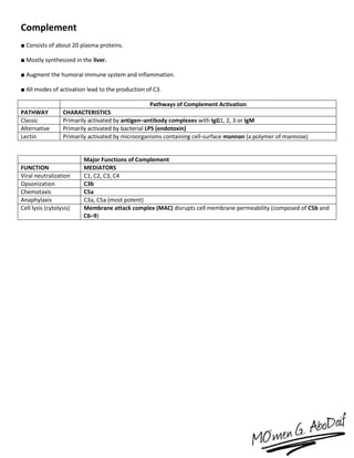
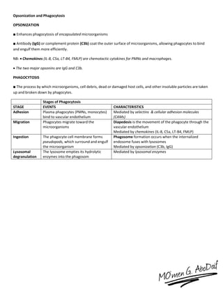
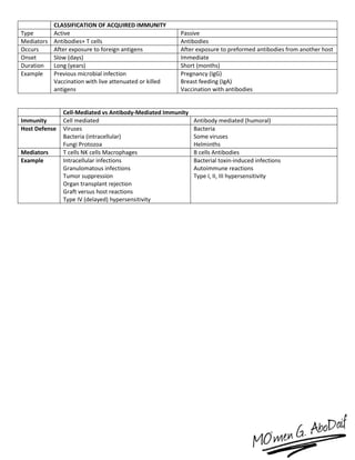
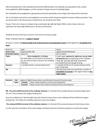
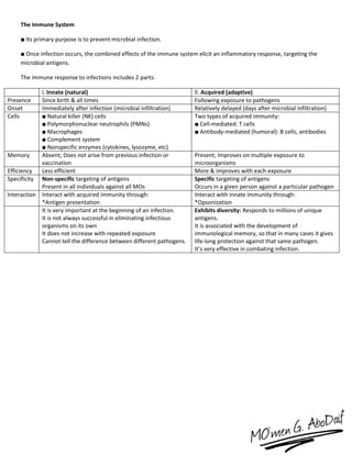
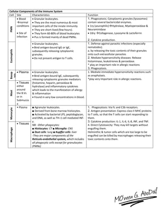
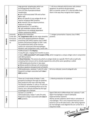
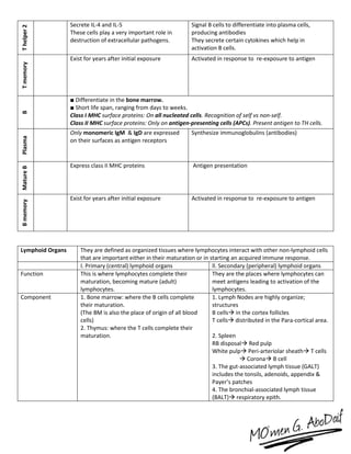
The immune system has both innate and acquired responses. The innate response is non-specific and provides immediate protection against infection. The acquired response is antigen-specific and develops over time through exposure to pathogens. It provides long-lasting immunity through immunological memory. The two main cell types of the acquired response are B cells and T cells. B cells produce antibodies while T cells help activate other immune cells. Together they provide a highly targeted defense against pathogens.
The immune system has evolved to protect the host from a universe of pathogenic microbes that are themselves constantly evolving. The immune system also helps the host eliminate toxic or allergenic substances that enter our body. It is a host defence system comprising many biological structures and processes within an organism that protects against disease. To function properly, an immune system must detect a wide variety of agents, known as pathogens, from viruses to parasitic worms, and distinguish them from the organism's own healthy tissue. The host uses both innate and adaptive mechanisms to detect and eliminate pathogenic foreign bodies. Both of these mechanisms include self-nonself discrimination.
The main parts of the immune system are:
• White Blood Cells
• Antibodies
• Complement System
• Lymphatic System
• Spleen
• Bone Marrow
• Thymus.
A brief covering basics of immunity understanding and also allowing students to understand with ease the concepts of innate immunity, adaptive immunity, Tcell, Bcell, MHC molecular genetics, and also cytokines and also its role in various disease.
Cell mediated immunity also known as T cell immunity. it is developed by cell mediated responses and it does not involve any antibodies. Cell mediated immunity is offered by T lymphocytes and it starts developing when T cells come in contact with the antigens. In the Cell mediated immunity T cell plays one of the important role for the process of crosstalk with other immune system as well as to signal B cells to produce the antibody mediated immune response. Primary function of cell mediated response-
1) Eliminate intracellular pathogens.
2)Eliminate tumor cells.
T cells regulate proliferation and activity of other cells of the immune system : B cells, macrophages, neutrophil, etc.
The immune response is how our body recognizes and defends itself against pathogens like bacteria, viruses, and substances that appear foreign and harmful.
Adaptive immunity is an immunity that occurs after exposure to an antigen either from a pathogen or a vaccination. This part of the immune system is activated when the innate immune response is insufficient to control an infection. In fact, without information from the innate immune system, the adaptive response could not be mobilized. There are two types of adaptive responses: the cell-mediated immune response, which is carried out by T cells, and the humoral immune response, which is controlled by activated B cells and antibodies.
By DR. MANPREET KAUR BEHL.
Description of classificaton of immune system, immune cells, HLA, MHC complexes, antigen presentation, t-cell responses and b-cell responses, antibody, isotype switching, hypersenstivity reactions etc.
introduction of adaptive immunity. classification of adaptive immunity, factor affecting it and mechanism of adaptive immunity comparison between adaptive immunity and innate immunity. characteristic of adaptive immunity . cell mediated immune responses immunoglobulins
types of immunoglobulins. functions of immunoglobulins, hypersensitivity reactions
As a periodontist, I have included the basics of immunity from the periodontist point of view that will help in understanding the immunological basis of periodontal disease...
When a pathogen enters the body, it’s confronted by elements of the innate immune system, which constitute the first line of defense.
Once breached, the adaptive response takes over, but it typically takes few days to be effective.
Immunity is the processes that occur to defend the body against foreign organisms or molecules.
Immunity includes:
Inflammation.
Complement activation.
Phagocytosis.
Antibody synthesis.
Effector T lymphocytes.
Embracing GenAI - A Strategic ImperativePeter Windle
Artificial Intelligence (AI) technologies such as Generative AI, Image Generators and Large Language Models have had a dramatic impact on teaching, learning and assessment over the past 18 months. The most immediate threat AI posed was to Academic Integrity with Higher Education Institutes (HEIs) focusing their efforts on combating the use of GenAI in assessment. Guidelines were developed for staff and students, policies put in place too. Innovative educators have forged paths in the use of Generative AI for teaching, learning and assessments leading to pockets of transformation springing up across HEIs, often with little or no top-down guidance, support or direction.
This Gasta posits a strategic approach to integrating AI into HEIs to prepare staff, students and the curriculum for an evolving world and workplace. We will highlight the advantages of working with these technologies beyond the realm of teaching, learning and assessment by considering prompt engineering skills, industry impact, curriculum changes, and the need for staff upskilling. In contrast, not engaging strategically with Generative AI poses risks, including falling behind peers, missed opportunities and failing to ensure our graduates remain employable. The rapid evolution of AI technologies necessitates a proactive and strategic approach if we are to remain relevant.
How to Make a Field invisible in Odoo 17Celine George
It is possible to hide or invisible some fields in odoo. Commonly using “invisible” attribute in the field definition to invisible the fields. This slide will show how to make a field invisible in odoo 17.
The Roman Empire A Historical Colossus.pdfkaushalkr1407
The Roman Empire, a vast and enduring power, stands as one of history's most remarkable civilizations, leaving an indelible imprint on the world. It emerged from the Roman Republic, transitioning into an imperial powerhouse under the leadership of Augustus Caesar in 27 BCE. This transformation marked the beginning of an era defined by unprecedented territorial expansion, architectural marvels, and profound cultural influence.
The empire's roots lie in the city of Rome, founded, according to legend, by Romulus in 753 BCE. Over centuries, Rome evolved from a small settlement to a formidable republic, characterized by a complex political system with elected officials and checks on power. However, internal strife, class conflicts, and military ambitions paved the way for the end of the Republic. Julius Caesar’s dictatorship and subsequent assassination in 44 BCE created a power vacuum, leading to a civil war. Octavian, later Augustus, emerged victorious, heralding the Roman Empire’s birth.
Under Augustus, the empire experienced the Pax Romana, a 200-year period of relative peace and stability. Augustus reformed the military, established efficient administrative systems, and initiated grand construction projects. The empire's borders expanded, encompassing territories from Britain to Egypt and from Spain to the Euphrates. Roman legions, renowned for their discipline and engineering prowess, secured and maintained these vast territories, building roads, fortifications, and cities that facilitated control and integration.
The Roman Empire’s society was hierarchical, with a rigid class system. At the top were the patricians, wealthy elites who held significant political power. Below them were the plebeians, free citizens with limited political influence, and the vast numbers of slaves who formed the backbone of the economy. The family unit was central, governed by the paterfamilias, the male head who held absolute authority.
Culturally, the Romans were eclectic, absorbing and adapting elements from the civilizations they encountered, particularly the Greeks. Roman art, literature, and philosophy reflected this synthesis, creating a rich cultural tapestry. Latin, the Roman language, became the lingua franca of the Western world, influencing numerous modern languages.
Roman architecture and engineering achievements were monumental. They perfected the arch, vault, and dome, constructing enduring structures like the Colosseum, Pantheon, and aqueducts. These engineering marvels not only showcased Roman ingenuity but also served practical purposes, from public entertainment to water supply.
2024.06.01 Introducing a competency framework for languag learning materials ...Sandy Millin
http://sandymillin.wordpress.com/iateflwebinar2024
Published classroom materials form the basis of syllabuses, drive teacher professional development, and have a potentially huge influence on learners, teachers and education systems. All teachers also create their own materials, whether a few sentences on a blackboard, a highly-structured fully-realised online course, or anything in between. Despite this, the knowledge and skills needed to create effective language learning materials are rarely part of teacher training, and are mostly learnt by trial and error.
Knowledge and skills frameworks, generally called competency frameworks, for ELT teachers, trainers and managers have existed for a few years now. However, until I created one for my MA dissertation, there wasn’t one drawing together what we need to know and do to be able to effectively produce language learning materials.
This webinar will introduce you to my framework, highlighting the key competencies I identified from my research. It will also show how anybody involved in language teaching (any language, not just English!), teacher training, managing schools or developing language learning materials can benefit from using the framework.
Francesca Gottschalk - How can education support child empowerment.pptxEduSkills OECD
Francesca Gottschalk from the OECD’s Centre for Educational Research and Innovation presents at the Ask an Expert Webinar: How can education support child empowerment?
Palestine last event orientationfvgnh .pptxRaedMohamed3
An EFL lesson about the current events in Palestine. It is intended to be for intermediate students who wish to increase their listening skills through a short lesson in power point.
Model Attribute Check Company Auto PropertyCeline George
In Odoo, the multi-company feature allows you to manage multiple companies within a single Odoo database instance. Each company can have its own configurations while still sharing common resources such as products, customers, and suppliers.
Instructions for Submissions thorugh G- Classroom.pptxJheel Barad
This presentation provides a briefing on how to upload submissions and documents in Google Classroom. It was prepared as part of an orientation for new Sainik School in-service teacher trainees. As a training officer, my goal is to ensure that you are comfortable and proficient with this essential tool for managing assignments and fostering student engagement.
Instructions for Submissions thorugh G- Classroom.pptx
The Immune System
1. The Immune System
■ Its primary purpose is to prevent microbial infection.
■ Once infection occurs, the combined effects of the immune system elicit an inflammatory response, targeting the
microbial antigens.
The immune response to infections includes 2 parts:
l. Innate (natural) ll. Acquired (adaptive)
Presence Since birth & all times Following exposure to pathogens
Onset Immediately after infection (microbial infiltration) Relatively delayed (days after microbial infiltration)
Cells ■ Natural killer (NK) cells
■ Polymorphonuclear neutrophils (PMNs)
■ Macrophages
■ Complement system
■ Nonspecific enzymes (cytokines, lysozyme, etc)
Two types of acquired immunity:
■ Cell-mediated: T cells
■ Antibody-mediated (humoral): B cells, antibodies
Memory Absent; Does not arise from previous infection or
vaccination
Present; Improves on multiple exposure to
microorganisms
Efficiency Less efficient More & improves with each exposure
Specificity Non-specific targeting of antigens
Present in all individuals against all MOs
Specific targeting of antigens
Occurs in a given person against a particular pathogen
Interaction Interact with acquired immunity through:
*Antigen presentation
Interact with innate immunity through:
*Opsonization
It is very important at the beginning of an infection.
It is not always successful in eliminating infectious
organisms on its own
It does not increase with repeated exposure
Cannot tell the difference between different pathogens.
Exhibits diversity: Responds to millions of unique
antigens.
It is associated with the development of
immunological memory, so that in many cases it gives
life-long protection against that same pathogen.
It’s very effective in combating infection.
2. Cellular Components of the Immune System
Cell Site Characteristics Function
PMN
Blood
normal
conditions
Site of
infection
Granular leukocytes.
They are the most numerous & most
important cells of the innate immunity
They are short-lived (few hours).
They form 60-80% of blood leukocytes
Pus is formed mainly of dead PMNs.
1. Phagocytosis; Cytoplasmic granules (lysosomes)
contain several bactericidal enzymes.
1ry (azurophilic)Hydrolase, Myeloperoxidase &
Neuraminidase
2dry Collagenase, Lysozyme & Lactoferrin
2. Cytokine production.
Eosinophils
Granular leukocytes.
Bind antigen-bound IgG or IgE,
subsequently releasing cytoplasmic
granules.
Do not present antigen to T cells.
1. Defense against parasitic infections (especially
nematodes).
a. by releasing the toxic contents of their granules
onto such extracellular parasites
2. Mediate hypersensitivity diseases: Release
histaminase, leukotrienes & peroxidase.
* play an important role in allergic reactions
3. Phagocytosis.
Basop
hils
Plasma Granular leukocytes.
Bind antigen-bound IgE, subsequently
releasing cytoplasmic granules mediators
(histamine, heparin, peroxidase &
hydrolase) and inflammatory cytokines
which leads to the manifestation of allergy
& inflammation
Found in very low concentrations in blood.
1. Mediate immediate hypersensitivity reactions such
as anaphylaxis.
*play very Important role in allergic reactions.
Mast
Tissues
either
around
the B.Vs
or in
Submucos
a
Monocyte
• Plasma ■ Agranular leukocytes.
■ Derived from bone marrow histiocytes.
■ Activated by bacterial LPS, peptidoglycan,
and DNA, as well as TH–1 cell mediated INF-
γ.
NB: -Other phagocytes:
■ Histiocytes: CT ■ Microglia: CNS
■ Dust cells: lung ■ Kupffer cells: liver
-They are major components of the
Reticulo-endothelial system, which includes
all phagocytic cells except for granulocytes
(PMNs)
1. Phagocytosis: Via Fc and C3b receptors.
2. Antigen presentation: Express class II MHC proteins
to T cells, so that the T cells can start responding to
them.
3. Cytokine production: IL-1, IL-6, IL-8, INF, and TNF.
4. Direct Cytotoxicity: They may kill targets without
engulfing them.
Helminthic & tumor cells which are too large to be
engulfed can be killed by macrophages releasing their
toxic contents onto them.
Macrophage
Tissues
3. NaturalKiller large granular lymphocytes which can
be distinguished from B & T cells
Form 10-15% of peripheral blood
lymphocytes
■ Lack a CD3-associated TCR and surface
IgM or IgD.
■ Are not specific to any antigen & do not
need to recognize MHC proteins.
■ No memory: Do not require previous
exposure to antigen.
■ Activated by IL-12 and INF-γ.
NB: IgG antibodies enhance NK cell
effectiveness via antibody-dependent
cellular cytotoxicity (ADCC).
1. Kill virus-infected cells & tumor cells (induce
apoptosis via perforins & granzymes).
(Kill in a manner similar to Tc cells but differ from
them in the way they recognize their targets)
Dendritic
primarily
in the skin
& mucous
membran
es
■ Agranular leukocytes.
NB: Langerhans cells are the major dendritic
cells of the gingival epithelium-Antigen-
presenting cells (APCs) express class II MHC
proteins and present antigen to CD4 T cells.
The predominant APCs of the immune
system are monocytes and macrophages,
dendritic cells (Langerhans cells), and B cells.
1. Antigen presentation: Express class II MHC
proteins.
T
Differentiate in the thymus.
Long life span, ranging from months to years.
Have a CD3-associated T-cell receptor (TCR), which recognizes a unique antigen only in conjunction
with MHC proteins.
Clonal Selection: The process by which an antigen binds to a specific TCR (T cell) or Ig (B cell),
activating that immune cell to clonally expand into cells of the same specificity is called.
CD8 lymphocytes function in two ways:
■ Release perforins (disrupt cell membranes).
■ Induce apoptosis (programmed cell death).
TCytotoxic
CD8 lymphocytes, cytotoxic T cells (TC cells)
Respond to antigen associated with class I
MHC proteins
Kill virus-infected, tumor & allograft cells
There are 2 main kinds of helper T cells.
grouped according to the type of cytokines
they produce & the main cells they help
The decision whether Tc cells or Th cells are
activated is not something which occurs by
chance, but is directly dictated by the type
of pathogen & its lifestyle.
1. Mainly production of cytokines
Thelper1
Secrete IL-2 and INF-
CD4 lymphocytes, helper T cells (TH cells)
Respond to antigen associated with class II
MHC proteins
Signal CD8 cells to differentiate into cytotoxic T cells
Signal macrophages in type IV (delayed)
hypersensitivity reactions
Mainly secrete cytokines which help in activation of
macrophages. This is very important because some
bacteria such as M.tuberculosis, after being ingested
by macrophages resist digestion & can survive for a
long time inside.
4. Thelper2 Secrete IL-4 and IL-5
These cells play a very important role in
destruction of extracellular pathogens.
Signal B cells to differentiate into plasma cells,
producing antibodies
They secrete certain cytokines which help in
activation B cells.
Tmemory
Exist for years after initial exposure Activated in response to re-exposure to antigen
B
■ Differentiate in the bone marrow.
■ Short life span, ranging from days to weeks.
Class I MHC surface proteins: On all nucleated cells. Recognition of self vs non-self.
Class II MHC surface proteins: Only on antigen-presenting cells (APCs). Present antigen to TH cells.
Plasma
Only monomeric IgM & IgD are expressed
on their surfaces as antigen receptors
Synthesize immunoglobulins (antibodies)
MatureB
Express class II MHC proteins Antigen presentation
Bmemory
Exist for years after initial exposure Activated in response to re-exposure to antigen
Lymphoid Organs They are defined as organized tissues where lymphocytes interact with other non-lymphoid cells
that are important either in their maturation or in starting an acquired immune response.
I. Primary (central) lymphoid organs ll. Secondary (peripheral) lymphoid organs
Function This is where lymphocytes complete their
maturation, becoming mature (adult)
lymphocytes.
They are the places where lymphocytes can
meet antigens leading to activation of the
lymphocytes.
Component 1. Bone marrow: where the B cells complete
their maturation.
(The BM is also the place of origin of all blood
cells)
2. Thymus: where the T cells complete their
maturation.
1. Lymph Nodes are highly organize;
structures
B cells in the cortex follicles
T cells distributed in the Para-cortical area.
2. Spleen
RB disposal Red pulp
White pulp Peri-arteriolar sheath T cells
Corona B cell
3. The gut-associated lymph tissue (GALT)
includes the tonsils, adenoids, appendix &
Payer’s patches
4. The bronchial-associated lymph tissue
(BALT) respiratory epith.
5. ANTIGENS
Most are proteins, but many are also polysaccharides, lipoproteins & nucleoproteins.
Immunogens: Molecules that react with antibodies to induce an immune response.
(All immunogens are antigens, but not all antigens are immunogens)
Immunogen: substance that can stimulate the immune system to produce an immune response (humoral and/or cell-
mediated) and reacts specially With the product of this response. The terms immunogen and antigen are used
interchangeably
Factors Affecting immunogenicity
1.Foreignness
For a molecule to be antigenic, it' must be foreign
to the host in which it is introduced.
The immune system can normally distinguish
between body components (Self) & Foreign
substance (Non-self) and is normally tolerated
(non-reactive) to self-antigen (Autotolerance)
4.Routeof
Administration
The route of administration of an antigen may
affect the type and intensity of the immune
response.
As a general rule, the subcutaneous and
intramuscular routes are the best in provoking an
immune response.
2.Molecular
Size
Usually, the larger the molecule, the stronger the
antigenicity. However, there are exceptions; e.g.
insulin is a small molecule but is immunogenic and
carbon particles are very large but are non-
immunogenic.
5.Dosage
There is an optimum dose at which any antigen is
most immunogenic.
Very low or very high doses may result in a state of
unresponsiveness (tolerance).
3.Chemical
Nature
The chemical complexity of a molecule contributes
significantly to its immunogenicity.
The more complex the molecule, the more
immunogenic it is.
The most potent immunogens are proteins.
6.HostFactors
The generation of an immune response is
genetically controlled. Thus, some antigens
stimulate an immune response in humans but not
in animals. Also, the immune system may not
respond to an antigen at the extremes of age (due
to immaturity or aging of the immune system).
7.Adjuvants
Adjuvant: A molecule that enhances the immune response to an antigen.
Added to vaccine to ↓ absorption and ↑ effectiveness
Elicits stronger T & B-cell response
Eliminates need for repeated boosters
NB: Freund’s adjuvant: ■ Inactivated M. tuberculosis suspended in lanolin and mineral oil
■ Functions as an immune-potentiator (booster)
■ Used for research as it is toxic in humans
Adjuvants are non-specific potentiators of the immune response.
The administration of an adjuvant together with an antigen enhances the immune response to that antigen.
One of the commonly used adjuvants is aluminum hydroxide which is added to diphtheria and tetanus toxoids
that are used for human immunization.
Aluminum hydroxide delays the absorption of toxoids & prolongs the period of their exposure to the immune
System
Hapten: An antigen that cannot elicit an immune response on its own (can’t activate TH cells); it must be bound to a
carrier protein.
Def: Low M.W substance which is incapable of inducing immune response alone
But when coupled with a carrier molecule (protein) it can act as an antigen
NB: Many drugs, such as penicillin, are haptens.
6. Super-antigen: Activates a large number of TH cells at one time. Eg, TSST.
Certain proteins secreted by some pathogen do not act like ordinary antigens.
They are not processed & presented to T cells like ordinary antigens, but have the ability to bind directly to the MHC II
molecule on the surface of the APC without entering the cell, and at the same time to the variable portion of the chain
of the TCR, acting as a clamp between both molecules
This type of binding to TCR is not very specific as in ordinary activation of T cells & consequently, very large numbers of
Th cells can be activated by one kind of Superantigen (That’s why these antigens are called superantigens)
The result is release of huge amount of cytokines, which is not beneficial to the host and even caused systemic toxicity
There is suppression of the normal acquired immune response and no memory cells are produced.
Superantigens are produced by many different bacteria and viruses and are effective at very low concentrations. Well
known examples of superantigens are staphylococcal enterotoxins and toxic shock syndrome-toxin.
The overproduction of cytokines in response to these toxins accounts for many of their toxic effects on the body.
Ordinary Antigen Superantigen
Processing inside APCs Yes No
Presentation by MHC molecules (Cleft) Yes No
Site of binding to MHC molecule peptide-binding cleft Outside peptide-binding cleft
Binding to TCR Variable portion of & chains Variable portion of chain
Specificity of TCR to it very specific Not very specific
Acquired immune response Stimulated Suppressed (due to toxicity)
Development of memory Yes No
Result of T cell stimulation Usually beneficial to host Usually harmful to host
Ex. H-antigen Enterotoxins (Staph. Aureus)
Dose Very low
Epitope (Antigenic Determinants): The specific antibody-binding site on an antigen.
Def: structural limited parts of the molecule to which the immune system react (does not recognize the antigen
molecule as a whole)
Composition: just 4 to 5 amino acids or monosaccharide residues; they are very small
Importance: They demine the specificity of the antigen.
NB: -The same antigen may possess different epitopes.
-Antigens that share one or more similar epitopes are known as Cross-reactive (Heterophil) antigen
7. Immunoglobulins (Antibodies)
■ Y-shaped glycoproteins secreted by plasma cells
■ Contain two identical light polypeptide chains ( or ), and two identical heavy polypeptide chains (, , , or )
linked by disulfide bonds.
Immunoglobulins Structure
■ The constant regions of the two heavy chains form the Fc site, which binds to APCs or C3b. They define the
immunoglobulin class (isotype).
■ The two variable regions of the heavy and light chains form the Fab sites, which are specific for binding antigen and
determine the idiotype.
■ Can be bound to plasma membrane of B cells, or free in extracellular fluid.
■ Functions: ■ Neutralize bacterial toxins and viruses.
■ Opsonization (enhances phagocytosis).
■ Activate complement via the classical pathway.
■ Inhibit microbial attachment to mucosal surfaces.
NB: IgA and IgM are the only antibodies that can exist as polymers, as a dimer, and a pentamer, respectively. Only the
polymeric forms contain a J chain, which initiates the polymerization process.
Secretory IgA (sIgA) differs from serum IgA in that it is more resistant to proteolytic degradation. It always exists as a
dimer.
ISOTYPE SUBUNITS LOCATION OF ACTION FUNCTION CHARACTERISTICS
Ig A 1 or 2 Blood plasma
(monomer)
Exocrine secretions
(dimer)
Prevents microbial attachment to
mucous membranes
2nd most abundant antibody
Ig D 1 B cells Uncertain Least abundant antibody
Ig E 1 Mast cells
Basophils
Eosinophils
Mediates type I hypersensitivity
reactions (anaphylaxis)
Main host defense against
parasites (especially
helminths)
Ig M 1 or 5 B cells (monomer)
Plasma (pentamer)
Main antimicrobial defense of
primary response
Activates complement
Opsonizes B cells
Largest antibody
Most potent activator of
complement
Has highest avidity
of all antibodies
Ig G 1 Plasma Main antimicrobial defense of
secondary response
Opsonizes bacteria
Activates complement
Neutralizes bacterial toxins & viruses
Most abundant antibody
Crosses the placenta
Has four subclasses
(IgG1, 2, 3, 4)
8. IgM and IgG are the only antibodies that can activate complement.
Cytokines
Cytokines are low M.W peptide or glycoprotein mediators that are produced by cells of the immune system & have an
effect on the behavior & properties of many cells
Although T cells are the major source of cytokines, many other cells can produce them
They have a wide range of functions; extending beyond the immune system (e.g. wound healing).
Many different and overlapping names have been given to the various cytokines:
Lymphokines: Cytokines produced by lymphocytes
Chemokines: Cytokines that are involved in the migration and activation of cells, especially phagocytic cells
Many cytokines are given the name Interleukin (IL), followed by a number (e.g. IL 2)
General Characteristics of Cytokines
1. They are highly potent, often acting at very low concentrations.
2. They are not specific to antigens that induce their production
3. They act rough high-affinity cell surface receptors.
4. Their action is transient.
5. They act mainly in an autocrine manner (affecting the cell which produced them) or in a paracrine manner (affecting
cells close by)
6. They are pleiotropic i.e. the same cytokine may have multiple effect
7. Different cytokines may have the same activity redundancy
8. They may act sequentially (Network interaction). They can also act together and increase the effect of one another
(Synergy) or act as antagonists
More than 100 cytokines have been identified so far. Classifying cytokines is difficult because many are produced by
more than one cell type & many have overlapping actions.
9. I. Cytokines that Mediate and Regulate Innate Immunity
These cytokines are produced mainly by cells of the innate immune system and their actions serve to mediate innate
immunity. However, some of them are produced by other cells as well and their actions can also affect cells of the
specific immune system.
Cytokine Type I Interferon (IFN)
It includes IFN- & IFN-
Interleukin-12 (IL-12)
Source IFN- is produced mainly by Monocytes / Macrophages
IFN- is produced mainly by Fibroblasts
Many other cell types also produce both.
mainly by Monocytes /
Macrophages
Stimulus Viral infection Bacterial constituents
Importance lt is very important in viral infections (Main cytokine fight virus)
Action:
1. Inhibition of viral replication: Type I IFN causes cells to synthesize a
number of enzymes that interfere with the translation of viral mRNA. This
antiviral action is a mainly paracrine, meaning that the viral-infected cell
secretes INF to protect neighboring cells not yet infected.
A cell that has responded to IFN is resistant to any viral infection & is said
to be in an Anti-viral State
2. Activation of NK cells: This is very important early in the course of
infection, before onset of the specific immune response.
3. Increase expression of MHC I molecules: this leads to more recognition
of viral peptides and more efficient killing of virally-infected cells by CD8 Tc
cells
4. Inhibition of cell proliferation
NB: -The first three actions of type I interferon act together to eradicate
viral infections, while the fourth action is important against tumours.
-IFN- is called type II interferon and is mentioned later
Actions:
1. Increase the cytotoxic
activity of NK cells: as it
stimulates NK cells to
secrete IFN-
2- Promotes the
differentiation of Th cells
into Th 1 cells. These in turn
produce IFN- which
activate macrophages
Thus, IL-12 provides an
important link between
macrophages & NK cells
&also between innate
immunity and specific
immunity
Tumour Necrosis Factor- (TNF-) Interleukin-1 (IL-1)
Source mainly by monocytes/macrophages
also by Th1 cells
mainly by Monocytes /
Macrophages
Stimulus Bacterial Lipopolysaccharides (endotoxin) Bacterial
Lipopolysaccharides
10. Importance The principal mediator of innate immune response against Gram- negative
bacteria
Actions:
1. Small quantities of TNF- act locally to recruit & activate neutrophils
and other cells to combat bacterial infection especially those caused by
Gram negative bacteria
2. Large quantities of TNF- enter the blood stream, causing systemic
effects such as fever, shock & even death. Many of the manifestations of
Septic (endotoxic) Shock are actually caused by over-secretion of TNF-
3. TNF- induce the production of IL-1 , and both cytokines have similar
pro-inflammatory actions:
a. Production of fever.
b. Promotion of local inflammation.
c. Induction of synthesis of acute phase proteins.
4. It synergizes with IFN in activation of macrophages
5. It has cytotoxic activity against some cells, such as tumour cells
It is considered an important
mediator in the host
inflammatory response in
innate immunity
Actions:
1. It has the same pro-
inflammatory actions as
TNF-a.
The Chemokines Interleukin-6 (IL-6)
Source Interleukin-8 is produced by monocytes/macrophages mainly by Monocytes /
Macrophages
also by Th2 cells & others
Stimulus
Importance Are chemotactic cytokines that are capable of stimulating leucocyte
movement in a certain direction;
Interleukin-8: is chemotactic for neutrophils.
Eotaxin: chemotactic for eosinophils
NB: TNF-, IL-1, IL-6 & the chemokines are known as the pro-
inflammatory cytokines
Actions:
1. It has pro-inflammatory
actions.
2. It is a growth factor for B
cells.
Inerteukin-10 (IL-10)
Source mainly by Macrophages, Th2
cells & others
Stimulus
Importance Action:
1. Regulatory Cytokine;
inhibiting certain cells.
11. II. Cytokines that Mediate and Regulate Specific Immunity
These cytokines are produced mainly by Th cells and mediate their actions.
Some are produced by Th1 cells, some by Th2 cells and some, by both
Th 1 Th 2
Cytokines produced IL-2 * FN- * TNF- &
GM-CSF & IL-3
IL-4 * IL-5 * IL-6 * IL-10 * TGF-
GM-CSF & IL-3
Development promoted by IL-12 * IFN- & Large dose of antigen IL-4 & Small dose of antigen
Development inhibited by IL-4 & IL-10 IFN-
Promote Cell-mediated immunity (Macrophages) Humoral Immunity (B cells)
Cytokine Interferon- (IFN-) Type II interferon or immune interferon Interleukin-2 (IL-2)
Source Mainly by Th1 cells & is considered the Hallmark of Th1 cells. e.g.
Production of IFN- defines a Th cell as a Th1 cell.
Also by NK cells.
by activated Th1 cells
Importance Actions:
1- Activation of macrophages (lt synergizes with TNF- in this action):
a. Promotes fusion of phagosomes containing the bacteria to lysosomes
containing anti-bacterial substances.
b. Induces synthesis of nitric oxide and other bactericidal substance
c. Induces macrophages to secrete their cytokines
2. Increases the expression of MHC l molecules, leading to better killing
of target cells by Tc cells
3. Increases the expression of MHC II molecules on APCs, leading to
better presentation of antigen to Th cells
4. Promotes the development of Th1 cells and inhibits the development
of Th2 cells.
5. Activates NK cells
It is mainly known as
autocrine & paracrine
growth factor for T cells
Actions:
1. Promotes proliferation of
T cells & B cells
2. Promotes cytokines
production by T cells.
3. Activates NK cells, so that
their killing ability is
enhanced NK cells activated
by IL-2 become
Lymphokine-activated killer
cells (LAK cells).
Interleukin-4 (IL-4) Interleukin-5 (IL-5)
Source By Th1 cells and is the Hallmark of these cells, i.e. Production of IL-4
defines a Th cells as a Th2 cells
Also by NK cells & Mast cells
Mainly by Th2 cells.
Importance Actions:
1. Helps activation & growth of B cells
2. Promotes product of Ig-E
3. Promotes growth & function of mast cells & eosinophils
(The above three actions promote the development of type I
hypersensitivity)
4. Promotes the development of Th2 cells and inhibits the development
of Th1 cells.
5. Suppresses the synthesis of the pro-inflammatory cytokines.
Actions:
Its main function is
1. Promoting growth &
differentiation of
eosinophils, thus playing an
important role in allergic
diseases & control of
helminthic infections.
Transforming growth factor- (TGF-) Tumour Necrosis Factor-
(T NF-)
Source By Th2 cells & macrophages. by Th1 cells
12. Importance Actions:
1. It was originally discovered as growth factor that promotes wound
healing.
2. It is an immunosuppression cytokine
Action:
Similar to TNF-
III. Cytokines that Stimulate Hematopoiesis
These are cytokines that support the production of all blood cells, or particular blood cells:
Cytokine Interleukin-3 (IL-3) Interleukin-7 (IL-7)
Source By both Th1 cells & Th2 cells by thymic & bone marrow stromal cells
Stimulus
Importance Actions:
1. Promotes formation of blood cells.
Actions:
1. Promotes production of T & B cells
Granulocyte-monocyte colony stimulating factor (GM-CSF)
Source By both Th1 cells & Th2 cells
Stimulus
Importance Actions:
1. Promotes development of granulocytes & monocytes
Therapeutic Uses of Cytokines
There is considerable interest in the possible use of cytokines as therapeutic agents, either to augment an immune
response or to inhibit inflammation (by using anti-inflammatory cytokines such as IL-10 & TGF-) However, the
administration of cytokines in therapeutic doses may lead to toxicity
Examples of Therapeutic Uses
Cytokine Treatment
Interferon-
(IFN- )
Has shown success in treatment of viral hepatitis
Is being tested as a possible treatment for many malignancies especially lymphomas and leukaemias
Interleukin-2
(IL-2)
Experimentally, its administration to normal or immune-deficient mice enhances immune responses,
but use in humans is limited by severe toxic side effects.
GM-CSF They can be used to treat cases of leucopenia and bone marrow depression
13. Complement
■ Consists of about 20 plasma proteins.
■ Mostly synthesized in the liver.
■ Augment the humoral immune system and inflammation.
■ All modes of activation lead to the production of C3.
Pathways of Complement Activation
PATHWAY CHARACTERISTICS
Classic Primarily activated by antigen–antibody complexes with IgG1, 2, 3 or IgM
Alternative Primarily activated by bacterial LPS (endotoxin)
Lectin Primarily activated by microorganisms containing cell-surface mannan (a polymer of mannose)
Major Functions of Complement
FUNCTION MEDIATORS
Viral neutralization C1, C2, C3, C4
Opsonization C3b
Chemotaxis C5a
Anaphylaxis C3a, C5a (most potent)
Cell lysis (cytolysis) Membrane attack complex (MAC) disrupts cell membrane permeability (composed of C5b and
C6–9)
14. Opsonization and Phagocytosis
OPSONIZATION
■ Enhances phagocytosis of encapsulated microorganisms
■ Antibody (IgG) or complement protein (C3b) coat the outer surface of microorganisms, allowing phagocytes to bind
and engulf them more efficiently.
NB: Chemokines (IL-8, C5a, LT-B4, FMLP) are chemotactic cytokines for PMNs and macrophages.
The two major opsonins are IgG and C3b.
PHAGOCYTOSIS
■ The process by which microorganisms, cell debris, dead or damaged host cells, and other insoluble particles are taken
up and broken down by phagocytes.
Stages of Phagocytosis
STAGE EVENTS CHARACTERISTICS
Adhesion Plasma phagocytes (PMNs, monocytes)
bind to vascular endothelium
Mediated by selectins & cellular adhesion molecules
(CAMs)
Migration Phagocytes migrate toward the
microorganisms
Diapedesis is the movement of the phagocyte through the
vascular endothelium
Mediated by chemokines (IL-8, C5a, LT-B4, FMLP)
Ingestion The phagocyte cell membrane forms
pseudopods, which surround and engulf
the microorganism
Phagosome formation occurs when the internalized
endosome fuses with lysosomes
Mediated by opsonization (C3b, IgG)
Lysosomal
degranulation
The lysosome empties its hydrolytic
enzymes into the phagosom
Mediated by lysosomal enzymes
15. CLASSIFICATION OF ACQUIRED IMMUNITY
Type Active Passive
Mediators Antibodies+ T cells Antibodies
Occurs After exposure to foreign antigens After exposure to preformed antibodies from another host
Onset Slow (days) Immediate
Duration Long (years) Short (months)
Example Previous microbial infection
Vaccination with live attenuated or killed
antigens
Pregnancy (IgG)
Breast feeding (IgA)
Vaccination with antibodies
Cell-Mediated vs Antibody-Mediated Immunity
Immunity Cell mediated Antibody mediated (humoral)
Host Defense Viruses
Bacteria (intracellular)
Fungi Protozoa
Bacteria
Some viruses
Helminths
Mediators T cells NK cells Macrophages B cells Antibodies
Example Intracellular infections
Granulomatous infections
Tumor suppression
Organ transplant rejection
Graft versus host reactions
Type IV (delayed) hypersensitivity
Bacterial toxin-induced infections
Autoimmune reactions
Type I, II, III hypersensitivity
16. After the proliferation of the activated B cells & their differentiation into antibody-secreting plasma cells, surface
immunoglobulins (BCR) disappear, and the secretion of large amounts of antibody begins.
Def: Antibodies (Immunoglobulins): glycoproteins that bind specifically to the antigen that induced their formation.
Site: In the blood, most of the immunoglobulins are present within the gamma globulin fraction of plasma proteins. They
are also present in the extravascular compartment, e.g. lymph & tissue fluids
Classes: There are 5 classes or isotypes of Igs; namely lgG, IgA, IgM, lgE & IgD. Within certain classes, there are
subclasses that show slight differences in structure and function
Antibody Structure (The basic structure is common to all classes of Igs)
Shape: antibody molecule is roughly Y-shaped
Structure: consists of 2 identical light (L) & 2 identical heavy (H) polypeptide chains linked together by Disulphide (S-S)
bond
Chain (1) Light Heavy
Amino acids approximately 200 approximately 400 (twice)
M.W (Kilo-Dalton) about 25 50-15 ( twice)
Types (K) or lambda (); 1 of them
NB: both types can be found in all classes of
Igs, but only one type is found in one antibody
molecule
5 main types: gamma (), alpha (), mu (), delta
() &epsilon () corresponding to the 5 isotypes
of lgs; lgG. lgA, lgM, lgD & lgE respectively.
So it determines the isotype of an Ig.
Domains 1 variable (VL) & 1 constant domain (CL) 1 variable (VH) & 3 or 4 constant domains (CH1,
Ch2, Ch3 & Ch4)
NB The two heavy chains are joined by a number of
Disulphide (S-S) bonds in the region known as the
hinge region.
Domains Def regions of light & heavy chains; each formed of around 110 amino acids
Types Variable Show a wide variation in amino acid composition
Constant Show a much more uniform (constant) amino acid sequence.
NB: -The amino (NH2)-terminal of the antibody molecule: It is formed of the variable domains of heavy & light chains
(VH, VL). They constitute the antigen-binding sites.
Since the antibody has 2 identical light chains & 2 identical heavy chains, each antibody will have 2identical antigen-
binding sites. This allows the antibody molecule to cross-link antigens.
-The carboxyl (COOH)-terminal of the antibody molecule: It is formed of the constant domains of both heavy chains.
It is the same for all members of the same isotype, and determines the functional properties at a particular isotype.
17. Hyper-variable regions
The variability in amino acid sequence in the variable domains of light & heavy chains is not spread evenly over their
entire length but is restricted to short segments. These segments show considerable variation & are termed hyper-
variable regions. The hyper-variable regions of heavy & light chains are folded & brought together, creating a single
hyper-variable surface or Paratope.
The Paratope is the antigen binding site; it is come complementary to & interacts with the epitope of the antigen
Proteolytic cleavage
If an antibody molecule is treated with a proteolytic enzyme, peptide bonds in the hinge region are broken. This produce
*2 identical Fab fragments (fragment antigen binding) which carry the antigen binding sites
*1 Fc fragment (fragment crystallizable; because it crystalizes easily); which is involved in the biological activities of the
antibody molecule such as complement fixation, placental transfer& attachment to various cells with Fc receptors. The
Fc fragment differs in antibodies of different isotypes; therefore different isotypes differ in their biological functions.
Functions of antibodies
1. Agglutination Binding of antibodies to a particulate antigen (e.g. bacteria) result in clumping of the pathogen
which prevents its dissemination & stimulates its removal by other mechanisms (e.g. phagocytosis)
2. Neutralization Inhibit the infectivity of a pathogen (viruses or bacteria) or the toxicity of a toxin molecule by
binding to them. Thereby preventing their attachment to their specific receptors on their target
cells.
3. Opsonization Phagocytic cells have Fc receptors their surface that can recognize & bind /fc portion of antibody
molecules coating a pathogen.
This facilitates the engulfment & subsequent intracellular killing of the pathogen by the phagocytic
cells
4. Complement
activation
Antibodies bound to the surface of a pathogen may activate proteins of the complement system.
Some of these complement proteins become deposited on the pathogen and also bind to
complement receptors on phagocytes, favoring the uptake & destruction of the pathogen by the
phagocyte.
This is another example for opsonization; Other complement proteins may lyse the pathogen
directly by forming pores in its membrane
5. Antibody-
dependent cell-
mediated
cytotoxicity
(ADCC)
It is the destruction of antibody-coated cells by NK cells. NK cells possess receptors for the Fc
portion of antibodies. An antibody bound to an antigen on a target cell can also bind to the NK cell
through its Fc portion facilitating adhesion of the NK cell to the target cell &triggering its cytotoxic
activity.
Other cells possessing Fc receptors, e.g. macrophages, may also exert ADCC
18. ISOTYPE SUBUNITS LOCATION OF ACTION FUNCTION (Biological activities) CHARACTERISTICS
Ig A 1 or 2
& a short J
chain that
joins them
together
and a
secretory
component
Blood plasma
(monomer);
represents about 15-
20% of total serum Igs
& its function is
uncertain.
Exocrine secretions
(dimer); (saliva, tears,
respiratory, GIT &
GUS)
produced by the
submucosal plasma
1. Neutralization; prevents microbial
attachment to mucous membranes
providing local immunity
2nd most abundant
antibody
The secretory component is
synthesized by local mucosal
cells. it facilitates the passage
of lgA through the epithelial
cells & protects the molecules
from proteolytic digestion
Ig D 1 Mature B cells
(less than 1% of
circulating Igs)
Uncertain
it acts as an antigen receptor (BCR)
Least abundant antibody
Ig E 1 Mast cells
Basophils
Eosinophils
( trace amounts in
serum)
1. Mediates type I hypersensitivity
reactions (anaphylaxis)
2. Triggers eosinophils to release
toxic substances on the surface of
the parasite
Main host defense against
parasites (especially
helminths)
Ig M 1 or 5
held
together
by
disulphide
bonds & 1 J
(joining)
chain
B cells (monomer);
forming BCR
Plasma (pentamer)
Because of its large
size, lgM is mainly
confined to the blood
(8-10%of circulating
Igs)
1. Main antimicrobial defense of
primary immune response
2. Opsonization of B cells
3. Activation of complement
4. Agglutination
The most efficient agglutinating &
complement-fixing antibody
Largest antibody
Most potent activator of
complement
Has highest avidity
of all antibodies
It is the only antibody made
to Thymus-independent
antigen e.g. ABO blood group
antigens of human RBCs
NOT crosses the placenta;
therefor, its presence in
newborn blood indicates
intrauterine infection
Ig G 1 Plasma
(The principal isotype
in blood (75% of
circulating lgs) &
extracellular fluids.
1. Main antimicrobial defense of
secondary immune response
2. Opsonization of bacteria
3. Activation of complement
4. Neutralization of bacterial toxins
& viruses
5. ADCC
Most abundant antibody
Crosses the placenta; ; lgG
interacts with Fc receptors in
the placenta and is, therefore,
the only lg that can pass the
placental barrier to the foetal
circulation.
Adv.: This provides passive
protection to the newborn
during the first few months of
life.
Dis: Anti-Rh antibodies are of
the IgG class.
Has four subclasses
(IgG1, 2, 3, 4) based on H
chain differences
19. Ig A Ig D Ig E Ig M Ig G
SUBUNIT
S
1 or 2
& a short J chain that joins
them together and a
secretory component
1 1 1 or 5
held together by
disulphide bonds & 1
J (joining) chain
1
LOCATIONOFACTION
Blood plasma
(monomer); represents
about 15-20% of total
serum Igs & its function is
uncertain.
Exocrine secretions
(dimer); (saliva, tears,
respiratory, GIT & GUS)
produced by the
submucosal plasma
Mature B cells
(less than 1% of
circulating Igs)
Mast cells
Basophils
Eosinophils
( trace
amounts in
serum)
B cells (monomer);
forming BCR
Plasma (pentamer)
Because of its large
size, lgM is mainly
confined to the
blood (8-10%of
circulating Igs)
Plasma
(The principal isotype in
blood (75% of circulating
lgs) & extracellular fluids.
FUNCTION(Biologicalactivities)
1. Neutralization; prevents
microbial attachment to
mucous membranes
providing local immunity
Uncertain
it acts as an
antigen
receptor (BCR)
1. Mediates
type I
hypersensitivit
y reactions
(anaphylaxis)
2. Triggers
eosinophils to
release toxic
substances on
the surface of
the parasite
1. Main antimicrobial
defense of primary
immune response
2. Opsonization of B
cells
3. Activation of
complement
4. Agglutination
The most efficient
agglutinating &
complement-fixing
antibody
1. Main antimicrobial
defense of secondary
immune response
2. Opsonization of
bacteria
3. Activation of
complement
4. Neutralization of
bacterial toxins & viruses
5. ADCC
CHARACTERISTICS
2nd most abundant
antibody
The secretory component
is synthesized by local
mucosal cells. it facilitates
the passage of lgA through
the epithelial cells &
protects the molecules
from proteolytic digestion
Least
abundant
antibody
Main host
defense
against
parasites
(especially
helminths)
Largest antibody
Most potent
activator of
complement
Has highest avidity
of all antibodies
It is the only
antibody made to
Thymus-
independent antigen
e.g. ABO blood group
antigens of human
RBCs
NOT crosses the
placenta; therefor,
its presence in
newborn blood
indicates
intrauterine
infection
Most abundant
antibody
Crosses the placenta; ;
lgG interacts with Fc
receptors in the placenta
and is, therefore, the only
lg that can pass the
placental barrier to the
foetal circulation.
Adv.: This provides
passive protection to the
newborn during the first
few months of life.
Dis: Anti-Rh antibodies
are of the IgG class.
Has four subclasses
(IgG1, 2, 3, 4) based on H
chain differences
20. Immunoglobulin Class Switching (Isotype Switching):
●During the immune response, plasma cells switch from producing lgM to- produce lgG or other immunoglobulin
classes.
●The immunoglobulin produced later (IgG, lgA or lgE) has the same specificity as the original IgM but differs in the
biological characteristics as
Class switching is mediated by a change in the constant domains of the heavy chain (CH) & there is no alteration in the L
chain or the variable domain of the heavy chain (VH).
●Class switching is dependent on cytokines released from T cells;
a. Under the effect of IL-4 alone, the expended B cell clones can differentiate & mature into IgE-secreting cells
b. TGF-β encourages cells to switch Ig classes to IgA, and then augments IgA production by these cells.
c. High rates of lgG secretion occurs from plasma cells under the combined influence of IL-4, IL-5 & IL-6
Primary & Secondary antibody response
Response Primary Secondary
Induction (lag) period Long (7-10 days) Short (few hours to few days)
Antibody level Low High (10 times greater)
Duration Short (antibodies decline rapidly) Long (months)
Ig class Predominantly Ig M Predominantly Ig G
Memory cells absent present
Definition 1st
exposure Subsequent exposure
Heterophil Antibodies
Antibodies produced in response to an antigen may cross-react with another one
Because of the similarity that may be found between different antigens.
Monoclonal Antibody
Highly specific antibodies against a single epitope produced by a single clone of B cells
They can be artificially produced to be used in diagnosis & therapy
Diagnostic applications
Monoclonal antibodies are widely used in different kinds of serological reactions for antigen detection, e.g.
1. Determination of lymphocyte markers (e.g. CD markers)
2. Detection of HLA antigens (Tissue typing).
21. 3. Detection & typing of viruses.
4. Hormonal assays
5. Detection of tumor antigens
Therapeutic applications
1. Anti-tumour therapy: the use of tumour specific monoclonal antibodies linked to cytotoxic drugs (magic bullet
therapy)
2. Immunosuppressive therapy in graft rejection: the use of monoclonal antibodies against CD3 on T cells.
3. Treatment of drug toxicity e.g. digitalis.
4. Passive immunotherapy in some viral diseases.
5. Prevention of Rh incompatibility; the use of monoclonal anti-Rh D.
22. Complement
Def.: a group of heat-labile proteins normally found in blood and tissue fluids (except urine and CSF}.
NB: These proteins are termed so because they are required to "complement" the bactericidal effect of antibodies
Production: mainly by the liver.
The basic complement proteins are termed C to C9, in addition to factor B, D, properdin & some complement regulatory
proteins
Most of them are normally found in an inactive form.
Activation of complement occurs through interaction of complement factors in a sequential manner one step after the
other
The product of one reaction forms the enzyme for the next, and so on.
This mode of activation is called complement activation cascade.
When they become activated, some complement factors are split into a small fragment (a) which is considered a by-
product, and a large fragment (b) which continues the activation process.
Complement Activation
There are 3 pathways for complement activation: the classicalgathway. Which is triggered by antibodies, and the lectin
and alternative pathways which are I initiated in the absence of aggpgpy. The early steps in all pathways involve a series
of cleavage reactions which end with the production of an enzyme, Ca convertase. that splits the third complement
component (C3) into C33 and C3,» Cm'becomes attached to microbial surface and initiates the late steps of the
complement cascade. Thus. the 3 pathways of complement activation differ in how they are initiated (early steps). but
they share the late steps and also perform the same elleclor functlons
lhli pathway is activat‘GFbytq:nticrzen-antibod"):i oomplexéfimmune complexes).
Only lgG or lgM can activate ( x comp e . e reaction starts by binding of the first complement component (C1) to the Fc
portion ol the antibody molecule attached to the antigen (3.9. a bacterial cell). This causes activation of C1 which acts as
an enzyme that cleaves 2 other complement proteins. C. and CZ, into small (a) and large (b) fragments- The resultant C",
Cat, is the C3 convertase of the classical pathway, that splits Ca into C38 and Cab. Cab adheres to th microbial surface to
start the l of complement amivfififigg
1'fififi§'§§_slzfl'5}7ci=e=a$§3&5"?(“£55nto Cs, and Ca, C50 binds to the terminal complement common 5 C. C7. Cs 3 -, C9
sequentially to form a complex. called This complex (Cameras) forms a hollow c war a ueoomes inse e. mo the target
cell membrane. Allowing free passage of water and solutes across the membrane which leads to cell death (osmotic
Iysis).
N.B.: Activation of complement via the classical pathway occurs in the following
mwd whe a plasma protein, mannose-binding tectin
(MBL), binds to‘-on microbial surface. MQL is structurally similar to C, an. activates C5, The subsequent steps are
essentially ‘the' same
23. as in the classical pathway.
3. The attemative pathwaya
3 is a n a l , d 03, is produced continuously by its spontaneous
Cleavage. Although much of this C35 is inactivated by hydrolysis, some attaches to the host cell surface or microbial
surface.
The alternative athway is triggered when Cy, on the surface 0 a microbe. Here . om» _ LEI-mus Wit mrcrera products.
such as an atom and mosan of east cell wal and is thus protected from degradation. 3b which OBCOfneS deposited on
host cell surface is prevented from binding stably by several regulatory proteins that are present on hostcells
t sent from microbes.
The microbe-bound Cab terms a this complex is the Q}.
zézéée ol lgnative pathway. _-Iso help in the generation and s ' ” - n n of the C conv is . IS convertase bfiilfifi 9¥E more 03
resulting in the attachment emf . the micro a surface. 3, attached to the microbial surface activates t - rest of the
complement components in the same order as in the classrcal pathway to produce MAC.
N.B.: Ci. C4 and C: are not activated in this pathway.
Antibodies have no role in the activation of complement via the alternative or Iectin pathways. Therefore, these
pathways are considered to be mechanisms of innate immunity, acting early before development of specific antibodies-
On the other hand, the classical pathway requires the presence of antibodies and.
u: tore 3 art ofthe ; ~u_d imunc em. 3 a ... a o
Table 10 : Como son between the 3_.-mlement . th .-:jb<,35g314§
Classical pathway Anomalivo pathway
Mtigen-antibody .Lectin binding 0 Microbial componenls
Initiation M
fiv,,,,,,,,,,,____, com, pathogen surface (e.g.endotoxin) H
Needed for initiatio
Role of antibodlos ' Ma 3 no role Have no role
(activationyt 01) L, 9 m________7(_
Roi l l t B, D
e 0 ac or Have no role x Have no role Have a role V
Winslow!!!“ Am anfl
24. Role of mannose- a X
h! Has no role Has a role Has no role
blndlnglectln ‘g t
The involved :i
l components 5: gtllsblflfl l ..8.6.7.l.9
25. Functions of Complement ' "5"
1. Direct cytolysis: Insertion of th A into the cell surface leads to killing of
many cells. e.g. bacterial and tumour cells. through osmotic I is (see Fig.
28. Chapter 16). m Hzo +5”
2. Opsonlzation: During complement activation. Cab becomes deposited on the surface of the pathogen (antigen).
Phagocytic cells recognize Cab bound to the pathogen via their Cab receptors. This facilitates the attachment and
subsequent uptake and killing of the Cab-coated pathogen by the phagocytic cell (see Fig. 28. Chapter 16).
3. immune complex clearance: C i, receptors are also found on RBCs. These recognize C3b bound to soluble immune
complexes. EWroaes bind the immune complexes via these receptors and transport them to organs rich in fixed
phagocytes (e.g. liver and spleen). Using their own C1» ant Fc receptors. these phagocytes remove the immune
complexes from the red cells. This helps clearance of soluble immune complexes from the circulation and prevents the
development of immune complex diseases.
4. Inflammatory response: During complement activtin t - . -roducts 03, and ($53 are produced. These moleculesflhave
the following important biological activities:
a- Degranulation of mast cells and basoehils to release mediators of inflammation.
b- Recruitment 0 phagocytic cells to the site of inflammation (chemotaxis) and stimulation of their phagocytic power
and intracellular killing.
1 clued;
Regulation of the Complement System 345 “9-533 46“
Complement tends to undergo spontaneous activation. especially by the alternative pathway. The activated
complement components can destroy any cell to which they bind. Hg§t cells are protected from such damage by 3
2331133;
oi complement-regulatory proteins, e. . 1 inh ior binds to an inactivates C, preventing further gleavage of C4 and C .
§ome of these proteins are associate w the host Qt sgficg. wags others are plasma proteins.
Deficiency of regulatory proteins results in ex ssive complement activation that causes inflammation and widespread cell
death.