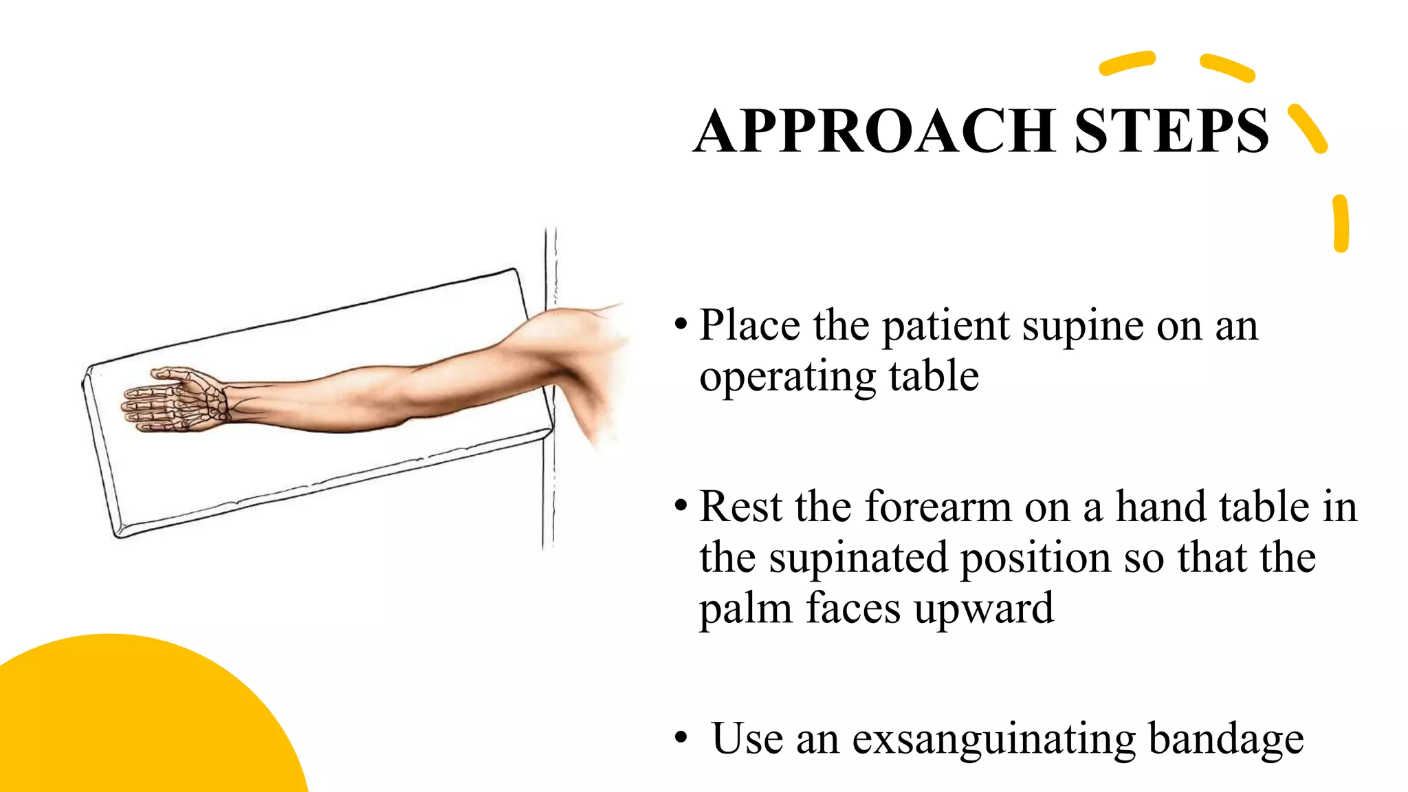
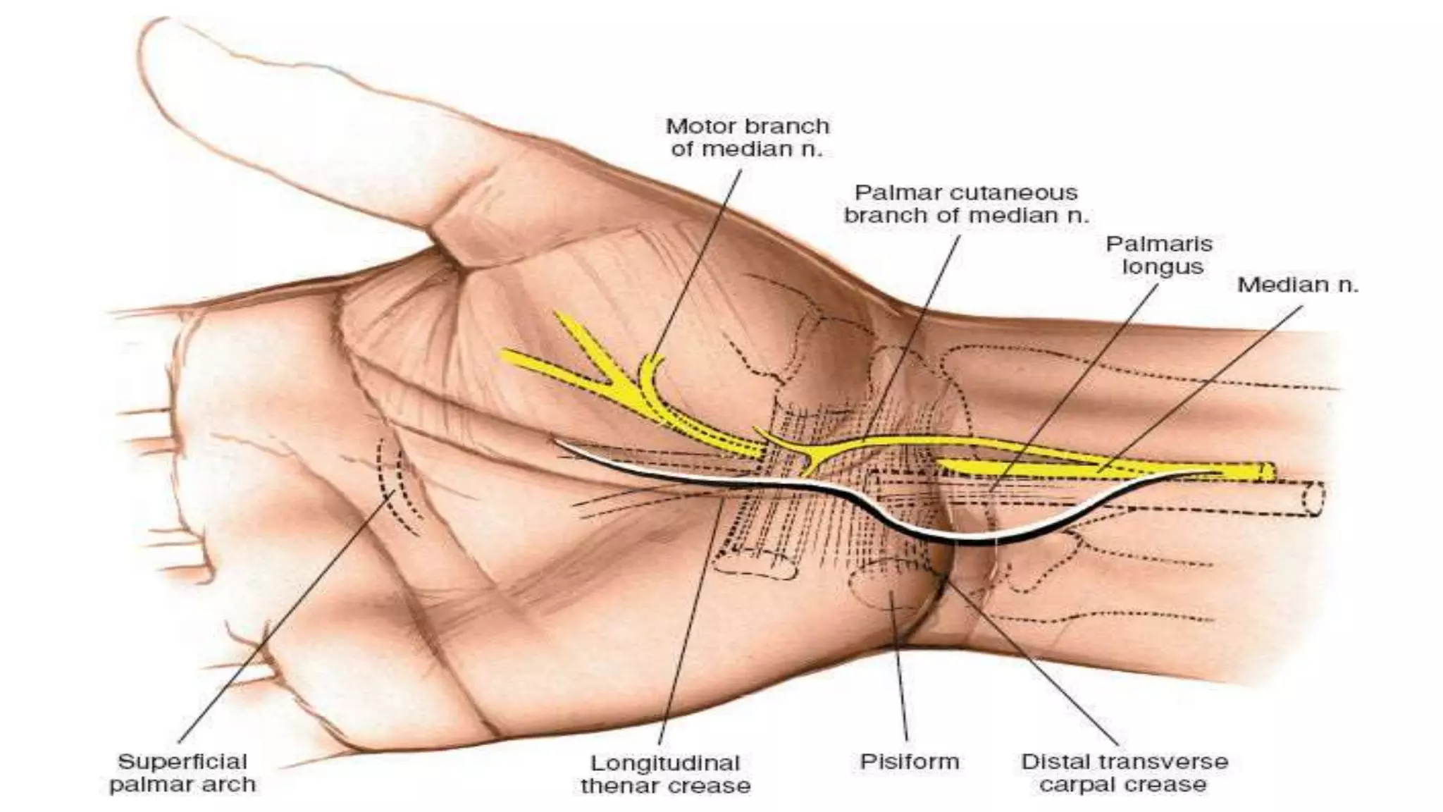
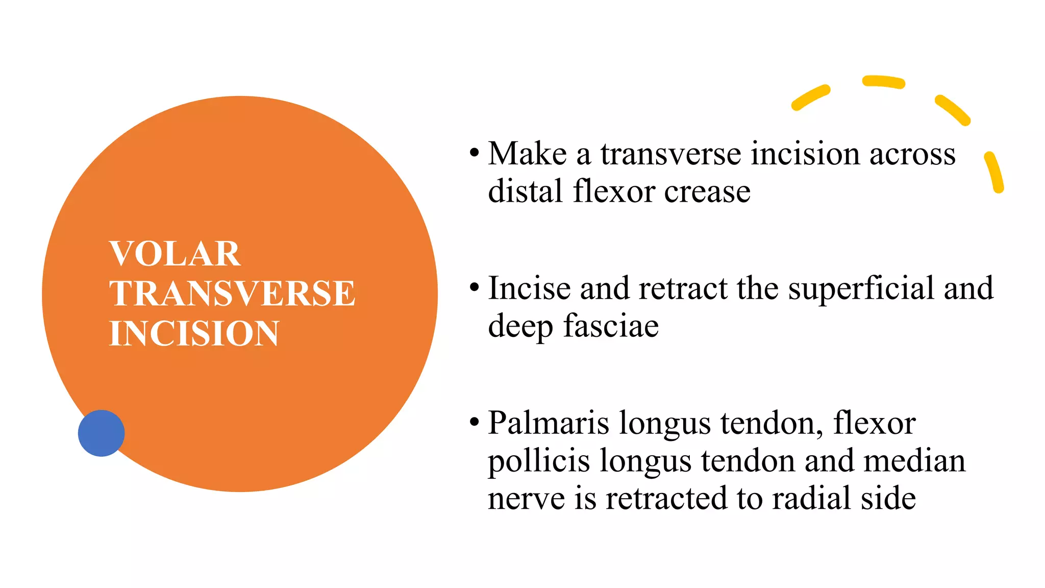
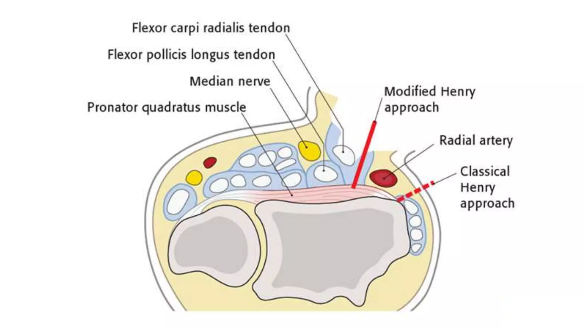
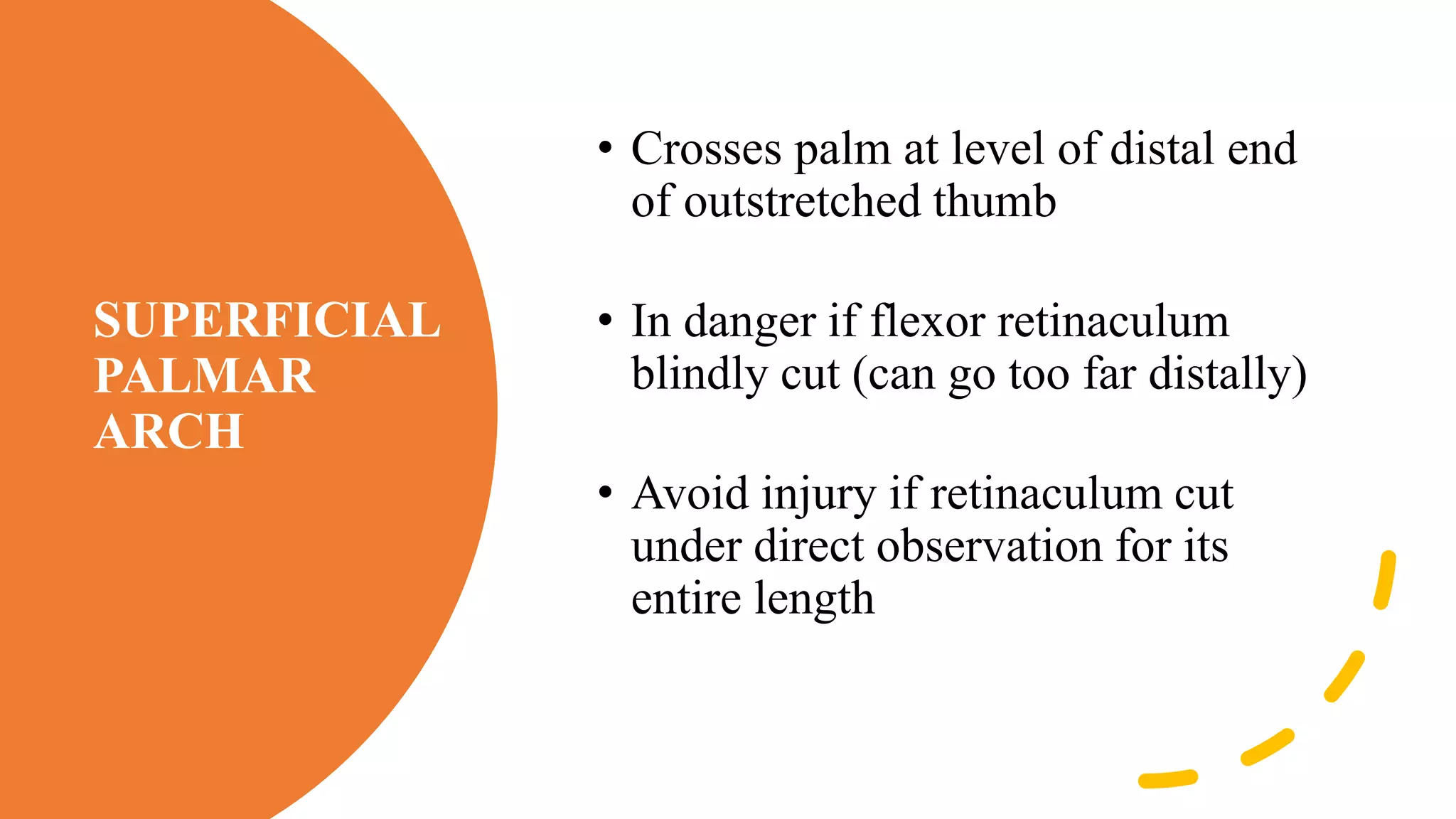
This document provides an overview of the volar approach to the wrist. It begins with an introduction and objectives. The indications for the volar approach include carpal tunnel release, tendon repair, and fixation of distal radius fractures. The approach steps are then described, including making a curved incision just ulnar to the thenar crease. Key structures at risk include the palmar cutaneous branch of the median nerve and the motor branch to the thenar muscles. Other volar approaches and an accompanying video are also mentioned.
3. OBJECTIVES
At the end of the class all the students
able to know
1.Indications
2.Volar approaches to the wrist in details
3.Structures at risk
4. INTRODUCTION
• The volar approach to the wrist is one of the most used approaches in
the wrist to address the distal end radius fracture fixation, carpal tunnel
release ,to tackle the carpal bones pathologies .
5. INDICATIONS
1. Decompression of median nerve
2. Flexor tendon synovectomy
3. Carpal tunnel tumor excision
4. Tendon repair
5. Drainage of sepsis tracking up from
the mid-palmar space
6. ORIF
6. APPROACH STEPS
• Place the patient supine on an
operating table
• Rest the forearm on a hand table in
the supinated position so that the
palm faces upward
• Use an exsanguinating bandage
7. • Begin the incision just to the
ulnar side of the thenar crease
• Curve it proximally, remaining
just to the ulnar side of the thenar
crease
• Then, curve the incision toward
the ulnar side of the forearm so
that the flexion crease is not
crossed transversely
8.
9. • The skin is retracted,
and the deep fascia and
tendon of the palmaris
longus are inspected
10. • The deep fascia is
incised The palmaris
longus is retracted
toward the ulna,
revealing the median
nerve as it enters the
carpal tunnel
11. The transverse carpal
ligament is released on the
ulnar side of the nerve to
avoid damage to the motor
branch of the thenar
muscle
12. The median nerve is
retracted radially and
the flexor tendons are
retracted toward the
ulna, revealing the
distal radius and joint
capsule. An incision
then is made into the
capsule to expose the
carpus.
13. VOLAR
TRANSVERSE
INCISION
• Make a transverse incision across
distal flexor crease
• Incise and retract the superficial and
deep fasciae
• Palmaris longus tendon, flexor
pollicis longus tendon and median
nerve is retracted to radial side
14. • Retract the flexor digitorum sublimis and profundus tendons to the
ulnar side
• Incise the joint capsule, exposing the distal end of the radius and the
lunate
15.
16. OTHER VOLAR APPROACHES
• The modified Henry approach
uses the plane between
flexor carpi radialis tendon and
the radial artery
17. • The classical Henry approach goes
between brachioradialis and the radial
artery, i.e., radial to the radial artery
• The main difference is in the modified
approach is ulnar to the radial artery
20. STRUCTURES AT RISK
PALMAR CUTANEOUS BRANCH OF MEDIAN NERVE
• Arises 5 cm proximal to wrist joint
• Runs ulnar to FCR before crossing flexor
retinaculum
• Greatest threat if the incision not curved ulnarly
21.
22. MOTOR BRANCH OF MEDIAN NERVE
• Risk minimized by incision made ulnar to median nerve
• The thenar motor branch (TMB) of the median nerve
(MN) - which is also referred to as the recurrent motor
branch of the median nerve
23. TMB motor innervation
to the thenar muscles
1.Opponens pollicis
2.Abductor pollicis brevis
3.Superficial part of flexor
pollicis brevis
24. SUPERFICIAL
PALMAR
ARCH
• Crosses palm at level of distal end
of outstretched thumb
• In danger if flexor retinaculum
blindly cut (can go too far distally)
• Avoid injury if retinaculum cut
under direct observation for its
entire length