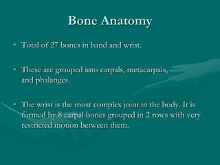
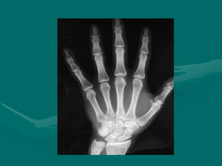
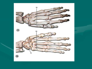
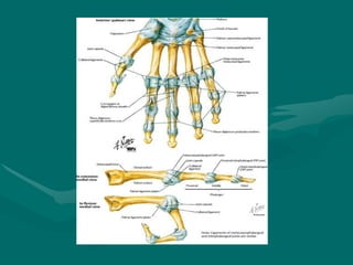
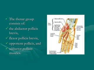
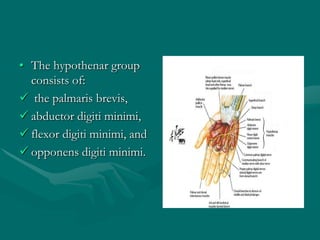
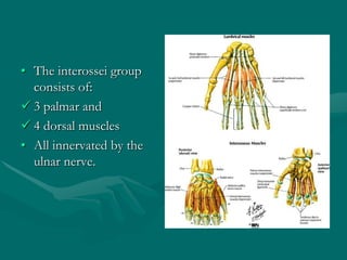
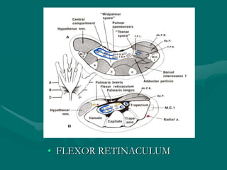
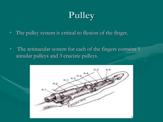
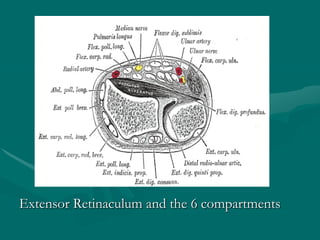
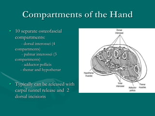
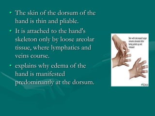
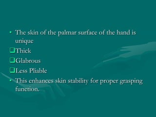
The hand and wrist have 27 bones organized into carpals, metacarpals, and phalanges. The wrist is a complex joint formed by 8 carpal bones with limited motion. Muscles are divided into intrinsic and extrinsic groups, with extrinsic muscles including wrist and digit flexors/extensors. Nerves include the median, ulnar, and radial nerves. The hand has a rich blood supply from the radial, ulnar, and deep palmar arches. Fascia including the flexor and extensor retinacula contain tendons. The hand's skin and pulley system enhance grasping.
These simplified slides by Dr. Sidra Arshad present an overview of the non-respiratory functions of the respiratory tract.
Learning objectives:
1. Enlist the non-respiratory functions of the respiratory tract
2. Briefly explain how these functions are carried out
3. Discuss the significance of dead space
4. Differentiate between minute ventilation and alveolar ventilation
5. Describe the cough and sneeze reflexes
Study Resources:
1. Chapter 39, Guyton and Hall Textbook of Medical Physiology, 14th edition
2. Chapter 34, Ganong’s Review of Medical Physiology, 26th edition
3. Chapter 17, Human Physiology by Lauralee Sherwood, 9th edition
4. Non-respiratory functions of the lungs https://academic.oup.com/bjaed/article/13/3/98/278874
These lecture slides, by Dr Sidra Arshad, offer a quick overview of physiological basis of a normal electrocardiogram.
Learning objectives:
1. Define an electrocardiogram (ECG) and electrocardiography
2. Describe how dipoles generated by the heart produce the waveforms of the ECG
3. Describe the components of a normal electrocardiogram of a typical bipolar leads (limb II)
4. Differentiate between intervals and segments
5. Enlist some common indications for obtaining an ECG
Study Resources:
1. Chapter 11, Guyton and Hall Textbook of Medical Physiology, 14th edition
2. Chapter 9, Human Physiology - From Cells to Systems, Lauralee Sherwood, 9th edition
3. Chapter 29, Ganong’s Review of Medical Physiology, 26th edition
4. Electrocardiogram, StatPearls - https://www.ncbi.nlm.nih.gov/books/NBK549803/
5. ECG in Medical Practice by ABM Abdullah, 4th edition
6. ECG Basics, http://www.nataliescasebook.com/tag/e-c-g-basics
Pulmonary Thromboembolism - etilogy, types, medical- Surgical and nursing man...VarunMahajani
Disruption of blood supply to lung alveoli due to blockage of one or more pulmonary blood vessels is called as Pulmonary thromboembolism. In this presentation we will discuss its causes, types and its management in depth.
Lung Cancer: Artificial Intelligence, Synergetics, Complex System Analysis, S...Oleg Kshivets
RESULTS: Overall life span (LS) was 2252.1±1742.5 days and cumulative 5-year survival (5YS) reached 73.2%, 10 years – 64.8%, 20 years – 42.5%. 513 LCP lived more than 5 years (LS=3124.6±1525.6 days), 148 LCP – more than 10 years (LS=5054.4±1504.1 days).199 LCP died because of LC (LS=562.7±374.5 days). 5YS of LCP after bi/lobectomies was significantly superior in comparison with LCP after pneumonectomies (78.1% vs.63.7%, P=0.00001 by log-rank test). AT significantly improved 5YS (66.3% vs. 34.8%) (P=0.00000 by log-rank test) only for LCP with N1-2. Cox modeling displayed that 5YS of LCP significantly depended on: phase transition (PT) early-invasive LC in terms of synergetics, PT N0—N12, cell ratio factors (ratio between cancer cells- CC and blood cells subpopulations), G1-3, histology, glucose, AT, blood cell circuit, prothrombin index, heparin tolerance, recalcification time (P=0.000-0.038). Neural networks, genetic algorithm selection and bootstrap simulation revealed relationships between 5YS and PT early-invasive LC (rank=1), PT N0—N12 (rank=2), thrombocytes/CC (3), erythrocytes/CC (4), eosinophils/CC (5), healthy cells/CC (6), lymphocytes/CC (7), segmented neutrophils/CC (8), stick neutrophils/CC (9), monocytes/CC (10); leucocytes/CC (11). Correct prediction of 5YS was 100% by neural networks computing (area under ROC curve=1.0; error=0.0).
CONCLUSIONS: 5YS of LCP after radical procedures significantly depended on: 1) PT early-invasive cancer; 2) PT N0--N12; 3) cell ratio factors; 4) blood cell circuit; 5) biochemical factors; 6) hemostasis system; 7) AT; 8) LC characteristics; 9) LC cell dynamics; 10) surgery type: lobectomy/pneumonectomy; 11) anthropometric data. Optimal diagnosis and treatment strategies for LC are: 1) screening and early detection of LC; 2) availability of experienced thoracic surgeons because of complexity of radical procedures; 3) aggressive en block surgery and adequate lymph node dissection for completeness; 4) precise prediction; 5) adjuvant chemoimmunoradiotherapy for LCP with unfavorable prognosis.
Explore natural remedies for syphilis treatment in Singapore. Discover alternative therapies, herbal remedies, and lifestyle changes that may complement conventional treatments. Learn about holistic approaches to managing syphilis symptoms and supporting overall health.
NVBDCP.pptx Nation vector borne disease control programSapna Thakur
NVBDCP was launched in 2003-2004 . Vector-Borne Disease: Disease that results from an infection transmitted to humans and other animals by blood-feeding arthropods, such as mosquitoes, ticks, and fleas. Examples of vector-borne diseases include Dengue fever, West Nile Virus, Lyme disease, and malaria.
ARTIFICIAL INTELLIGENCE IN HEALTHCARE.pdfAnujkumaranit
Artificial intelligence (AI) refers to the simulation of human intelligence processes by machines, especially computer systems. It encompasses tasks such as learning, reasoning, problem-solving, perception, and language understanding. AI technologies are revolutionizing various fields, from healthcare to finance, by enabling machines to perform tasks that typically require human intelligence.
New Directions in Targeted Therapeutic Approaches for Older Adults With Mantl...i3 Health
i3 Health is pleased to make the speaker slides from this activity available for use as a non-accredited self-study or teaching resource.
This slide deck presented by Dr. Kami Maddocks, Professor-Clinical in the Division of Hematology and
Associate Division Director for Ambulatory Operations
The Ohio State University Comprehensive Cancer Center, will provide insight into new directions in targeted therapeutic approaches for older adults with mantle cell lymphoma.
STATEMENT OF NEED
Mantle cell lymphoma (MCL) is a rare, aggressive B-cell non-Hodgkin lymphoma (NHL) accounting for 5% to 7% of all lymphomas. Its prognosis ranges from indolent disease that does not require treatment for years to very aggressive disease, which is associated with poor survival (Silkenstedt et al, 2021). Typically, MCL is diagnosed at advanced stage and in older patients who cannot tolerate intensive therapy (NCCN, 2022). Although recent advances have slightly increased remission rates, recurrence and relapse remain very common, leading to a median overall survival between 3 and 6 years (LLS, 2021). Though there are several effective options, progress is still needed towards establishing an accepted frontline approach for MCL (Castellino et al, 2022). Treatment selection and management of MCL are complicated by the heterogeneity of prognosis, advanced age and comorbidities of patients, and lack of an established standard approach for treatment, making it vital that clinicians be familiar with the latest research and advances in this area. In this activity chaired by Michael Wang, MD, Professor in the Department of Lymphoma & Myeloma at MD Anderson Cancer Center, expert faculty will discuss prognostic factors informing treatment, the promising results of recent trials in new therapeutic approaches, and the implications of treatment resistance in therapeutic selection for MCL.
Target Audience
Hematology/oncology fellows, attending faculty, and other health care professionals involved in the treatment of patients with mantle cell lymphoma (MCL).
Learning Objectives
1.) Identify clinical and biological prognostic factors that can guide treatment decision making for older adults with MCL
2.) Evaluate emerging data on targeted therapeutic approaches for treatment-naive and relapsed/refractory MCL and their applicability to older adults
3.) Assess mechanisms of resistance to targeted therapies for MCL and their implications for treatment selection
Title: Sense of Taste
Presenter: Dr. Faiza, Assistant Professor of Physiology
Qualifications:
MBBS (Best Graduate, AIMC Lahore)
FCPS Physiology
ICMT, CHPE, DHPE (STMU)
MPH (GC University, Faisalabad)
MBA (Virtual University of Pakistan)
Learning Objectives:
Describe the structure and function of taste buds.
Describe the relationship between the taste threshold and taste index of common substances.
Explain the chemical basis and signal transduction of taste perception for each type of primary taste sensation.
Recognize different abnormalities of taste perception and their causes.
Key Topics:
Significance of Taste Sensation:
Differentiation between pleasant and harmful food
Influence on behavior
Selection of food based on metabolic needs
Receptors of Taste:
Taste buds on the tongue
Influence of sense of smell, texture of food, and pain stimulation (e.g., by pepper)
Primary and Secondary Taste Sensations:
Primary taste sensations: Sweet, Sour, Salty, Bitter, Umami
Chemical basis and signal transduction mechanisms for each taste
Taste Threshold and Index:
Taste threshold values for Sweet (sucrose), Salty (NaCl), Sour (HCl), and Bitter (Quinine)
Taste index relationship: Inversely proportional to taste threshold
Taste Blindness:
Inability to taste certain substances, particularly thiourea compounds
Example: Phenylthiocarbamide
Structure and Function of Taste Buds:
Composition: Epithelial cells, Sustentacular/Supporting cells, Taste cells, Basal cells
Features: Taste pores, Taste hairs/microvilli, and Taste nerve fibers
Location of Taste Buds:
Found in papillae of the tongue (Fungiform, Circumvallate, Foliate)
Also present on the palate, tonsillar pillars, epiglottis, and proximal esophagus
Mechanism of Taste Stimulation:
Interaction of taste substances with receptors on microvilli
Signal transduction pathways for Umami, Sweet, Bitter, Sour, and Salty tastes
Taste Sensitivity and Adaptation:
Decrease in sensitivity with age
Rapid adaptation of taste sensation
Role of Saliva in Taste:
Dissolution of tastants to reach receptors
Washing away the stimulus
Taste Preferences and Aversions:
Mechanisms behind taste preference and aversion
Influence of receptors and neural pathways
Impact of Sensory Nerve Damage:
Degeneration of taste buds if the sensory nerve fiber is cut
Abnormalities of Taste Detection:
Conditions: Ageusia, Hypogeusia, Dysgeusia (parageusia)
Causes: Nerve damage, neurological disorders, infections, poor oral hygiene, adverse drug effects, deficiencies, aging, tobacco use, altered neurotransmitter levels
Neurotransmitters and Taste Threshold:
Effects of serotonin (5-HT) and norepinephrine (NE) on taste sensitivity
Supertasters:
25% of the population with heightened sensitivity to taste, especially bitterness
Increased number of fungiform papillae
Surat @ℂall @Girls ꧁❤8527049040❤꧂@ℂall @Girls Service Vip Top Model Safe
Hand anatomy new
1. Wrist and Hand Anatomy
Moderator: Dr. Asif Sultan
Presented by: Qazi Manaan
2. Overview
• The anatomy of the hand is complex, intricate,
and fascinating.
• Absolutely essential for our everyday functional
living.
3. Bone Anatomy
• Total of 27 bones in hand and wrist.
• These are grouped into carpals, metacarpals,
and phalanges.
• The wrist is the most complex joint in the body. It is
formed by 8 carpal bones grouped in 2 rows with very
restricted motion between them.
4.
5. • All carpal bones participate in wrist function
o except for the pisiform.
• The scaphoid serves as link between each row;
therefore, it is vulnerable to fractures.
• The distal row of carpal bones is strongly attached
to the base of the second and third metacarpals,
forming a fixed unit.
6. • The hand contains 5 metacarpal bone.
• First Metacarpal articulates proximally with the
trapezium.
• The other 4 metacarpals articulate with the
trapezoid, capitate, and hamate at the base.
• The hand contains 14 phalanges.
9. • All 4 distal carpal bones articulate with the
metacarpals at the carpometacarpal (CMC) joints.
• At the metacarpophalangeal joints, lateral motion is
limited by the collateral ligaments
10. • At the CMC joint, the volar plate is part of the joint
capsule that attaches only to the proximal phalanx,
allowing hyperextension.
• At the interphalangeal joints:
extension is limited by the volar plate, which attaches
to the phalanges at each side of the joint.
Radial and ulnar motion is restricted by collateral
ligaments.
13. • The muscles of the hand are divided into:
Intrinsic and
Extrinsic group.
14. Extrinsic extensors
• All extensors are extrinsic and supplied by radial
nerve.
• Except for the interosseous-lumbrical complex.
• Group contains:
3 wrist extensors
A larger group of thumb and digit extensors.
15. • The main extensors at wrist are:
The extensor carpi radialis brevis (ECRB)
extensor carpi radialis longus (ECRL)
extensor carpi ulnaris (ECU)
16. • At the digits, extension occurs due to:
The extensor digitorum communis,
extensor indicis proprius, and
extensor digiti minimi
17. • Extension at the thumb is bought about by:
The abductor pollicis longus,
extensor pollicis brevis, and
extensor pollicis longus
18. Extrinsic Flexors
• 3 wrist flexors
• A larger group of thumb and digit flexors
• Innervated by the median nerve
• Except for the FCU, and the FDP to the small and ring
finger, which are innervated by the ulnar nerve.
19. • The main Flexors at the wrist joint are:
The flexor carpi radialis
the flexor carpi ulnaris and
the palmaris longus
20. • The digital flexors include:
Flexor Digitorum Superficialis
Flexor Digitorum Profundus
Flexor Pollicis Longus
21. Intrinsics
• Situated totally within the hand
• Divided into 4 groups:
the thenar,
hypothenar,
lumbrical, and
interossei muscles.
22. • The thenar group
consists of:
the abductor pollicis
brevis,
flexor pollicis brevis,
opponens pollicis, and
adductor pollicis
muscles.
23. • The hypothenar group
consists of:
the palmaris brevis,
abductor digiti minimi,
flexor digiti minimi, and
opponens digiti minimi.
24. • The lumbrical muscles
contribute to
the flexion of the MCP
joints and
extension of the
interphalangeal joints.
25. • The interossei group
consists of:
3 palmar and
4 dorsal muscles
• All innervated by the
ulnar nerve.
30. Median nerve
• Crucial in the gripping mechanism by the thumb.
• It originates from the lateral and medial cords of the brachial
plexus (C5-T1).
• In the forearm, the motor branches supply the pronator teres,
flexor carpi radialis, palmaris longus, and flexor digitorum
superficialis muscles.
• The anterior interosseus branch innervates the flexor pollicis
longus, flexor digitorum profundus (radial part), and pronator
quadratus muscles.
31. • Palmar cutaneous branch provides sensation at the
thenar eminence. Also sensory digital branches
provide sensation to the radial three and a half
fingers and corresponding palm area
• As the median nerve passes through the carpal
tunnel, the recurrent motor branch innervates the
thenar muscles
• It also innervates the index and middle finger
lumbrical muscles
37. Ulnar nerve
• Controls fine movement of the fingers
• It originates at the medial cord of the
brachial plexus (C8-T1)
• Sensory to the:
hypothenar eminence
The palmar and dorsal ulnar one and a
half fingers
38. • The deep motor branch passes through the Guyon canal in
company with the ulnar artery. It innervates the
hypothenar muscles
all interossei,
the 2 ulnar lumbricals,
the adductor pollicis, and
the deep head of the flexor pollicis brevis
43. Radial nerve
• Innervates the wrist extensors.
• Originates from the posterior cord of the brachial
plexus (C6-8).
• At the elbow, motor branches innervate the
brachioradialis and extensor carpi radialis longus
muscles.
44. • At the proximal forearm, the radial nerve divides into
the superficial and deep branches.
• The deep posterior interosseous branch innervates all
the muscles in the extensor compartment.
• The superficial branch provides sensation at the radial
aspect of the dorsum of the hand, the dorsum of the
thumb, and the dorsum of the radial three and a half
digits proximal to the distal interphalangeal joints.
48. Radial Artery
• Course
• A superficial branch arises at the level of the wrist and
contributes to the superficial palmar arch.
• In the palm it gives off:
Princeps pollicis artery and
Radialis Indicis artery, before terminating in the deep
palmar arch.
49.
50. Ulnar Artery
• It travels into the hand through the Guyon
canal, where it divides into:
• the deep palmar branch and
• the superficial palmar branch.
51. Superficial Palmar Arch
• Lies directly deep to the palmar fascia.
• It gives rise to the:
volar common digital arteries and
multiple branches to intrinsic muscles and skin.
• Distally,the common digital arteries bifurcate into the
proper digital arteries.
52. Deep Palmar Arch
• Lies at the base of the metacarpals, deep to
the flexor tendons.
• From its convexity it gives off three
palmar metacarpal arteries.
• Dorsally it gives off three perforating
arteries.
• Recurrent branches arise from the
concavity to supply the carpal bones
53. Dorsal Carpal Arch
• Formed by:
the posterior interosseous artery and
a dorsal perforating branch of the anterior interosseous
artery.
• Dorsal metacarpal arteries arise from a dorsal carpal
arch
55. Veins
• Veins generally follow
the deep arterial system
as venae comitantes.
• A superficial venous
system also exists at the
dorsum of the hand
and contributes to the
cephalic and basilic
veins in the upper
extremity.
56. Deep Fascia
• The Flexor Retinaculum
• The Palmar Fascia
• Fibrous flexor sheaths in fingers
• The Extensor Retinaculum
58. • The Palmar fascia consists of resistant fibrous
tissue arranged in longitudinal, transverse,
oblique, and vertical fibers.
• The transverse fibers are concentrated in the
mid palm and web spaces and serve as pulleys
for the flexor tendons proximal to the digital
pulleys.
59. •The digits contain 2 fascial
bands of clinical importance:
the Grayson ligament and
the Cleland ligament
60. Pulley
• The pulley system is critical to flexion of the finger.
• The retinacular system for each of the fingers contains 5
annular pulleys and 3 cruciate pulleys.
61. • The thumb has 2 annular pulleys and 1
oblique pulley.
• The system supplies mechanical advantage
by maintaining the flexor tendons close to
the joint's axis of motion. In doing so, the
pulleys prevent bowstringing.
.
63. Compartments of the Hand
• 10 separate osteofascial
compartments:
- dorsal interossei (4
compartments)
- palmar interossei (3
compartments)
- adductor pollicis
- thenar and hypothenar
• Typically can be released with
carpal tunnel release and 2
dorsal incisions
66. • The skin of the dorsum of the
hand is thin and pliable.
• It is attached to the hand's
skeleton only by loose areolar
tissue, where lymphatics and
veins course.
• explains why edema of the
hand is manifested
predominantly at the dorsum.
67. • The skin of the palmar surface of the hand is
unique
Thick
Glabrous
Less Pliable
• This enhances skin stability for proper grasping
function.
68. • The skin is most firmly anchored to the deep
structures at the palmar creases.
• Blood supply is through numerous small, vertical
branches from the common digital vessels.
• High concentration of sensory nerve organs
essential to the hand's normal function.