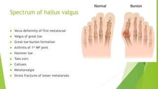
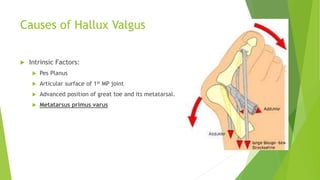
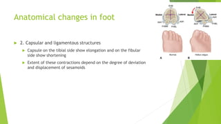
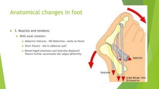
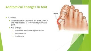
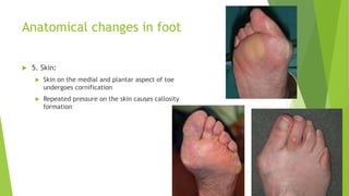
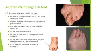
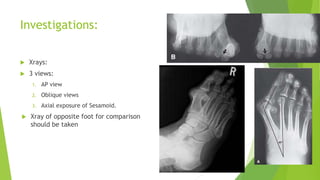
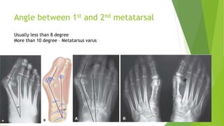
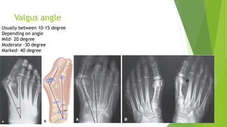
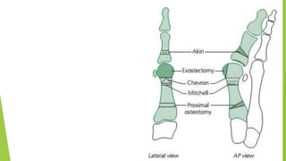
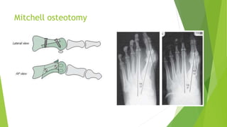
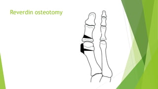
Hallux Valgus
Dr. Ankit Madharia
Junior resident
Department of Orthopaedics
Introduction
 Hallux valgus means lateral deviation of great toe
 Commonest of foot deformities
 Not a single disorder; but a complex deformity of the first ray
 Frequently accompanied by deformity and symptoms in lesser toes
Spectrum of hallux valgus
 Varus deformity of first metatarsal
 Valgus of great toe
 Great toe bunion formation
 Arthritis of 1st MP joint
 Hammer toe
 Toes corn
 Calluses
 Metatarsalgia
 Stress fractures of lesser metatarsals
Anatomy
 The head is grooved inferiorly
by medial and lateral sesamoid
bones in the tendons of Flexor
hallucis brevis
 The proximal phalynx is round
on three sides but flat
inferiorly, even concave
inferiorly for the flexor hallucis
longus tendon
Causes of Hallux Valgus
 Congenital Hallus valgus :
 Very rare
 Metatarsus primus varus is common at birth
 Concealed supernumerary phalynx may be the cause
 Shortened second metatarsal
 Hereditory :
 Johnston(1956) : Transmitted as autosomal dominance with incomplete
penetration
 Age:
 14-16 in girls and little later in boys
 Symptomatic hallux valgus is more common after 40 years of age, mostly
bilateral.
Causes of Hallux Valgus
 Sex:
 Women > Men
 Women : Men = 50: 1
 Predisposed by pointed compressive high heeled shoes
Causes of Hallux Valgus
 Shoes:
 Has been considered as extrinsic cause of Hallux Valgus
 In Chinese population, Hallux valgus was more common due to special
compressive shoe wearing habits.
 Tight fitting show at toes have been proven as cause of bunion, corns.
“Shoes as at present worn, serve but to
deform the toes and cover the feet with
corns” – Camper (1781)
Causes of Hallux Valgus
 Intrinsic Factors:
 Pes Planus
 Articular surface of 1st MP joint
 Advanced position of great toe and its metatarsal.
 Metatarsus primus varus
Causes of Hallux Valgus
 Intrinsic Factors:
 Pes Planus
 Articular surface of 1st MP joint
 Advanced position of great toe and its metatarsal.
 Metatarsus primus varus
Causes of Hallux Valgus
 Intrinsic Factors:
 Pes Planus
 Articular surface of 1st MP joint
 Advanced position of great toe and its metatarsal.
 Metatarsus primus varus
Causes of Hallux Valgus
 Intrinsic Factors:
 Pes Planus
 Articular surface of 1st MP joint
 Advanced position of great toe and its metatarsal.
 Metatarsus primus varus
Causes of Hallux Valgus
 Miscellaneous causes:
 Muscular imbalance: Poliomyelitis, Spastic CP
 Trauma : Malunited # - secondary deformity
 Congenitally absent, Amputated or hammered second toe
Pathogenesis
 1st metatarsal inclines medially
 Proximal phalynx deflects in opposite direction
 Joint knuckles towards the midline –
prominence of forefoot at the tibial border
 Great toe pronates – Nail and hallux slants
medially, pulp towards second toe
 Great toe may ride over 2nd digit or slip under
it
 Lesser toe – crowded together, clawed,
hammered
 Bunionette:- deformity at outer border of
forefoot
 Forefoot is splayed
Anatomical changes in foot
 In a full blown hallux valgus, several changes take place in and around
the first metatarso-phalyngeal joint. They Involve
1. Articular Bones
2. Capsular and ligamentous structure
3. Muscle and tendon
4. Bursa
5. Skin
Anatomical changes in foot
 1. Articular bones
 Mild cases: Outward deviation of proximal phalynx is the sole feature
 Severe deformity: Axial rotation of proximal phalynx
: Subluxation of MP joint.
 Crista on the undersurface of 1st Metatarsal smoothens out, effaced due
to migration of sesamoid.
Anatomical changes in foot
 In more advanced cases, the interior of metatarsal head is
cystic due to proliferation of marrow connective tissue in
response to denuded hyaline surface
 Sesamoids: Lateral migration of Sesamoids is evidence of
Hallux valgus
In these new incongruent location, sesamoids wear out, loose
hyaline cartilage, become mushroomed, forms spurs and
fragments
Incarcerted in the first inter metatarsal space, the fibular
sesamoid may serve as a wedge and push the 1st metatarsal into
greater varus
Rarely, there is bony union between sesamoids and the
metatarsal head.
Anatomical changes in foot
 2. Capsular and ligamentous structures
 Capsule on the tibial side show elongation and on the fibular
side show shortening
 Extent of these contractions depend on the degree of deviation
and displacement of sesamoids
Anatomical changes in foot
 3. Muscles and tendons:
 With axial rotation:
 Abductor halluces – NO Abduction, works as flexor
 Short flexors – Aid in adductor pull’
 Bowstringed extensors and laterally displaced
flexors further accentuste the valgus deformity
Anatomical changes in foot
 4. Bursa:
 Adventitious bursa occurs on the dorsal, plantar
and medial aspect of 1st metatarso phalyngeal
joint.
 May Undergo
1. Suppurative bursitis with regional cellulitis
2. Sinus formation
3. lymphangitis
Anatomical changes in foot
 5. Skin:
 Skin on the medial and plantar aspect of toe
undergoes cornification
 Repeated pressure on the skin causes callosity
formation
Anatomical changes in foot
 6. Changes affecting the lesser toes
 Relative or real plantar descent of the central
metatarsal heads
 Proximal phalynx subluxates dorsally with PIP
joint in flexion
 Skin over these knuckled IP joint develops
callosities
 2nd toe is usually hammered.
 Splaying of foot: Side to side span of foot is
increased.
 5th metatarsal inclines fibularwards, with its
head presenting as lateral eminence
 Bursa over this eminence is known as
Bunionette.
Clinical features
 Symptoms:
 Pain in foot
 Difficulty in being fitted with shoes
 Gait changes
 Corns
 Keratosis
 Cosmetic deformity
Pain in foot
 Bursitis:
 Bursa develops between skin and medial eminence
 Complications like inflammation result in regional cellulitis,
Rupture and pus discharge through sinus
 May also lead to osteomyelitis of the adjacent bones
 Arthritic pain:
 Movement of toes are limited with crepitus and painful movement
 Metatarsalgia:
 Most distressing and disabling
 Frequently associated with hallux valgus
Investigations:
 Xrays:
 3 views:
1. AP view
2. Oblique views
3. Axial exposure of Sesamoid.
 Xray of opposite foot for comparison
should be taken
Angle between 1st and 2nd metatarsal
Usually less than 8 degree
More than 10 degree – Metatarsus varus
Valgus angle
Usually between 10-15 degree
Depending on angle
Mild- 20 degree
Moderate -30 degree
Marked- 40 degree
Classification of hallux valgus
 Pigott (1960) classified HV into 3 types based on congruity of 1st MP
joint
 Type 1: Congrous joint
 Type 2: Deviated non congruous joint
 Type 3: Subluxated joint
Classification of hallux valgus
 Mann and conghlin(1993) classified HV into 3 types based on Hallux
valgus angle
 Mild: Angle < 20 degree, intermetatarsal angle usually less than 11 degree
 Moderate: Angle 20 - 40 degree, intermetatarsal angle between 11 and 18
degree
 Severe: Angle > 40 degree, intermetatarsal angle > 16-18 degree
Classification of hallux valgus
 From surgical point of view , it can be classified as
1. Simple hallux valgus
1. Without sagittal groove
2. With sagittal grove
2. Hallux valgus with axial rotation
1. Reducible
2. Irreducible
3. Hallux valgus with metatarsus primus varus
1. Mobile/ hypermobile first metatarsal
2. Fixed varus
4. Hallux varus with degenerative arthritis of joint
5. Hallux valgus with mixed deformities
Treatment of hallux valgus
 Conservative Management:
 Young and asymptomatic patients
• Proper fitting shoes with wide deep toe boxes
• Night splinting and other orthosis
 Once the deformity is established, it is difficult
to check the progression of disease by
conservative measures.
Surgical Treatment
 Indications of operative treatment
 Failure of non operative measures
 Persistent pain that interferes with daily work
 Severe deformity and pain unlikely to respond to conservative
measures
Surgical
procedures
Soft tissue
procedures
Modifies
McBride
bunionectomy
Combined bony
and soft tissue
procedures
Keller resection
arthroplasty
Modified Keller
technique
Bony procedures
Metatarsal
osteotomy
Distal
metatarsal
osteotomy
Mitchell
osteotomy
Distal chevron
osteotomy
Reverdin
osteotomy
Proximal
metatarsal
osteotomy
Proximal
crescentic
osteotomy
Proximal
chevron
osteotomy
Osteotomy of
medial
cuneiform
Proximal
phalynx
osteotomy
Arthrodesis of
1st MP joint
Arthrodesis of
1st metatarso
cuneiform joint
Keller resection arthroplasty
Mitchell osteotomy
Chevron osteotomy
Reverdin osteotomy
Proximal crescentic osteotomy
Medial cuneiform osteotomy
Proximal phalynx osteotomy (akin)
Mann Algorithm for selection of appropriate
operative procedure in treatment of hallux
valgus
 Hallux valgus < 25 degree
 Congruent joint
 Chevron osteotomy
 Mitchell Osteotomy
 Incongruent joint
 Distal soft tissue realignment
 Chevron osteotomy
 Mitchell Osteotomy
Mann Algorithm for selection of appropriate
operative procedure in treatment of hallux
valgus
 Hallux valgus 25 - 40 degree
 Congruent joint
 Chevron osteotomy with akin procedure
 Mitchell Osteotomy
 Incongruent joint
 Distal soft tissue realignment with proximal metatarsal osteotomy
 Mitchell Osteotomy
Mann Algorithm for selection of appropriate
operative procedure in treatment of hallux
valgus
 Severe Hallux valgus >40 degree
 Congruent joint
 Double osteotomy
 Akin and chevron osteotomy
 Akin and 1st metatarsal proximal osteotomy
 Akin and 1st cuneiform opening wedge osteotomy
 Incongruent joint
 Distal soft tissue realignment with Proximal osteotomy
 First metatarsal crescentic osteotomy
 1st cuneiform open wedge osteotomy
 Hypermobile 1st Metatarso-cuneiform joint
 Distal soft tissue realignment and fusion 1st metatarso-cuneiform joint
Conclusion
 Hallux valgus is the most common deformity of foot
 Commonly seen an adolescent females and becomes symptomatic in
middle age
 Can be treated conservatively if diagnosed early
 Surgery is the only option after the deformity develops.
Thankyou

Hallux valgus

  • 1.
    Hallux Valgus Dr. AnkitMadharia Junior resident Department of Orthopaedics
  • 2.
    Introduction  Hallux valgusmeans lateral deviation of great toe  Commonest of foot deformities  Not a single disorder; but a complex deformity of the first ray  Frequently accompanied by deformity and symptoms in lesser toes
  • 3.
    Spectrum of halluxvalgus  Varus deformity of first metatarsal  Valgus of great toe  Great toe bunion formation  Arthritis of 1st MP joint  Hammer toe  Toes corn  Calluses  Metatarsalgia  Stress fractures of lesser metatarsals
  • 4.
    Anatomy  The headis grooved inferiorly by medial and lateral sesamoid bones in the tendons of Flexor hallucis brevis  The proximal phalynx is round on three sides but flat inferiorly, even concave inferiorly for the flexor hallucis longus tendon
  • 5.
    Causes of HalluxValgus  Congenital Hallus valgus :  Very rare  Metatarsus primus varus is common at birth  Concealed supernumerary phalynx may be the cause  Shortened second metatarsal  Hereditory :  Johnston(1956) : Transmitted as autosomal dominance with incomplete penetration  Age:  14-16 in girls and little later in boys  Symptomatic hallux valgus is more common after 40 years of age, mostly bilateral.
  • 6.
    Causes of HalluxValgus  Sex:  Women > Men  Women : Men = 50: 1  Predisposed by pointed compressive high heeled shoes
  • 7.
    Causes of HalluxValgus  Shoes:  Has been considered as extrinsic cause of Hallux Valgus  In Chinese population, Hallux valgus was more common due to special compressive shoe wearing habits.  Tight fitting show at toes have been proven as cause of bunion, corns. “Shoes as at present worn, serve but to deform the toes and cover the feet with corns” – Camper (1781)
  • 8.
    Causes of HalluxValgus  Intrinsic Factors:  Pes Planus  Articular surface of 1st MP joint  Advanced position of great toe and its metatarsal.  Metatarsus primus varus
  • 9.
    Causes of HalluxValgus  Intrinsic Factors:  Pes Planus  Articular surface of 1st MP joint  Advanced position of great toe and its metatarsal.  Metatarsus primus varus
  • 10.
    Causes of HalluxValgus  Intrinsic Factors:  Pes Planus  Articular surface of 1st MP joint  Advanced position of great toe and its metatarsal.  Metatarsus primus varus
  • 11.
    Causes of HalluxValgus  Intrinsic Factors:  Pes Planus  Articular surface of 1st MP joint  Advanced position of great toe and its metatarsal.  Metatarsus primus varus
  • 12.
    Causes of HalluxValgus  Miscellaneous causes:  Muscular imbalance: Poliomyelitis, Spastic CP  Trauma : Malunited # - secondary deformity  Congenitally absent, Amputated or hammered second toe
  • 13.
    Pathogenesis  1st metatarsalinclines medially  Proximal phalynx deflects in opposite direction  Joint knuckles towards the midline – prominence of forefoot at the tibial border  Great toe pronates – Nail and hallux slants medially, pulp towards second toe  Great toe may ride over 2nd digit or slip under it  Lesser toe – crowded together, clawed, hammered  Bunionette:- deformity at outer border of forefoot  Forefoot is splayed
  • 14.
    Anatomical changes infoot  In a full blown hallux valgus, several changes take place in and around the first metatarso-phalyngeal joint. They Involve 1. Articular Bones 2. Capsular and ligamentous structure 3. Muscle and tendon 4. Bursa 5. Skin
  • 15.
    Anatomical changes infoot  1. Articular bones  Mild cases: Outward deviation of proximal phalynx is the sole feature  Severe deformity: Axial rotation of proximal phalynx : Subluxation of MP joint.  Crista on the undersurface of 1st Metatarsal smoothens out, effaced due to migration of sesamoid.
  • 16.
    Anatomical changes infoot  In more advanced cases, the interior of metatarsal head is cystic due to proliferation of marrow connective tissue in response to denuded hyaline surface  Sesamoids: Lateral migration of Sesamoids is evidence of Hallux valgus In these new incongruent location, sesamoids wear out, loose hyaline cartilage, become mushroomed, forms spurs and fragments Incarcerted in the first inter metatarsal space, the fibular sesamoid may serve as a wedge and push the 1st metatarsal into greater varus Rarely, there is bony union between sesamoids and the metatarsal head.
  • 17.
    Anatomical changes infoot  2. Capsular and ligamentous structures  Capsule on the tibial side show elongation and on the fibular side show shortening  Extent of these contractions depend on the degree of deviation and displacement of sesamoids
  • 18.
    Anatomical changes infoot  3. Muscles and tendons:  With axial rotation:  Abductor halluces – NO Abduction, works as flexor  Short flexors – Aid in adductor pull’  Bowstringed extensors and laterally displaced flexors further accentuste the valgus deformity
  • 19.
    Anatomical changes infoot  4. Bursa:  Adventitious bursa occurs on the dorsal, plantar and medial aspect of 1st metatarso phalyngeal joint.  May Undergo 1. Suppurative bursitis with regional cellulitis 2. Sinus formation 3. lymphangitis
  • 20.
    Anatomical changes infoot  5. Skin:  Skin on the medial and plantar aspect of toe undergoes cornification  Repeated pressure on the skin causes callosity formation
  • 21.
    Anatomical changes infoot  6. Changes affecting the lesser toes  Relative or real plantar descent of the central metatarsal heads  Proximal phalynx subluxates dorsally with PIP joint in flexion  Skin over these knuckled IP joint develops callosities  2nd toe is usually hammered.  Splaying of foot: Side to side span of foot is increased.  5th metatarsal inclines fibularwards, with its head presenting as lateral eminence  Bursa over this eminence is known as Bunionette.
  • 22.
    Clinical features  Symptoms: Pain in foot  Difficulty in being fitted with shoes  Gait changes  Corns  Keratosis  Cosmetic deformity
  • 23.
    Pain in foot Bursitis:  Bursa develops between skin and medial eminence  Complications like inflammation result in regional cellulitis, Rupture and pus discharge through sinus  May also lead to osteomyelitis of the adjacent bones  Arthritic pain:  Movement of toes are limited with crepitus and painful movement  Metatarsalgia:  Most distressing and disabling  Frequently associated with hallux valgus
  • 24.
    Investigations:  Xrays:  3views: 1. AP view 2. Oblique views 3. Axial exposure of Sesamoid.  Xray of opposite foot for comparison should be taken
  • 25.
    Angle between 1stand 2nd metatarsal Usually less than 8 degree More than 10 degree – Metatarsus varus
  • 26.
    Valgus angle Usually between10-15 degree Depending on angle Mild- 20 degree Moderate -30 degree Marked- 40 degree
  • 27.
    Classification of halluxvalgus  Pigott (1960) classified HV into 3 types based on congruity of 1st MP joint  Type 1: Congrous joint  Type 2: Deviated non congruous joint  Type 3: Subluxated joint
  • 28.
    Classification of halluxvalgus  Mann and conghlin(1993) classified HV into 3 types based on Hallux valgus angle  Mild: Angle < 20 degree, intermetatarsal angle usually less than 11 degree  Moderate: Angle 20 - 40 degree, intermetatarsal angle between 11 and 18 degree  Severe: Angle > 40 degree, intermetatarsal angle > 16-18 degree
  • 29.
    Classification of halluxvalgus  From surgical point of view , it can be classified as 1. Simple hallux valgus 1. Without sagittal groove 2. With sagittal grove 2. Hallux valgus with axial rotation 1. Reducible 2. Irreducible 3. Hallux valgus with metatarsus primus varus 1. Mobile/ hypermobile first metatarsal 2. Fixed varus 4. Hallux varus with degenerative arthritis of joint 5. Hallux valgus with mixed deformities
  • 30.
    Treatment of halluxvalgus  Conservative Management:  Young and asymptomatic patients • Proper fitting shoes with wide deep toe boxes • Night splinting and other orthosis  Once the deformity is established, it is difficult to check the progression of disease by conservative measures.
  • 31.
    Surgical Treatment  Indicationsof operative treatment  Failure of non operative measures  Persistent pain that interferes with daily work  Severe deformity and pain unlikely to respond to conservative measures
  • 32.
    Surgical procedures Soft tissue procedures Modifies McBride bunionectomy Combined bony andsoft tissue procedures Keller resection arthroplasty Modified Keller technique Bony procedures Metatarsal osteotomy Distal metatarsal osteotomy Mitchell osteotomy Distal chevron osteotomy Reverdin osteotomy Proximal metatarsal osteotomy Proximal crescentic osteotomy Proximal chevron osteotomy Osteotomy of medial cuneiform Proximal phalynx osteotomy Arthrodesis of 1st MP joint Arthrodesis of 1st metatarso cuneiform joint
  • 34.
  • 35.
  • 36.
  • 37.
  • 38.
  • 39.
  • 40.
  • 41.
    Mann Algorithm forselection of appropriate operative procedure in treatment of hallux valgus  Hallux valgus < 25 degree  Congruent joint  Chevron osteotomy  Mitchell Osteotomy  Incongruent joint  Distal soft tissue realignment  Chevron osteotomy  Mitchell Osteotomy
  • 42.
    Mann Algorithm forselection of appropriate operative procedure in treatment of hallux valgus  Hallux valgus 25 - 40 degree  Congruent joint  Chevron osteotomy with akin procedure  Mitchell Osteotomy  Incongruent joint  Distal soft tissue realignment with proximal metatarsal osteotomy  Mitchell Osteotomy
  • 43.
    Mann Algorithm forselection of appropriate operative procedure in treatment of hallux valgus  Severe Hallux valgus >40 degree  Congruent joint  Double osteotomy  Akin and chevron osteotomy  Akin and 1st metatarsal proximal osteotomy  Akin and 1st cuneiform opening wedge osteotomy  Incongruent joint  Distal soft tissue realignment with Proximal osteotomy  First metatarsal crescentic osteotomy  1st cuneiform open wedge osteotomy  Hypermobile 1st Metatarso-cuneiform joint  Distal soft tissue realignment and fusion 1st metatarso-cuneiform joint
  • 44.
    Conclusion  Hallux valgusis the most common deformity of foot  Commonly seen an adolescent females and becomes symptomatic in middle age  Can be treated conservatively if diagnosed early  Surgery is the only option after the deformity develops.
  • 45.

Editor's Notes

  • #4 Insert images for each
  • #5 The first metatarsal is large and strong and plays an important role in supporting the weight of the body
  • #9  Pes planus: Owing to eversion of calcaneus , the Abductor hallucis shifts towards the outer border of the foot Plantarly depressed tarsal bones bear down on the AbH muscle , and causes it to stretch and lose its tone. Weakened abductors no longer counterbalance the adductors halluces Pes planus causes the abductirs to be non functional
  • #10 Distal metatarsal Articular angle : 10-15 degree Phalyngeal articular angle. 7-10 degree
  • #11 Presence of comparatively long first ray is considered as the cause of HV
  • #12 MPV predisposed and provokes symptomatic HV Oblique setting of medial cuneiform –metatarsal joint is seen in cases of HV Point of debate
  • #38 Close wedge intracapsular osteotomy of 1st distal metatarsal