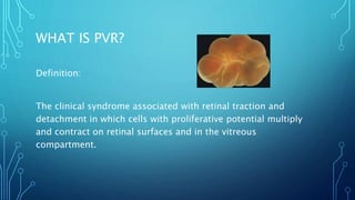
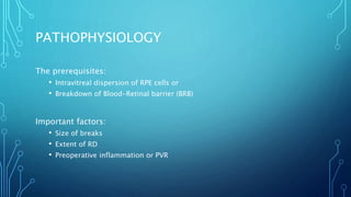
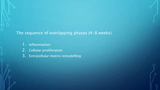
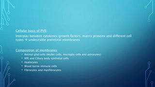
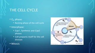
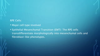
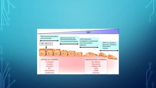
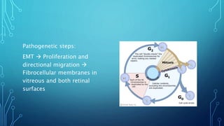
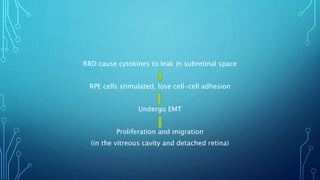
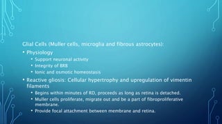
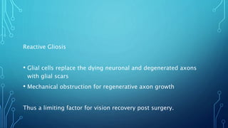
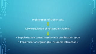
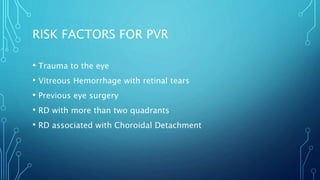
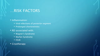
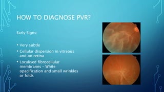
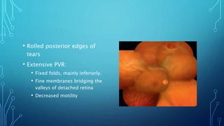
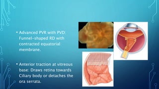
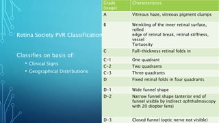
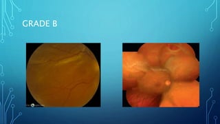
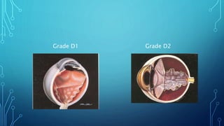
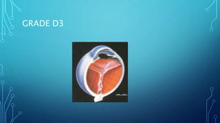
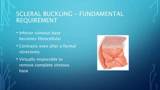
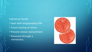
SlideShare a Scribd company logo
PROLIFERATIVE
VITREORETINOPATHY
DR. PAVAN SHROFF
WHAT IS PVR?
Definition:
The clinical syndrome associated with retinal traction and
detachment in which cells with proliferative potential multiply
and contract on retinal surfaces and in the vitreous
compartment.
• Vitreoretinal wound-healing response
• Nonangiogenic and fibrotic
• Seen in:
• Rhegmatogenous Retinal Detachment (RRD)
• Surgical intervention (most common scenario)
• Trauma
• Longstanding inflammation
• Incidence: 5-10% of all RRD
• Higher incidence, rapid and aggressive in children
• Most common cause of failure of RRD surgeries
PATHOPHYSIOLOGY
The prerequisites:
• Intravitreal dispersion of RPE cells or
• Breakdown of Blood-Retinal barrier (BRB)
Important factors:
• Size of breaks
• Extent of RD
• Preoperative inflammation or PVR
The sequence of overlapping phases (4-8 weeks)
1. Inflammation
2. Cellular proliferation
3. Extracellular matrix remodelling
Cellular basis of PVR:
Interplay between cytokines/growth factors, matrix proteins and different cell
types  undesirable preretinal membranes
Composition of membranes:
• Retinal glial cells (Muller cells, microglia cells and astrocytes)
• RPE and Ciliary body epithelial cells
• Hyalocytes
• Blood borne immune cells
• Fibrocytes and myofibrocytes
THE CELL CYCLE
• G0 phase:
• Resting phase of the cell cycle
• Interphase:
• Gap1, Synthesis and Gap2
phases
• Cell prepares itself for the cell
division
• Mitosis
RPE Cells:
• Major cell type involved
• Epithelial Mesenchymal Transition (EMT): The RPE cells
transdifferentiate morphologically into mesenchymal cells and
fibroblast-like phenotypes.
Pathogenetic steps:
EMT  Proliferation and
directional migration 
Fibrocellular membranes in
vitreous and both retinal
surfaces
RRD cause cytokines to leak in subretinal space
RPE cells stimulated, lose cell-cell adhesion
Undergo EMT
Proliferation and migration
(in the vitreous cavity and detached retina)
Glial Cells (Muller cells, microglia and fibrous astrocytes):
• Physiology
• Support neuronal activity
• Integrity of BRB
• Ionic and osmotic homeostasis
• Reactive gliosis: Cellular hypertrophy and upregulation of vimentin
filaments
• Begins within minutes of RD, proceeds as long as retina is detached.
• Muller cells proliferate, migrate out and be a part of fibroproliferative
membrane.
• Provide focal attachment between membrane and retina.
Reactive Gliosis
• Glial cells replace the dying neuronal and degenerated axons
with glial scars
• Mechanical obstruction for regenerative axon growth
Thus a limiting factor for vision recovery post surgery.
Proliferation of Muller cells
Downregulation of Potassium channels
• Depolarisation causes reentry into proliferation cycle
• Impairment of regular glial-neuronal interactions
Blood borne cells:
• Macrophages and circulating fibrocytes are precursors of
myofibroblasts
• Hyalocytes have a role in synthesis of ECM and modulation of
inflammation.
• Hyalocytes also have contractile properties
GROWTH FACTORS, CYTOKINES AND
INTERLEUKINS
• Platelet Derived Growth Factor
(PDGF)
• Transforming Growth Factor-B
(TGF-B)
• Insulin-like Growth Factors (IGF)
• Monocyte Chemotactic Protein-1
(MCP-1)
• Basic Fibroblast Growth Factor
(bFGF)
• Hepatocyte Growth Factor (HGF)
• Connective Tissue Growth Factor
(CTGF)
• Epidermal Growth Factor (EGF)
• Vascular Endothelial Growth
Factor (VEGF)
• Interleukins and TNF-alpha
ECM Remodelling and Myofibroblasts:
• ECM – A dynamic, complex array of collagens, glycoproteins, GAGs
and proteoglycans
• Functions:
• Mechanical and structural support
• Interacts with cytokines and cytoskeleton – transmits biological signals.
• Re-modelling is a healing response detrimental to the retinal
function.
HOW REMODELLING HAPPENS
1. Structural proteins
2. Adhesive proteins
3. Antiadhesive proteins
Structural Proteins:
• Collagens of various types.
• Produced by RPE cells, glial cells and fibroblast-like cells
Adhesion protein:
• Fibronectin, expression is low in normal retina.
• Promotes adhesion amongst ECM cells.
• Also enhances EMT of RPE cells.
Antiadhesive protein:
• Thrombospondin functions contrary to Fibronectin
• Facilitate the detachment of activated RPE cells and migrate to wounded
areas.
The loss of Balance:
• Normally, MMPs and Tissue Inhibitor of MMP (TIMPs) function in
balance.
• In pathology, RPE cells, glial cells and fibroblasts overproduce
MMP-2 and MMP-9.
Myofibroblasts:
• Characterized by smooth-muscle actin and generate Tractional
force by contraction
• Transmembrane integrins at the myofibroblast surface link
actin microfilaments
• Cytokines, PDGF, IGF and TNF-B mediate the ECM contraction.
BIOMARKERS AND GENETIC PROFILING
• MMP concentration in vitreous is raised
• Chemokine CXCL-1 correlates with the grade of PVR
• Inflammation associated proteins:
• Alpha 1-Antitrypsin
• Apolipoprotein A-IV
• Transferrin
• Kininogen-1 – Serum biomarker of PVR
• Laser flare photometry – Risk estimation for PVR development.
• Genetic association – TNF locus and PVR
RISK FACTORS FOR PVR
• Trauma to the eye
• Vitreous Hemorrhage with retinal tears
• Previous eye surgery
• RD with more than two quadrants
• RD associated with Choroidal Detachment
…RISK FACTORS
• Inflammation:
• Viral infections of posterior segment
• Prolonged chorioretinitis
• RD associated with:
• Wagner’s Syndrome
• Marfan Syndrome
• FEVR
• Cryotherapy
PREDICTION OF PVR
• Asaria et al presented a formula based on risk factors
Preoperative Grade B PVR 1.85+
Preoperative Grade C PVR 2.88+
Aphakia 2.92+
Each quadrant of RD 1.23+
Anterior Uveitis 1.77+
Vitreous Haemorrhage 0.83+
Previous Cryotherapy 1.23+
Score of >6.33+ is a high risk for PDR
HOW TO DIAGNOSE PVR?
Early Signs:
• Very subtle
• Cellular dispersion in vitreous
and on retina
• Localised fibrocellular
membranes – White
opacification and small wrinkles
or folds
• Rolled posterior edges of
tears
• Extensive PVR:
• Fixed folds, mainly inferiorly.
• Fine membranes bridging the
valleys of detached retina
• Decreased motility
• Advanced PVR with PVD:
Funnel-shaped RD with
contracted equatorial
membrane.
• Anterior traction at vitreous
base: Draws retina towards
Ciliary body or detaches the
ora serrata.
• Preoperative identification of PVR may result in modification of
surgical techniques
• Recognition of PVR post-operatively:
• At 4-12 weeks after surgery
• Allows timely intervention and avoid substantial visual loss
CLASSIFICATION OF PVR
• Classifying PVR allows:
• Cross-comparison of severity of a disease
• Assessment of effects of various therapies in clinical trials
• No classification of PVR is without flaws
• Most commonly used classification system: Retina Society PVR
Classification – 1983 AD.
Retina Society PVR Classification:
Classifies on basis of:
• Clinical Signs
• Geographical Distributions
Grade
(stage)
Characteristics
A Vitreous haze, vitreous pigment clumps
B Wrinkling of the inner retinal surface,
rolled
edge of retinal break, retinal stiffness,
vessel
Tortuosity
C Full-thickness retinal folds in
C-1 One quadrant
C-2 Two quadrants
C-3 Three quadrants
D Fixed retinal folds in four quadrants
D-1 Wide funnel shape
D-2 Narrow funnel shape (anterior end of
funnel visible by indirect ophthalmoscopy
with 20 diopter lens)
D-3 Closed funnel (optic nerve not visible)
GRADE B
GRADE C
Grade D1 Grade D2
GRADE D3
Drawbacks:
• Ignores antero-posterior epiretinal proliferation and hence
anterior traction.
• Ignores degree of cellular proliferative activity at the time of
grading.
• Inactive Grade D may have a better prognosis than a very active Grade C
PVR
Revised Classification of PVR
(1991)
• Includes location, extent and
severity of PVR
• More useful, mainly for
clinical trials
Grade Features
A Vitreous haze, vitreous pigment
clumps, pigment clusters on inferior
retina
B Wrinkling of the inner retinal surface,
retinal
stiffness, vessel tortuosity, rolled and
irregular edge of retinal break,
decreased mobility of vitreous
CP 1-12 Posterior to equator, focal, diffuse or
circumferential full-thickness folds,
subretinal Strands
CA 1-
12
Anterior to equator, focal, diffuse, or
circumferential full-thickness folds,
subretinal strands, anterior
displacement, condensed vitreous
Type Location (in
relation to
equator)
Features
Focal Posterior Star fold posterior to vitreous base
Diffuse Posterior Confluent star folds posterior to vitreous base; optic
disc may not be visible
Subretinal Posterior/Anteri
or
Proliferation under the retina; annular strand near
disc; linear strands; motheaten-appearing Sheets,
Napkin ring around disc
Circumferent
ial
Anterior Contraction along posterior edge of vitreous base
with central displacement of the retina; peripheral
retina stretched; posterior retina in radial folds
Anterior Anterior Vitreous base pulled anteriorly by proliferative tissue;
peripheral retinal trough; displacement ciliary
processes may be stretched, may be covered by
CAN PVR BE PREVENTED?
• Most eyes with PVR will have undergone some treatment for RD, 1-3 months ago
• PVR develops almost regardless of the technique of surgery used.
• Identify the eyes at risk and keep a close watch
• Intraoperative complications:
• Choroidal haemorrhage
• Retained vitreous haemorrhage
• Intense photocoagulation
• Heavy cryotherapy
Laser may be preferred over cryotherapy.
PVR in eyes with no intervention yet:
• At early subtle stages – Vitrectomy to prevent the progression
to the full syndrome
• Better to combine scleral buckling with vitrectomy rather than
treating with either of them.
• Longer acting vitreous substitutes should be preferred.
SURGERY FOR PVR
• Timely surgery is of utmost importance in cases with PVR
• Urgent if macula is attached and vision salvageable
• Aim of surgery:
• Close any open retinal breaks
• Permanently support the retina and relieve the tractions
• Should be achieved without causing prolonged inflammation or
cellular access to the retinal surface.
SCLERAL BUCKLING – FUNDAMENTAL
REQUIREMENT
• Inferior vitreous base
becomes fibrocellular
• Contracts even after a formal
vitrectomy
• Virtually impossible to
remove complete vitreous
base
HOW BUCKLE HELPS?
• Supports vitreous base against the traction
• Prevents leakage from new or small missed retinal breaks in
periphery
• Inactive PVR may not need a vitrectomy.
• For chorioretinal adhesion, laser is better but cannot be used in
a residual SRF.
VITRECTOMY
• Meticulous vitrectomy and Silicone oil (SO) tamponade has been reported
comparable to combined procedure
• Vitrectomy is essential for removal of cellular and inflammatory material and
fibroblastic membranes
• Efficiency in surgery due to advances in technology.
• Better view
• Faster core vitrectomy
• Safer cutting and aspiration near retinal surface
• Use of Perfluorocarbon (PFCL) fluid:
• Displaces SRF anteriorly
• Flattens posterior retina
• Highlights membranes
• Stabilizes the retina
• Relaxing retinotomy may be resorted to if needed.
• Persistent retinal elevation after fluid-air exchange indicates presence of
traction.
Subretinal bands:
• Seen with longstanding PVR
• Cause tenting of retina
• Prevent retinal reattachment
• Removed through a
retinotomy.
Fluid-air exchange:
• Done to achieve totally flat
retina
• With or without the aid of PFCL
• Carefully aspirate the egressing
SRF
• Avoid spreading the mobilized
pigment cells on retinal surface
• Persistent retinal elevation
indicates traction
Role of Silicone Oil:
• Less postoperative inflammation
• Quicker rehabilitation
• Fewer reoperations
• Heavy SO gives better inferior tamponade
Silicone oil removal:
• Wound-healing sequence of PVR takes around 3 months, hence
SO should be kept in-situ for 3 months
• Delayed removal of up to 18 months – No improvement in
functional outcomes
COMPLICATIONS OF SURGERY
• Possibility of substantial vision loss despite anatomical success
• Intraoperative bleeding while dissecting membranes and retinotomy
• Regrowth of membranes (Perisilicone proliferation) and recurrent
detachment (33-50%).
• Commonest: Inferior RD with a new or reopened retinal break
• Reoperate: If fluid extends towards posterior pole and threatens
macula.
• Macular pucker (5-15%): Peeled if significant visual potential
• Retinotomy and retinectomy edges may fibrose and retract
back to posterior pole
• Hypotony due to interference with Ciliary body secretory
function
• Rubeosis iridis: Recurrent persisting RD with intraocular
inflammation
MEDICAL ADJUNCTIVE THERAPY
• Systemic Prednisolone, subtenon’s injection of Triamcinolone to
control inflammation
• Beneficial dose persists after intraoperative use of Triamcinolone
• Studies on PDGF and Connective Tissue Growth Factor (CTGF) are in
preliminary stages.
• Antineoplastic drugs:
• 5-FU and Daunorubicin have been studied, less success and fear of potential
toxicity to normal neuronal cells
RESULTS OF SURGERY
• Anatomical success: Retinal reattachment for at least 6 months.
• Earlier, Scleral buckling reattached 50% of milder cases
• With all the techniques at disposal, 90% cases are anatomically
reattached
• Many eyes need more than one surgery due to cellular
proliferation and retinal traction
• 360 degree extensive laser and long term SO may improve
success rates
• Functional success: Any improvement in vision.
• Macula detached for more than a few days: Unlikely to recover more than 10-20%
Stable visual results:
• Reattachment attained for 6 years after surgery
• Eyes who needed only one surgery
No significant difference was seen in reattachment rates and visual acuity gain with C3F8
and SO, at 6 years follow-up.
Dilemma of SO removal:
• Settled eyes were likely to gain >/= 3 lines with improvement
in quality of vision
• 19% eyes developed recurrent RD (twice that of eyes where SO
was not removed)
WHEN NOT TO OPERATE?
Second eye has good vision and no disease, and the affected eye has
• Chronic RD and no hope of macular redemption
• Extensive Intraretinal gliosis
• Inferior retinal shortening with posterior retinal breaks
• Failure after SO injection
If second eye is lost already and affected eye has poor prognosis,
surgery may still be attempted for ambulatory vision
Decision-making is still flexible and depends a lot on:
• Patients and relative having thorough understanding of
expectations and possible outcomes
• Prudence of the surgeon after considering factors involved for
each patient.
Thank you.

More Related Content

What's hot

Anophthalmic socket
Anophthalmic socketAnophthalmic socket
Anophthalmic socket
Sivateja Challa
 
Vitrectomy Principles
Vitrectomy PrinciplesVitrectomy Principles
Vitrectomy Principles
Dr. Shah Noor Hassan
 
Accommodating IOLs
Accommodating IOLsAccommodating IOLs
Accommodating IOLs
Visionary Ophthamology
 
Mgmt of pcr
Mgmt of pcrMgmt of pcr
Mgmt of pcr
Poonam Shrestha
 
Scleral fixation technique
Scleral fixation techniqueScleral fixation technique
Scleral fixation technique
Panit Cherdchu
 
Principles of strabismus surgery
Principles of strabismus surgeryPrinciples of strabismus surgery
Principles of strabismus surgery
punnukmc
 
Complications of trabeculectomy
Complications of trabeculectomyComplications of trabeculectomy
Complications of trabeculectomy
Sumeet Agrawal
 
Peripheral fundus & its disorders
Peripheral fundus & its disordersPeripheral fundus & its disorders
Peripheral fundus & its disorders
Rohit Rao
 
Multifocal iols
Multifocal iolsMultifocal iols
Multifocal iols
SSSIHMS-PG
 
Vitreous substitutes
Vitreous substitutesVitreous substitutes
Vitreous substitutes
sri kiran eye institue
 
Pigment Dispersion Syndrome & Pigmentary Glaucoma
Pigment Dispersion Syndrome & Pigmentary GlaucomaPigment Dispersion Syndrome & Pigmentary Glaucoma
Pigment Dispersion Syndrome & Pigmentary Glaucoma
Marc Japitana
 
Glaucoma drainage devices
Glaucoma drainage devicesGlaucoma drainage devices
Glaucoma drainage devices
Dinesh Madduri
 
Phakic iol ppt
Phakic iol pptPhakic iol ppt
Phakic iol ppt
DrArino John
 
Retinoschisis
RetinoschisisRetinoschisis
Retinoschisis
Othman Al-Abbadi
 
Minimally invasive glaucoma surgery
Minimally invasive glaucoma surgery Minimally invasive glaucoma surgery
Minimally invasive glaucoma surgery
aditisingh77985
 
Diabetic retinopathy Trials
Diabetic retinopathy TrialsDiabetic retinopathy Trials
Diabetic retinopathy Trials
Karan Bhatia
 
Contracted eye socket reconstruction
Contracted eye socket reconstructionContracted eye socket reconstruction
Contracted eye socket reconstruction
Mohammed Aljodah
 
Disc anomalies, pits and treatment of associated
Disc anomalies, pits and treatment of associatedDisc anomalies, pits and treatment of associated
Disc anomalies, pits and treatment of associated
abhishek ghelani
 
Hereditary macular dystrophies
Hereditary macular  dystrophiesHereditary macular  dystrophies
Hereditary macular dystrophies
Shruti Laddha
 

What's hot (20)

Anophthalmic socket
Anophthalmic socketAnophthalmic socket
Anophthalmic socket
 
Vitrectomy Principles
Vitrectomy PrinciplesVitrectomy Principles
Vitrectomy Principles
 
Accommodating IOLs
Accommodating IOLsAccommodating IOLs
Accommodating IOLs
 
Mgmt of pcr
Mgmt of pcrMgmt of pcr
Mgmt of pcr
 
Scleral fixation technique
Scleral fixation techniqueScleral fixation technique
Scleral fixation technique
 
Principles of strabismus surgery
Principles of strabismus surgeryPrinciples of strabismus surgery
Principles of strabismus surgery
 
Complications of trabeculectomy
Complications of trabeculectomyComplications of trabeculectomy
Complications of trabeculectomy
 
Peripheral fundus & its disorders
Peripheral fundus & its disordersPeripheral fundus & its disorders
Peripheral fundus & its disorders
 
Multifocal iols
Multifocal iolsMultifocal iols
Multifocal iols
 
Vitreous substitutes
Vitreous substitutesVitreous substitutes
Vitreous substitutes
 
Pigment Dispersion Syndrome & Pigmentary Glaucoma
Pigment Dispersion Syndrome & Pigmentary GlaucomaPigment Dispersion Syndrome & Pigmentary Glaucoma
Pigment Dispersion Syndrome & Pigmentary Glaucoma
 
Glaucoma drainage devices
Glaucoma drainage devicesGlaucoma drainage devices
Glaucoma drainage devices
 
Phakic iol ppt
Phakic iol pptPhakic iol ppt
Phakic iol ppt
 
Newer IOLs
Newer IOLsNewer IOLs
Newer IOLs
 
Retinoschisis
RetinoschisisRetinoschisis
Retinoschisis
 
Minimally invasive glaucoma surgery
Minimally invasive glaucoma surgery Minimally invasive glaucoma surgery
Minimally invasive glaucoma surgery
 
Diabetic retinopathy Trials
Diabetic retinopathy TrialsDiabetic retinopathy Trials
Diabetic retinopathy Trials
 
Contracted eye socket reconstruction
Contracted eye socket reconstructionContracted eye socket reconstruction
Contracted eye socket reconstruction
 
Disc anomalies, pits and treatment of associated
Disc anomalies, pits and treatment of associatedDisc anomalies, pits and treatment of associated
Disc anomalies, pits and treatment of associated
 
Hereditary macular dystrophies
Hereditary macular  dystrophiesHereditary macular  dystrophies
Hereditary macular dystrophies
 

Similar to Proliferative vitreoretinopathy

Radiation of eye AND ORBIT
Radiation of eye AND ORBITRadiation of eye AND ORBIT
Radiation of eye AND ORBIT
Kiran Ramakrishna
 
Diabetic retinopathy
Diabetic retinopathyDiabetic retinopathy
Diabetic retinopathy
sumit singh maharjan
 
Retinopathy of prematurity
Retinopathy of prematurityRetinopathy of prematurity
Retinopathy of prematurity
erameshita
 
Surgery for proliferative diabetic retinopathy
Surgery for proliferative diabetic retinopathySurgery for proliferative diabetic retinopathy
Surgery for proliferative diabetic retinopathy
Amit Srivastava
 
Pathology of Retina and Vitreous
Pathology of Retina and VitreousPathology of Retina and Vitreous
Pathology of Retina and Vitreous
University of the Philippines
 
Retinoblastoma
RetinoblastomaRetinoblastoma
Retinoblastoma
MEDICS india
 
Retinal detachment
Retinal detachmentRetinal detachment
Retinal detachment
MEDICS india
 
Retinoblastoma
RetinoblastomaRetinoblastoma
Retinoblastoma
Dr. Mahziba Rahman
 
Retinoblastoma: A Beginner's Guide....
Retinoblastoma: A Beginner's Guide....Retinoblastoma: A Beginner's Guide....
Retinopathy of prematurity
Retinopathy of prematurityRetinopathy of prematurity
Retinopathy of prematurity
sumit singh maharjan
 
Management of diabetic retinopathy
Management of diabetic retinopathyManagement of diabetic retinopathy
Management of diabetic retinopathy
Bipin Bista
 
Diabetic retinopathy management
Diabetic retinopathy management  Diabetic retinopathy management
Diabetic retinopathy management
DINESH and SONALEE
 
Vitreo macular interface disorder
Vitreo macular interface disorderVitreo macular interface disorder
Vitreo macular interface disorder
dipusarkar2
 
Intermediate uveitis
Intermediate uveitisIntermediate uveitis
Intermediate uveitis
Barun Garg
 
retinopathy of prematurity
retinopathy of prematurityretinopathy of prematurity
retinopathy of prematurity
Maruthi Upputuri
 
Preoperative evaluation for LASIK & PRK
Preoperative evaluation for LASIK & PRKPreoperative evaluation for LASIK & PRK
Preoperative evaluation for LASIK & PRK
Basrah Teaching Hspital
 
Diabetic macular odema update 2016
Diabetic macular odema update 2016Diabetic macular odema update 2016
Diabetic macular odema update 2016
DINESH and SONALEE
 
Diabetic Retinopathy
Diabetic RetinopathyDiabetic Retinopathy
Diabetic Retinopathy
Fahmida Hoque
 
❤ !
❤ !❤ !

Similar to Proliferative vitreoretinopathy (20)

Radiation of eye AND ORBIT
Radiation of eye AND ORBITRadiation of eye AND ORBIT
Radiation of eye AND ORBIT
 
Diabetic retinopathy
Diabetic retinopathyDiabetic retinopathy
Diabetic retinopathy
 
Retinopathy of prematurity
Retinopathy of prematurityRetinopathy of prematurity
Retinopathy of prematurity
 
Surgery for proliferative diabetic retinopathy
Surgery for proliferative diabetic retinopathySurgery for proliferative diabetic retinopathy
Surgery for proliferative diabetic retinopathy
 
Pathology of Retina and Vitreous
Pathology of Retina and VitreousPathology of Retina and Vitreous
Pathology of Retina and Vitreous
 
Retinoblastoma
RetinoblastomaRetinoblastoma
Retinoblastoma
 
Retinal detachment
Retinal detachmentRetinal detachment
Retinal detachment
 
Retinoblastoma
RetinoblastomaRetinoblastoma
Retinoblastoma
 
Retinoblastoma: A Beginner's Guide....
Retinoblastoma: A Beginner's Guide....Retinoblastoma: A Beginner's Guide....
Retinoblastoma: A Beginner's Guide....
 
Diabetic Retinopathy
Diabetic RetinopathyDiabetic Retinopathy
Diabetic Retinopathy
 
Retinopathy of prematurity
Retinopathy of prematurityRetinopathy of prematurity
Retinopathy of prematurity
 
Management of diabetic retinopathy
Management of diabetic retinopathyManagement of diabetic retinopathy
Management of diabetic retinopathy
 
Diabetic retinopathy management
Diabetic retinopathy management  Diabetic retinopathy management
Diabetic retinopathy management
 
Vitreo macular interface disorder
Vitreo macular interface disorderVitreo macular interface disorder
Vitreo macular interface disorder
 
Intermediate uveitis
Intermediate uveitisIntermediate uveitis
Intermediate uveitis
 
retinopathy of prematurity
retinopathy of prematurityretinopathy of prematurity
retinopathy of prematurity
 
Preoperative evaluation for LASIK & PRK
Preoperative evaluation for LASIK & PRKPreoperative evaluation for LASIK & PRK
Preoperative evaluation for LASIK & PRK
 
Diabetic macular odema update 2016
Diabetic macular odema update 2016Diabetic macular odema update 2016
Diabetic macular odema update 2016
 
Diabetic Retinopathy
Diabetic RetinopathyDiabetic Retinopathy
Diabetic Retinopathy
 
❤ !
❤ !❤ !
❤ !
 

Recently uploaded

heat stroke and heat exhaustion in children
heat stroke and heat exhaustion in childrenheat stroke and heat exhaustion in children
heat stroke and heat exhaustion in children
SumeraAhmad5
 
The Normal Electrocardiogram - Part I of II
The Normal Electrocardiogram - Part I of IIThe Normal Electrocardiogram - Part I of II
The Normal Electrocardiogram - Part I of II
MedicoseAcademics
 
Phone Us ❤85270-49040❤ #ℂall #gIRLS In Surat By Surat @ℂall @Girls Hotel With...
Phone Us ❤85270-49040❤ #ℂall #gIRLS In Surat By Surat @ℂall @Girls Hotel With...Phone Us ❤85270-49040❤ #ℂall #gIRLS In Surat By Surat @ℂall @Girls Hotel With...
Phone Us ❤85270-49040❤ #ℂall #gIRLS In Surat By Surat @ℂall @Girls Hotel With...
Savita Shen $i11
 
Ozempic: Preoperative Management of Patients on GLP-1 Receptor Agonists
Ozempic: Preoperative Management of Patients on GLP-1 Receptor Agonists  Ozempic: Preoperative Management of Patients on GLP-1 Receptor Agonists
Ozempic: Preoperative Management of Patients on GLP-1 Receptor Agonists
Saeid Safari
 
Hemodialysis: Chapter 3, Dialysis Water Unit - Dr.Gawad
Hemodialysis: Chapter 3, Dialysis Water Unit - Dr.GawadHemodialysis: Chapter 3, Dialysis Water Unit - Dr.Gawad
Hemodialysis: Chapter 3, Dialysis Water Unit - Dr.Gawad
NephroTube - Dr.Gawad
 
Alcohol_Dr. Jeenal Mistry MD Pharmacology.pdf
Alcohol_Dr. Jeenal Mistry MD Pharmacology.pdfAlcohol_Dr. Jeenal Mistry MD Pharmacology.pdf
Alcohol_Dr. Jeenal Mistry MD Pharmacology.pdf
Dr Jeenal Mistry
 
Ophthalmology Clinical Tests for OSCE exam
Ophthalmology Clinical Tests for OSCE examOphthalmology Clinical Tests for OSCE exam
Ophthalmology Clinical Tests for OSCE exam
KafrELShiekh University
 
Cervical & Brachial Plexus By Dr. RIG.pptx
Cervical & Brachial Plexus By Dr. RIG.pptxCervical & Brachial Plexus By Dr. RIG.pptx
Cervical & Brachial Plexus By Dr. RIG.pptx
Dr. Rabia Inam Gandapore
 
Are There Any Natural Remedies To Treat Syphilis.pdf
Are There Any Natural Remedies To Treat Syphilis.pdfAre There Any Natural Remedies To Treat Syphilis.pdf
Are There Any Natural Remedies To Treat Syphilis.pdf
Little Cross Family Clinic
 
MANAGEMENT OF ATRIOVENTRICULAR CONDUCTION BLOCK.pdf
MANAGEMENT OF ATRIOVENTRICULAR CONDUCTION BLOCK.pdfMANAGEMENT OF ATRIOVENTRICULAR CONDUCTION BLOCK.pdf
MANAGEMENT OF ATRIOVENTRICULAR CONDUCTION BLOCK.pdf
Jim Jacob Roy
 
24 Upakrama.pptx class ppt useful in all
24 Upakrama.pptx class ppt useful in all24 Upakrama.pptx class ppt useful in all
24 Upakrama.pptx class ppt useful in all
DrSathishMS1
 
TEST BANK for Operations Management, 14th Edition by William J. Stevenson, Ve...
TEST BANK for Operations Management, 14th Edition by William J. Stevenson, Ve...TEST BANK for Operations Management, 14th Edition by William J. Stevenson, Ve...
TEST BANK for Operations Management, 14th Edition by William J. Stevenson, Ve...
kevinkariuki227
 
THOA 2.ppt Human Organ Transplantation Act
THOA 2.ppt Human Organ Transplantation ActTHOA 2.ppt Human Organ Transplantation Act
THOA 2.ppt Human Organ Transplantation Act
DrSathishMS1
 
HOT NEW PRODUCT! BIG SALES FAST SHIPPING NOW FROM CHINA!! EU KU DB BK substit...
HOT NEW PRODUCT! BIG SALES FAST SHIPPING NOW FROM CHINA!! EU KU DB BK substit...HOT NEW PRODUCT! BIG SALES FAST SHIPPING NOW FROM CHINA!! EU KU DB BK substit...
HOT NEW PRODUCT! BIG SALES FAST SHIPPING NOW FROM CHINA!! EU KU DB BK substit...
GL Anaacs
 
Prix Galien International 2024 Forum Program
Prix Galien International 2024 Forum ProgramPrix Galien International 2024 Forum Program
Prix Galien International 2024 Forum Program
Levi Shapiro
 
For Better Surat #ℂall #Girl Service ❤85270-49040❤ Surat #ℂall #Girls
For Better Surat #ℂall #Girl Service ❤85270-49040❤ Surat #ℂall #GirlsFor Better Surat #ℂall #Girl Service ❤85270-49040❤ Surat #ℂall #Girls
For Better Surat #ℂall #Girl Service ❤85270-49040❤ Surat #ℂall #Girls
Savita Shen $i11
 
BRACHYTHERAPY OVERVIEW AND APPLICATORS
BRACHYTHERAPY OVERVIEW  AND  APPLICATORSBRACHYTHERAPY OVERVIEW  AND  APPLICATORS
BRACHYTHERAPY OVERVIEW AND APPLICATORS
Krishan Murari
 
New Directions in Targeted Therapeutic Approaches for Older Adults With Mantl...
New Directions in Targeted Therapeutic Approaches for Older Adults With Mantl...New Directions in Targeted Therapeutic Approaches for Older Adults With Mantl...
New Directions in Targeted Therapeutic Approaches for Older Adults With Mantl...
i3 Health
 
Physiology of Chemical Sensation of smell.pdf
Physiology of Chemical Sensation of smell.pdfPhysiology of Chemical Sensation of smell.pdf
Physiology of Chemical Sensation of smell.pdf
MedicoseAcademics
 
Maxilla, Mandible & Hyoid Bone & Clinical Correlations by Dr. RIG.pptx
Maxilla, Mandible & Hyoid Bone & Clinical Correlations by Dr. RIG.pptxMaxilla, Mandible & Hyoid Bone & Clinical Correlations by Dr. RIG.pptx
Maxilla, Mandible & Hyoid Bone & Clinical Correlations by Dr. RIG.pptx
Dr. Rabia Inam Gandapore
 

Recently uploaded (20)

heat stroke and heat exhaustion in children
heat stroke and heat exhaustion in childrenheat stroke and heat exhaustion in children
heat stroke and heat exhaustion in children
 
The Normal Electrocardiogram - Part I of II
The Normal Electrocardiogram - Part I of IIThe Normal Electrocardiogram - Part I of II
The Normal Electrocardiogram - Part I of II
 
Phone Us ❤85270-49040❤ #ℂall #gIRLS In Surat By Surat @ℂall @Girls Hotel With...
Phone Us ❤85270-49040❤ #ℂall #gIRLS In Surat By Surat @ℂall @Girls Hotel With...Phone Us ❤85270-49040❤ #ℂall #gIRLS In Surat By Surat @ℂall @Girls Hotel With...
Phone Us ❤85270-49040❤ #ℂall #gIRLS In Surat By Surat @ℂall @Girls Hotel With...
 
Ozempic: Preoperative Management of Patients on GLP-1 Receptor Agonists
Ozempic: Preoperative Management of Patients on GLP-1 Receptor Agonists  Ozempic: Preoperative Management of Patients on GLP-1 Receptor Agonists
Ozempic: Preoperative Management of Patients on GLP-1 Receptor Agonists
 
Hemodialysis: Chapter 3, Dialysis Water Unit - Dr.Gawad
Hemodialysis: Chapter 3, Dialysis Water Unit - Dr.GawadHemodialysis: Chapter 3, Dialysis Water Unit - Dr.Gawad
Hemodialysis: Chapter 3, Dialysis Water Unit - Dr.Gawad
 
Alcohol_Dr. Jeenal Mistry MD Pharmacology.pdf
Alcohol_Dr. Jeenal Mistry MD Pharmacology.pdfAlcohol_Dr. Jeenal Mistry MD Pharmacology.pdf
Alcohol_Dr. Jeenal Mistry MD Pharmacology.pdf
 
Ophthalmology Clinical Tests for OSCE exam
Ophthalmology Clinical Tests for OSCE examOphthalmology Clinical Tests for OSCE exam
Ophthalmology Clinical Tests for OSCE exam
 
Cervical & Brachial Plexus By Dr. RIG.pptx
Cervical & Brachial Plexus By Dr. RIG.pptxCervical & Brachial Plexus By Dr. RIG.pptx
Cervical & Brachial Plexus By Dr. RIG.pptx
 
Are There Any Natural Remedies To Treat Syphilis.pdf
Are There Any Natural Remedies To Treat Syphilis.pdfAre There Any Natural Remedies To Treat Syphilis.pdf
Are There Any Natural Remedies To Treat Syphilis.pdf
 
MANAGEMENT OF ATRIOVENTRICULAR CONDUCTION BLOCK.pdf
MANAGEMENT OF ATRIOVENTRICULAR CONDUCTION BLOCK.pdfMANAGEMENT OF ATRIOVENTRICULAR CONDUCTION BLOCK.pdf
MANAGEMENT OF ATRIOVENTRICULAR CONDUCTION BLOCK.pdf
 
24 Upakrama.pptx class ppt useful in all
24 Upakrama.pptx class ppt useful in all24 Upakrama.pptx class ppt useful in all
24 Upakrama.pptx class ppt useful in all
 
TEST BANK for Operations Management, 14th Edition by William J. Stevenson, Ve...
TEST BANK for Operations Management, 14th Edition by William J. Stevenson, Ve...TEST BANK for Operations Management, 14th Edition by William J. Stevenson, Ve...
TEST BANK for Operations Management, 14th Edition by William J. Stevenson, Ve...
 
THOA 2.ppt Human Organ Transplantation Act
THOA 2.ppt Human Organ Transplantation ActTHOA 2.ppt Human Organ Transplantation Act
THOA 2.ppt Human Organ Transplantation Act
 
HOT NEW PRODUCT! BIG SALES FAST SHIPPING NOW FROM CHINA!! EU KU DB BK substit...
HOT NEW PRODUCT! BIG SALES FAST SHIPPING NOW FROM CHINA!! EU KU DB BK substit...HOT NEW PRODUCT! BIG SALES FAST SHIPPING NOW FROM CHINA!! EU KU DB BK substit...
HOT NEW PRODUCT! BIG SALES FAST SHIPPING NOW FROM CHINA!! EU KU DB BK substit...
 
Prix Galien International 2024 Forum Program
Prix Galien International 2024 Forum ProgramPrix Galien International 2024 Forum Program
Prix Galien International 2024 Forum Program
 
For Better Surat #ℂall #Girl Service ❤85270-49040❤ Surat #ℂall #Girls
For Better Surat #ℂall #Girl Service ❤85270-49040❤ Surat #ℂall #GirlsFor Better Surat #ℂall #Girl Service ❤85270-49040❤ Surat #ℂall #Girls
For Better Surat #ℂall #Girl Service ❤85270-49040❤ Surat #ℂall #Girls
 
BRACHYTHERAPY OVERVIEW AND APPLICATORS
BRACHYTHERAPY OVERVIEW  AND  APPLICATORSBRACHYTHERAPY OVERVIEW  AND  APPLICATORS
BRACHYTHERAPY OVERVIEW AND APPLICATORS
 
New Directions in Targeted Therapeutic Approaches for Older Adults With Mantl...
New Directions in Targeted Therapeutic Approaches for Older Adults With Mantl...New Directions in Targeted Therapeutic Approaches for Older Adults With Mantl...
New Directions in Targeted Therapeutic Approaches for Older Adults With Mantl...
 
Physiology of Chemical Sensation of smell.pdf
Physiology of Chemical Sensation of smell.pdfPhysiology of Chemical Sensation of smell.pdf
Physiology of Chemical Sensation of smell.pdf
 
Maxilla, Mandible & Hyoid Bone & Clinical Correlations by Dr. RIG.pptx
Maxilla, Mandible & Hyoid Bone & Clinical Correlations by Dr. RIG.pptxMaxilla, Mandible & Hyoid Bone & Clinical Correlations by Dr. RIG.pptx
Maxilla, Mandible & Hyoid Bone & Clinical Correlations by Dr. RIG.pptx
 

Proliferative vitreoretinopathy

  • 2. WHAT IS PVR? Definition: The clinical syndrome associated with retinal traction and detachment in which cells with proliferative potential multiply and contract on retinal surfaces and in the vitreous compartment.
  • 3. • Vitreoretinal wound-healing response • Nonangiogenic and fibrotic • Seen in: • Rhegmatogenous Retinal Detachment (RRD) • Surgical intervention (most common scenario) • Trauma • Longstanding inflammation
  • 4. • Incidence: 5-10% of all RRD • Higher incidence, rapid and aggressive in children • Most common cause of failure of RRD surgeries
  • 5. PATHOPHYSIOLOGY The prerequisites: • Intravitreal dispersion of RPE cells or • Breakdown of Blood-Retinal barrier (BRB) Important factors: • Size of breaks • Extent of RD • Preoperative inflammation or PVR
  • 6. The sequence of overlapping phases (4-8 weeks) 1. Inflammation 2. Cellular proliferation 3. Extracellular matrix remodelling
  • 7. Cellular basis of PVR: Interplay between cytokines/growth factors, matrix proteins and different cell types  undesirable preretinal membranes Composition of membranes: • Retinal glial cells (Muller cells, microglia cells and astrocytes) • RPE and Ciliary body epithelial cells • Hyalocytes • Blood borne immune cells • Fibrocytes and myofibrocytes
  • 8. THE CELL CYCLE • G0 phase: • Resting phase of the cell cycle • Interphase: • Gap1, Synthesis and Gap2 phases • Cell prepares itself for the cell division • Mitosis
  • 9. RPE Cells: • Major cell type involved • Epithelial Mesenchymal Transition (EMT): The RPE cells transdifferentiate morphologically into mesenchymal cells and fibroblast-like phenotypes.
  • 10.
  • 11. Pathogenetic steps: EMT  Proliferation and directional migration  Fibrocellular membranes in vitreous and both retinal surfaces
  • 12. RRD cause cytokines to leak in subretinal space RPE cells stimulated, lose cell-cell adhesion Undergo EMT Proliferation and migration (in the vitreous cavity and detached retina)
  • 13. Glial Cells (Muller cells, microglia and fibrous astrocytes): • Physiology • Support neuronal activity • Integrity of BRB • Ionic and osmotic homeostasis • Reactive gliosis: Cellular hypertrophy and upregulation of vimentin filaments • Begins within minutes of RD, proceeds as long as retina is detached. • Muller cells proliferate, migrate out and be a part of fibroproliferative membrane. • Provide focal attachment between membrane and retina.
  • 14. Reactive Gliosis • Glial cells replace the dying neuronal and degenerated axons with glial scars • Mechanical obstruction for regenerative axon growth Thus a limiting factor for vision recovery post surgery.
  • 15. Proliferation of Muller cells Downregulation of Potassium channels • Depolarisation causes reentry into proliferation cycle • Impairment of regular glial-neuronal interactions
  • 16. Blood borne cells: • Macrophages and circulating fibrocytes are precursors of myofibroblasts • Hyalocytes have a role in synthesis of ECM and modulation of inflammation. • Hyalocytes also have contractile properties
  • 17. GROWTH FACTORS, CYTOKINES AND INTERLEUKINS • Platelet Derived Growth Factor (PDGF) • Transforming Growth Factor-B (TGF-B) • Insulin-like Growth Factors (IGF) • Monocyte Chemotactic Protein-1 (MCP-1) • Basic Fibroblast Growth Factor (bFGF) • Hepatocyte Growth Factor (HGF) • Connective Tissue Growth Factor (CTGF) • Epidermal Growth Factor (EGF) • Vascular Endothelial Growth Factor (VEGF) • Interleukins and TNF-alpha
  • 18. ECM Remodelling and Myofibroblasts: • ECM – A dynamic, complex array of collagens, glycoproteins, GAGs and proteoglycans • Functions: • Mechanical and structural support • Interacts with cytokines and cytoskeleton – transmits biological signals. • Re-modelling is a healing response detrimental to the retinal function.
  • 19. HOW REMODELLING HAPPENS 1. Structural proteins 2. Adhesive proteins 3. Antiadhesive proteins Structural Proteins: • Collagens of various types. • Produced by RPE cells, glial cells and fibroblast-like cells
  • 20. Adhesion protein: • Fibronectin, expression is low in normal retina. • Promotes adhesion amongst ECM cells. • Also enhances EMT of RPE cells. Antiadhesive protein: • Thrombospondin functions contrary to Fibronectin • Facilitate the detachment of activated RPE cells and migrate to wounded areas.
  • 21. The loss of Balance: • Normally, MMPs and Tissue Inhibitor of MMP (TIMPs) function in balance. • In pathology, RPE cells, glial cells and fibroblasts overproduce MMP-2 and MMP-9.
  • 22. Myofibroblasts: • Characterized by smooth-muscle actin and generate Tractional force by contraction • Transmembrane integrins at the myofibroblast surface link actin microfilaments • Cytokines, PDGF, IGF and TNF-B mediate the ECM contraction.
  • 23. BIOMARKERS AND GENETIC PROFILING • MMP concentration in vitreous is raised • Chemokine CXCL-1 correlates with the grade of PVR • Inflammation associated proteins: • Alpha 1-Antitrypsin • Apolipoprotein A-IV • Transferrin • Kininogen-1 – Serum biomarker of PVR • Laser flare photometry – Risk estimation for PVR development. • Genetic association – TNF locus and PVR
  • 24. RISK FACTORS FOR PVR • Trauma to the eye • Vitreous Hemorrhage with retinal tears • Previous eye surgery • RD with more than two quadrants • RD associated with Choroidal Detachment
  • 25. …RISK FACTORS • Inflammation: • Viral infections of posterior segment • Prolonged chorioretinitis • RD associated with: • Wagner’s Syndrome • Marfan Syndrome • FEVR • Cryotherapy
  • 26. PREDICTION OF PVR • Asaria et al presented a formula based on risk factors Preoperative Grade B PVR 1.85+ Preoperative Grade C PVR 2.88+ Aphakia 2.92+ Each quadrant of RD 1.23+ Anterior Uveitis 1.77+ Vitreous Haemorrhage 0.83+ Previous Cryotherapy 1.23+ Score of >6.33+ is a high risk for PDR
  • 27. HOW TO DIAGNOSE PVR? Early Signs: • Very subtle • Cellular dispersion in vitreous and on retina • Localised fibrocellular membranes – White opacification and small wrinkles or folds
  • 28. • Rolled posterior edges of tears • Extensive PVR: • Fixed folds, mainly inferiorly. • Fine membranes bridging the valleys of detached retina • Decreased motility
  • 29. • Advanced PVR with PVD: Funnel-shaped RD with contracted equatorial membrane. • Anterior traction at vitreous base: Draws retina towards Ciliary body or detaches the ora serrata.
  • 30. • Preoperative identification of PVR may result in modification of surgical techniques • Recognition of PVR post-operatively: • At 4-12 weeks after surgery • Allows timely intervention and avoid substantial visual loss
  • 31. CLASSIFICATION OF PVR • Classifying PVR allows: • Cross-comparison of severity of a disease • Assessment of effects of various therapies in clinical trials • No classification of PVR is without flaws • Most commonly used classification system: Retina Society PVR Classification – 1983 AD.
  • 32. Retina Society PVR Classification: Classifies on basis of: • Clinical Signs • Geographical Distributions Grade (stage) Characteristics A Vitreous haze, vitreous pigment clumps B Wrinkling of the inner retinal surface, rolled edge of retinal break, retinal stiffness, vessel Tortuosity C Full-thickness retinal folds in C-1 One quadrant C-2 Two quadrants C-3 Three quadrants D Fixed retinal folds in four quadrants D-1 Wide funnel shape D-2 Narrow funnel shape (anterior end of funnel visible by indirect ophthalmoscopy with 20 diopter lens) D-3 Closed funnel (optic nerve not visible)
  • 37. Drawbacks: • Ignores antero-posterior epiretinal proliferation and hence anterior traction. • Ignores degree of cellular proliferative activity at the time of grading. • Inactive Grade D may have a better prognosis than a very active Grade C PVR
  • 38. Revised Classification of PVR (1991) • Includes location, extent and severity of PVR • More useful, mainly for clinical trials Grade Features A Vitreous haze, vitreous pigment clumps, pigment clusters on inferior retina B Wrinkling of the inner retinal surface, retinal stiffness, vessel tortuosity, rolled and irregular edge of retinal break, decreased mobility of vitreous CP 1-12 Posterior to equator, focal, diffuse or circumferential full-thickness folds, subretinal Strands CA 1- 12 Anterior to equator, focal, diffuse, or circumferential full-thickness folds, subretinal strands, anterior displacement, condensed vitreous
  • 39. Type Location (in relation to equator) Features Focal Posterior Star fold posterior to vitreous base Diffuse Posterior Confluent star folds posterior to vitreous base; optic disc may not be visible Subretinal Posterior/Anteri or Proliferation under the retina; annular strand near disc; linear strands; motheaten-appearing Sheets, Napkin ring around disc Circumferent ial Anterior Contraction along posterior edge of vitreous base with central displacement of the retina; peripheral retina stretched; posterior retina in radial folds Anterior Anterior Vitreous base pulled anteriorly by proliferative tissue; peripheral retinal trough; displacement ciliary processes may be stretched, may be covered by
  • 40. CAN PVR BE PREVENTED? • Most eyes with PVR will have undergone some treatment for RD, 1-3 months ago • PVR develops almost regardless of the technique of surgery used. • Identify the eyes at risk and keep a close watch • Intraoperative complications: • Choroidal haemorrhage • Retained vitreous haemorrhage • Intense photocoagulation • Heavy cryotherapy Laser may be preferred over cryotherapy.
  • 41. PVR in eyes with no intervention yet: • At early subtle stages – Vitrectomy to prevent the progression to the full syndrome • Better to combine scleral buckling with vitrectomy rather than treating with either of them. • Longer acting vitreous substitutes should be preferred.
  • 42. SURGERY FOR PVR • Timely surgery is of utmost importance in cases with PVR • Urgent if macula is attached and vision salvageable • Aim of surgery: • Close any open retinal breaks • Permanently support the retina and relieve the tractions • Should be achieved without causing prolonged inflammation or cellular access to the retinal surface.
  • 43. SCLERAL BUCKLING – FUNDAMENTAL REQUIREMENT • Inferior vitreous base becomes fibrocellular • Contracts even after a formal vitrectomy • Virtually impossible to remove complete vitreous base
  • 44. HOW BUCKLE HELPS? • Supports vitreous base against the traction • Prevents leakage from new or small missed retinal breaks in periphery • Inactive PVR may not need a vitrectomy. • For chorioretinal adhesion, laser is better but cannot be used in a residual SRF.
  • 45. VITRECTOMY • Meticulous vitrectomy and Silicone oil (SO) tamponade has been reported comparable to combined procedure • Vitrectomy is essential for removal of cellular and inflammatory material and fibroblastic membranes • Efficiency in surgery due to advances in technology. • Better view • Faster core vitrectomy • Safer cutting and aspiration near retinal surface
  • 46.
  • 47. • Use of Perfluorocarbon (PFCL) fluid: • Displaces SRF anteriorly • Flattens posterior retina • Highlights membranes • Stabilizes the retina • Relaxing retinotomy may be resorted to if needed. • Persistent retinal elevation after fluid-air exchange indicates presence of traction.
  • 48.
  • 49.
  • 50. Subretinal bands: • Seen with longstanding PVR • Cause tenting of retina • Prevent retinal reattachment • Removed through a retinotomy.
  • 51. Fluid-air exchange: • Done to achieve totally flat retina • With or without the aid of PFCL • Carefully aspirate the egressing SRF • Avoid spreading the mobilized pigment cells on retinal surface • Persistent retinal elevation indicates traction
  • 52. Role of Silicone Oil: • Less postoperative inflammation • Quicker rehabilitation • Fewer reoperations • Heavy SO gives better inferior tamponade
  • 53. Silicone oil removal: • Wound-healing sequence of PVR takes around 3 months, hence SO should be kept in-situ for 3 months • Delayed removal of up to 18 months – No improvement in functional outcomes
  • 54. COMPLICATIONS OF SURGERY • Possibility of substantial vision loss despite anatomical success • Intraoperative bleeding while dissecting membranes and retinotomy • Regrowth of membranes (Perisilicone proliferation) and recurrent detachment (33-50%). • Commonest: Inferior RD with a new or reopened retinal break • Reoperate: If fluid extends towards posterior pole and threatens macula. • Macular pucker (5-15%): Peeled if significant visual potential
  • 55. • Retinotomy and retinectomy edges may fibrose and retract back to posterior pole
  • 56. • Hypotony due to interference with Ciliary body secretory function • Rubeosis iridis: Recurrent persisting RD with intraocular inflammation
  • 57. MEDICAL ADJUNCTIVE THERAPY • Systemic Prednisolone, subtenon’s injection of Triamcinolone to control inflammation • Beneficial dose persists after intraoperative use of Triamcinolone • Studies on PDGF and Connective Tissue Growth Factor (CTGF) are in preliminary stages. • Antineoplastic drugs: • 5-FU and Daunorubicin have been studied, less success and fear of potential toxicity to normal neuronal cells
  • 58. RESULTS OF SURGERY • Anatomical success: Retinal reattachment for at least 6 months. • Earlier, Scleral buckling reattached 50% of milder cases • With all the techniques at disposal, 90% cases are anatomically reattached • Many eyes need more than one surgery due to cellular proliferation and retinal traction • 360 degree extensive laser and long term SO may improve success rates
  • 59. • Functional success: Any improvement in vision. • Macula detached for more than a few days: Unlikely to recover more than 10-20% Stable visual results: • Reattachment attained for 6 years after surgery • Eyes who needed only one surgery No significant difference was seen in reattachment rates and visual acuity gain with C3F8 and SO, at 6 years follow-up.
  • 60. Dilemma of SO removal: • Settled eyes were likely to gain >/= 3 lines with improvement in quality of vision • 19% eyes developed recurrent RD (twice that of eyes where SO was not removed)
  • 61. WHEN NOT TO OPERATE? Second eye has good vision and no disease, and the affected eye has • Chronic RD and no hope of macular redemption • Extensive Intraretinal gliosis • Inferior retinal shortening with posterior retinal breaks • Failure after SO injection If second eye is lost already and affected eye has poor prognosis, surgery may still be attempted for ambulatory vision
  • 62. Decision-making is still flexible and depends a lot on: • Patients and relative having thorough understanding of expectations and possible outcomes • Prudence of the surgeon after considering factors involved for each patient.

Editor's Notes

  1. Similar to general wound healing process….