

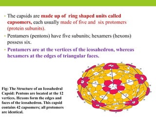
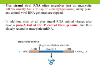
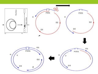
![TMV genome:
• The TMV genome consists of a 6.3-6.5 kb single-stranded (ss) RNA.
• The 5’ terminus has a methylated nucleotide cap (7 methylguanosine) and
the 3’-terminus has a tRNA-like structure.
• The genome contain 4 open reading frames.
• The 2 genes encode a replicase (with methyltransferase [MT] and RNA
helicase [Hel]), an RNA-dependent RNA polymerase, also-called
movement protein (MP) and a capsid protein (CP).](https://image.slidesharecdn.com/virusstructureandclassification-210220045819/85/Virus-structure-and-classification-33-320.jpg)
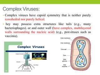
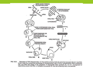
![Replication:
• Following entry into its host via mechanical inoculation, TMV uncoats
itself to release its viral [+]RNA strand.
• Then the viral genome transcribes and produce multiple mRNAs.
• The resulting mRNAs encode several proteins, including the coat protein
and an RNA-dependent RNA polymerase (RdRp).
• Thus TMV can replicate its own genome by RNA-dependent RNA
polymerase.
• After the coat protein and RNA genome of TMV have been synthesized,
they spontaneously assemble into complete TMV virions in a highly
organized process.](https://image.slidesharecdn.com/virusstructureandclassification-210220045819/85/Virus-structure-and-classification-34-320.jpg)


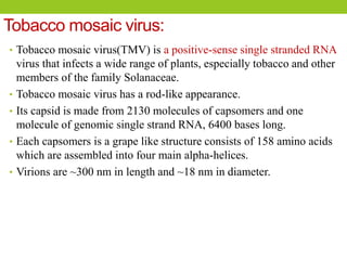
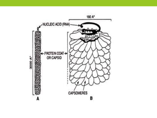
(1) The document discusses the structure and classification of viruses, focusing on plant viruses tobacco mosaic virus (TMV) and cauliflower mosaic virus (CaMV). (2) TMV is a helical, positive-sense RNA virus that infects tobacco and other plants and causes characteristic mosaic and mottling symptoms, while CaMV is a double-stranded DNA virus that infects cruciferous plants like cauliflower. (3) Both viruses have capsids that protect their genetic material and mechanisms for replication within host plant cells, while CaMV also has an envelope and uses reverse transcription.