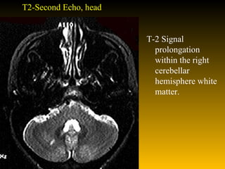
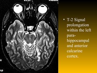
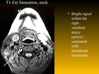
Vertebral artery dissection
HISTORY Date: September, 1995  The patient is a 29 year old right handed women with headache and monocular diplopia.  Several days prior to evaluation she developed a constant pressure sensation in her right lower neck region which was painful. This was unlike her usual headaches where she develops occipital throbbing, scotoma, and photophobia but no nausea or phonophobia. She underwent cervical manipulation by her chiropractor which caused worsened neck pain and new headache. The pain persisted and she returned the day of admission for repeat manipulation. During high cervical manipulation she developed a sudden change in her visual acuity and worsening of her neck pain to "11 out of 10". She described a loss of vision to her right; for example, she noticed that faces did not have a left eye. On further questioning she stated she saw objects as double, even if she covered an eye.  She denied Lhermitte's phenomenon, appendicular numbness or weakness, bowel or bladder disturbance, dysarthria, vertigo and ataxia.
Past Medical History   Classic migraine headaches: onset teenager, worse with menses  Medications: Oral contraceptives  Ibuprofen  Family History: Negative for connective tissue disorders
Physical Examination T 37.8 BP 110/60, RR 16, HR 80 Gen: Well appearing woman HEENT: No neck stiffness, bruits or vessel tenderness  Chest: Clear CV: 1/6 SEM at apex Ab: benign  Ext: normal skin and joints  Alert, oriented and attentive; no aphasia or dysarthria  CN: Right homonymous superior quadrantanopsia of macular vision. Peripheral field in this quadrant was normal without red desaturation. With either eye open alone, a pen appeared as two parallel lines, regardless of the angle of orientation Remainder of her cranial nerves were normal.  Gait:no ataxia, normal tandem walk and arm swings  Motor: normal bulk, tone, strength; fast finger movements symmetrical and no pronator drift Sense: symmetrical fine touch and pin prick  Coordination: normal finger-nose-finger and heel-knee-shin testing Reflexes:2+/symmetric; toes down going
Investigations WBC 7.1 K/uL Hgb 10.8 g/dL HCT 31.6 % MCV 96 fL Plt 251 K/ul PT 11.6 s INR 0.8  PTT 25.7 s  Na 142 mmol/L K 4.4 mmol/L Cl 109 mmol/L CO2 27 mmol/L BUN 5 mg/dL Creat 0.7 mg/dL Glucose 94 mg/dL
T2-Second Echo, head T-2 Signal prolongation within the right cerebellar hemisphere white matter.
T-2 Signal prolongation within the left para-hippocampal and anterior calcarine cortex.
T1-Fat Saturation, neck Bright signal within the right vertebral artery (arrow) consistent with intramural hematoma.
Discussion Case 96-2 represents a case of vertebral artery dissection with distal posterior circulation embolization and stroke. MRI scans revealed a right cerebellar hemisphere infarct (fig.1) and a mesial temporal infarct of the dominant hemisphere (fig 2). MRI of the neck with fat suppression reveals a classic cresentic lesion with signal characteristics of blood within the wall of the right vertebral artery (fig 3).
Discussion Whether the high cervical manipulation caused the right vertebral dissection or dislodged a clot from an already dissected vertebral artery is not clear. However, the frequent association of recent manipulation and posterior circulation stroke in young people suggests a causal relationship (1) . It may be that patients with spontaneous dissections seek manipulation for their neck pain and the manipulation dislodges a clot from the denuded endothelium. Since there is no clear difference between musculoskeletal pain and the pain from dissection (2), all patients with neck pain who undergo cervical manipulation are at risk for stroke. These patients should be consented for this procedure with the risks and benefits clearly explained.
Discussion The diagnosis of arterial dissection is becoming more frequent probably due to increased awareness of the disorder and the use of MR technology. MR imaging using fat saturation ("fat sat") suppresses the fat signal that surrounds the extracranial carotid and vertebral arteries revealing an intramural bright signal representing intramural hematoma. This is well illustrated in figure 3 (arrow). Using traditional T-1 weighted imaging, the intramural hematoma signal would merge with the surrounding fat signal reducing the likelihood of detection. Conventional angiography is the gold standard for dissection; however, if the diagnosis can be made noninvasively either with MR or ultrasound, the patient would not have to undergo conventional angiography.
Treatment We did anticoagulate the patient but not until a lumbar puncture was performed to rule out subarachnoid hemorrhage. Subarachnoid hemorrhage is associated with vertebral dissection (4,5) and is probably a function of the extent of vascular dissection. If the dissection remains extracranial (V3 section) it may cause TIA, stroke and/or neck pain. However, if the dissection continues intracranially, the thinner adventitia of intracranial vessels may allow complete transmural dissection and subarachnoid hemorrhage. Heparin therapy may not be the best initial management of such patients.
Followup This patient still experiences (at 6 months) monocular diplopia presumably of cortical origin. This disorder has been reported only rarely. Monocular diplopia of cortical origin can be distinguished from monocular diplopia caused by lens or retinal pathology primarily by the later producing symptoms in only one eye. Cases of cortical diplopia have been previously reported (6,7) and can occur from a variety of disorders. In this patient's case she had evidence of posterior temporal infarction within the parahippocampal gyrus
Thank You !

Vertebral artery dissection

  • 1.
  • 2.
    HISTORY Date: September,1995 The patient is a 29 year old right handed women with headache and monocular diplopia. Several days prior to evaluation she developed a constant pressure sensation in her right lower neck region which was painful. This was unlike her usual headaches where she develops occipital throbbing, scotoma, and photophobia but no nausea or phonophobia. She underwent cervical manipulation by her chiropractor which caused worsened neck pain and new headache. The pain persisted and she returned the day of admission for repeat manipulation. During high cervical manipulation she developed a sudden change in her visual acuity and worsening of her neck pain to "11 out of 10". She described a loss of vision to her right; for example, she noticed that faces did not have a left eye. On further questioning she stated she saw objects as double, even if she covered an eye. She denied Lhermitte's phenomenon, appendicular numbness or weakness, bowel or bladder disturbance, dysarthria, vertigo and ataxia.
  • 3.
    Past Medical History Classic migraine headaches: onset teenager, worse with menses Medications: Oral contraceptives Ibuprofen Family History: Negative for connective tissue disorders
  • 4.
    Physical Examination T37.8 BP 110/60, RR 16, HR 80 Gen: Well appearing woman HEENT: No neck stiffness, bruits or vessel tenderness Chest: Clear CV: 1/6 SEM at apex Ab: benign Ext: normal skin and joints Alert, oriented and attentive; no aphasia or dysarthria CN: Right homonymous superior quadrantanopsia of macular vision. Peripheral field in this quadrant was normal without red desaturation. With either eye open alone, a pen appeared as two parallel lines, regardless of the angle of orientation Remainder of her cranial nerves were normal. Gait:no ataxia, normal tandem walk and arm swings Motor: normal bulk, tone, strength; fast finger movements symmetrical and no pronator drift Sense: symmetrical fine touch and pin prick Coordination: normal finger-nose-finger and heel-knee-shin testing Reflexes:2+/symmetric; toes down going
  • 5.
    Investigations WBC 7.1K/uL Hgb 10.8 g/dL HCT 31.6 % MCV 96 fL Plt 251 K/ul PT 11.6 s INR 0.8 PTT 25.7 s Na 142 mmol/L K 4.4 mmol/L Cl 109 mmol/L CO2 27 mmol/L BUN 5 mg/dL Creat 0.7 mg/dL Glucose 94 mg/dL
  • 6.
    T2-Second Echo, headT-2 Signal prolongation within the right cerebellar hemisphere white matter.
  • 7.
    T-2 Signal prolongationwithin the left para-hippocampal and anterior calcarine cortex.
  • 8.
    T1-Fat Saturation, neckBright signal within the right vertebral artery (arrow) consistent with intramural hematoma.
  • 9.
    Discussion Case 96-2represents a case of vertebral artery dissection with distal posterior circulation embolization and stroke. MRI scans revealed a right cerebellar hemisphere infarct (fig.1) and a mesial temporal infarct of the dominant hemisphere (fig 2). MRI of the neck with fat suppression reveals a classic cresentic lesion with signal characteristics of blood within the wall of the right vertebral artery (fig 3).
  • 10.
    Discussion Whether thehigh cervical manipulation caused the right vertebral dissection or dislodged a clot from an already dissected vertebral artery is not clear. However, the frequent association of recent manipulation and posterior circulation stroke in young people suggests a causal relationship (1) . It may be that patients with spontaneous dissections seek manipulation for their neck pain and the manipulation dislodges a clot from the denuded endothelium. Since there is no clear difference between musculoskeletal pain and the pain from dissection (2), all patients with neck pain who undergo cervical manipulation are at risk for stroke. These patients should be consented for this procedure with the risks and benefits clearly explained.
  • 11.
    Discussion The diagnosisof arterial dissection is becoming more frequent probably due to increased awareness of the disorder and the use of MR technology. MR imaging using fat saturation ("fat sat") suppresses the fat signal that surrounds the extracranial carotid and vertebral arteries revealing an intramural bright signal representing intramural hematoma. This is well illustrated in figure 3 (arrow). Using traditional T-1 weighted imaging, the intramural hematoma signal would merge with the surrounding fat signal reducing the likelihood of detection. Conventional angiography is the gold standard for dissection; however, if the diagnosis can be made noninvasively either with MR or ultrasound, the patient would not have to undergo conventional angiography.
  • 12.
    Treatment We didanticoagulate the patient but not until a lumbar puncture was performed to rule out subarachnoid hemorrhage. Subarachnoid hemorrhage is associated with vertebral dissection (4,5) and is probably a function of the extent of vascular dissection. If the dissection remains extracranial (V3 section) it may cause TIA, stroke and/or neck pain. However, if the dissection continues intracranially, the thinner adventitia of intracranial vessels may allow complete transmural dissection and subarachnoid hemorrhage. Heparin therapy may not be the best initial management of such patients.
  • 13.
    Followup This patientstill experiences (at 6 months) monocular diplopia presumably of cortical origin. This disorder has been reported only rarely. Monocular diplopia of cortical origin can be distinguished from monocular diplopia caused by lens or retinal pathology primarily by the later producing symptoms in only one eye. Cases of cortical diplopia have been previously reported (6,7) and can occur from a variety of disorders. In this patient's case she had evidence of posterior temporal infarction within the parahippocampal gyrus
  • 14.