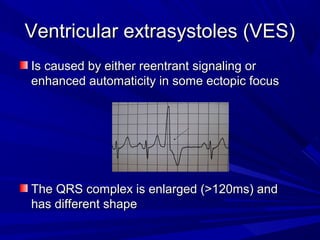
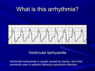
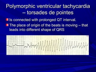
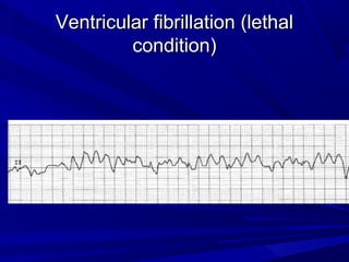
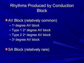
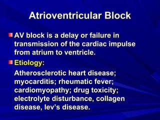
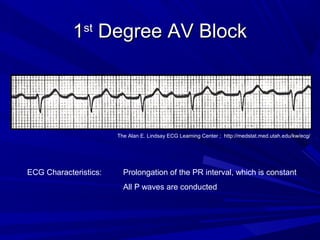
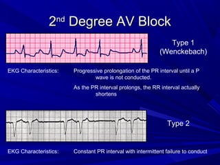
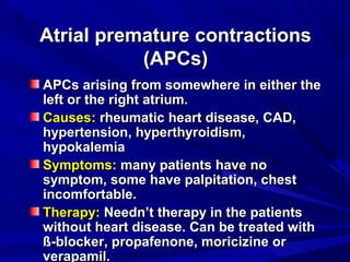
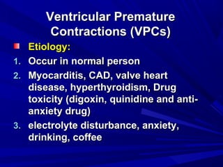
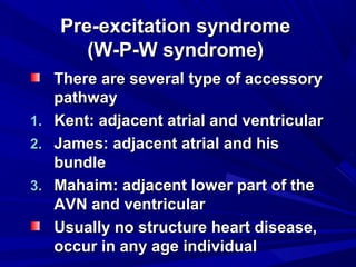
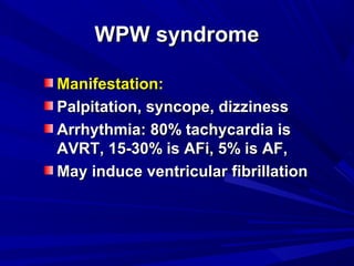
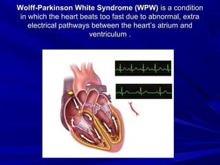
Ventricular ConductionVentricular Conduction
DisordersDisorders
vinay vermavinay verma
512512
Ventricular extrasystoles (VES)Ventricular extrasystoles (VES)
Is caused by either reentrant signaling orIs caused by either reentrant signaling or
enhanced automaticity in some ectopic focusenhanced automaticity in some ectopic focus
The QRS complex is enlarged (>120ms) andThe QRS complex is enlarged (>120ms) and
has different shapehas different shape
Coupling of VESCoupling of VES
Premature ventricular beats occurring after every normal beat arePremature ventricular beats occurring after every normal beat are
termed ventricular bigeminy, if 2 normal QRS complexes are follovedtermed ventricular bigeminy, if 2 normal QRS complexes are folloved
by VES, we speak of ventricular trigeminy.by VES, we speak of ventricular trigeminy.
Two VES grouped together are called a couplet, three a triplet. RunsTwo VES grouped together are called a couplet, three a triplet. Runs
longer than 3 VES is referred as ventricular tachycardialonger than 3 VES is referred as ventricular tachycardia
What is this arrhythmia?What is this arrhythmia?
Ventricular tachycardia
Ventricular tachycardia is usually caused by reentry, and most
commonly seen in patients following myocardial infarction.
Polymorphic ventricular tachycardiaPolymorphic ventricular tachycardia
– torsades de pointes– torsades de pointes
Is connected with prolonged QT interval.Is connected with prolonged QT interval.
The place of origin of the beats is moving – thatThe place of origin of the beats is moving – that
leads into different shape of QRSleads into different shape of QRS
Ventricular fibrillation (lethalVentricular fibrillation (lethal
condition)condition)
Conduction BlockConduction Block
Rhythms Produced by ConductionRhythms Produced by Conduction
BlockBlock
AV Block (relatively common)AV Block (relatively common)
– 11stst
degree AV blockdegree AV block
– Type 1 2Type 1 2ndnd
degree AV blockdegree AV block
– Type 2 2Type 2 2ndnd
degree AV blockdegree AV block
– 33rdrd
degree AV blockdegree AV block
SA Block (relatively rare)SA Block (relatively rare)
Atrioventricular BlockAtrioventricular Block
AV block is a delay or failure inAV block is a delay or failure in
transmission of the cardiac impulsetransmission of the cardiac impulse
from atrium to ventricle.from atrium to ventricle.
Etiology:Etiology:
Atherosclerotic heart disease;Atherosclerotic heart disease;
myocarditis; rheumatic fever;myocarditis; rheumatic fever;
cardiomyopathy; drug toxicity;cardiomyopathy; drug toxicity;
electrolyte disturbance, collagenelectrolyte disturbance, collagen
disease, lev’s disease.disease, lev’s disease.
11stst
Degree AV BlockDegree AV Block
ECG Characteristics: Prolongation of the PR interval, which is constant
All P waves are conducted
The Alan E. Lindsay ECG Learning Center ; http://medstat.med.utah.edu/kw/ecg/
22ndnd
Degree AV BlockDegree AV Block
Type 1
(Wenckebach)
EKG Characteristics: Progressive prolongation of the PR interval until a P
wave is not conducted.
As the PR interval prolongs, the RR interval actually
shortens
EKG Characteristics: Constant PR interval with intermittent failure to conduct
Type 2
33rdrd
Degree (Complete) AV BlockDegree (Complete) AV Block
EKG Characteristics: No relationship between P waves and QRS complexes
Relatively constant PP intervals and RR intervals
Greater number of P waves than QRS complexes
www.uptodate.com
SA arrest with compensatory AV activitySA arrest with compensatory AV activity
When the activity of SA node is stopped, AV node takes over the role of
pacemaker.
Very similar type of arrhythmia is SA block: Pacing in SA node is generated, but
not conducted to the myocardium+
Intraventricular BlockIntraventricular Block
Intraventricular conduction system:Intraventricular conduction system:
1.1. Right bundle branchRight bundle branch
2.2. Left bundle branchLeft bundle branch
3.3. Left anterior fascicularLeft anterior fascicular
4.4. Left posterior fascicularLeft posterior fascicular
Intraventricular BlockIntraventricular Block
Etiology:Etiology:
Myocarditis, valve disease,Myocarditis, valve disease,
cardiomyopathy, CAD, hypertension,cardiomyopathy, CAD, hypertension,
pulmonary heart disease, drug toxicity,pulmonary heart disease, drug toxicity,
Lenegre disease, Lev’s disease et al.Lenegre disease, Lev’s disease et al.
Manifestation:Manifestation:
Single fascicular or bifascicular blockSingle fascicular or bifascicular block
is asymptom; tri-fascicular block mayis asymptom; tri-fascicular block may
have dizziness; palpitation, syncopehave dizziness; palpitation, syncope
and Adams-stokes syndromeand Adams-stokes syndrome
Premature contractionsPremature contractions
The term “premature contractions”The term “premature contractions”
are used to describe non sinus beats.are used to describe non sinus beats.
Common arrhythmiaCommon arrhythmia
The morbidity rate is 3-5%The morbidity rate is 3-5%
Atrial premature contractionsAtrial premature contractions
(APCs)(APCs)
APCs arising from somewhere in either theAPCs arising from somewhere in either the
left or the right atrium.left or the right atrium.
Causes:Causes: rheumatic heart disease, CAD,rheumatic heart disease, CAD,
hypertension, hyperthyroidism,hypertension, hyperthyroidism,
hypokalemiahypokalemia
Symptoms:Symptoms: many patients have nomany patients have no
symptom, some have palpitation, chestsymptom, some have palpitation, chest
incomfortable.incomfortable.
Therapy:Therapy: Needn’t therapy in the patientsNeedn’t therapy in the patients
without heart disease. Can be treated withwithout heart disease. Can be treated with
ß-blocker, propafenone, moricizine orß-blocker, propafenone, moricizine or
verapamil.verapamil.
Ventricular PrematureVentricular Premature
Contractions (VPCs)Contractions (VPCs)
Etiology:Etiology:
1.1. Occur in normal personOccur in normal person
2.2. Myocarditis, CAD, valve heartMyocarditis, CAD, valve heart
disease, hyperthyroidism, Drugdisease, hyperthyroidism, Drug
toxicity (digoxin, quinidine and anti-toxicity (digoxin, quinidine and anti-
anxiety drug)anxiety drug)
3.3. electrolyte disturbance, anxiety,electrolyte disturbance, anxiety,
drinking,drinking, coffeecoffee
Pre-excitation syndromePre-excitation syndrome
(W-P-W syndrome)(W-P-W syndrome)
There are several type of accessoryThere are several type of accessory
pathwaypathway
1.1. Kent: adjacent atrial and ventricularKent: adjacent atrial and ventricular
2.2. James: adjacent atrial and hisJames: adjacent atrial and his
bundlebundle
3.3. Mahaim: adjacent lower part of theMahaim: adjacent lower part of the
AVN and ventricularAVN and ventricular
Usually no structure heart disease,Usually no structure heart disease,
occur in any age individualoccur in any age individual
WPW syndromeWPW syndrome
Manifestation:Manifestation:
Palpitation, syncope, dizzinessPalpitation, syncope, dizziness
Arrhythmia: 80% tachycardia isArrhythmia: 80% tachycardia is
AVRT, 15-30% is AFi, 5% is AF,AVRT, 15-30% is AFi, 5% is AF,
May induce ventricular fibrillationMay induce ventricular fibrillation
Wolff-Parkinson White Syndrome (WPW)Wolff-Parkinson White Syndrome (WPW) is a conditionis a condition
in which the heart beats too fast due to abnormal, extrain which the heart beats too fast due to abnormal, extra
electrical pathways between the heart’selectrical pathways between the heart’s atriumatrium andand
ventriculumventriculum ..
Thank you for your attentionThank you for your attention

Venticular conduction disorders by dr vinay verma

  • 1.
  • 2.
    Ventricular extrasystoles (VES)Ventricularextrasystoles (VES) Is caused by either reentrant signaling orIs caused by either reentrant signaling or enhanced automaticity in some ectopic focusenhanced automaticity in some ectopic focus The QRS complex is enlarged (>120ms) andThe QRS complex is enlarged (>120ms) and has different shapehas different shape
  • 3.
    Coupling of VESCouplingof VES Premature ventricular beats occurring after every normal beat arePremature ventricular beats occurring after every normal beat are termed ventricular bigeminy, if 2 normal QRS complexes are follovedtermed ventricular bigeminy, if 2 normal QRS complexes are folloved by VES, we speak of ventricular trigeminy.by VES, we speak of ventricular trigeminy. Two VES grouped together are called a couplet, three a triplet. RunsTwo VES grouped together are called a couplet, three a triplet. Runs longer than 3 VES is referred as ventricular tachycardialonger than 3 VES is referred as ventricular tachycardia
  • 4.
    What is thisarrhythmia?What is this arrhythmia? Ventricular tachycardia Ventricular tachycardia is usually caused by reentry, and most commonly seen in patients following myocardial infarction.
  • 5.
    Polymorphic ventricular tachycardiaPolymorphicventricular tachycardia – torsades de pointes– torsades de pointes Is connected with prolonged QT interval.Is connected with prolonged QT interval. The place of origin of the beats is moving – thatThe place of origin of the beats is moving – that leads into different shape of QRSleads into different shape of QRS
  • 6.
    Ventricular fibrillation (lethalVentricularfibrillation (lethal condition)condition)
  • 7.
  • 8.
    Rhythms Produced byConductionRhythms Produced by Conduction BlockBlock AV Block (relatively common)AV Block (relatively common) – 11stst degree AV blockdegree AV block – Type 1 2Type 1 2ndnd degree AV blockdegree AV block – Type 2 2Type 2 2ndnd degree AV blockdegree AV block – 33rdrd degree AV blockdegree AV block SA Block (relatively rare)SA Block (relatively rare)
  • 9.
    Atrioventricular BlockAtrioventricular Block AVblock is a delay or failure inAV block is a delay or failure in transmission of the cardiac impulsetransmission of the cardiac impulse from atrium to ventricle.from atrium to ventricle. Etiology:Etiology: Atherosclerotic heart disease;Atherosclerotic heart disease; myocarditis; rheumatic fever;myocarditis; rheumatic fever; cardiomyopathy; drug toxicity;cardiomyopathy; drug toxicity; electrolyte disturbance, collagenelectrolyte disturbance, collagen disease, lev’s disease.disease, lev’s disease.
  • 10.
    11stst Degree AV BlockDegreeAV Block ECG Characteristics: Prolongation of the PR interval, which is constant All P waves are conducted The Alan E. Lindsay ECG Learning Center ; http://medstat.med.utah.edu/kw/ecg/
  • 11.
    22ndnd Degree AV BlockDegreeAV Block Type 1 (Wenckebach) EKG Characteristics: Progressive prolongation of the PR interval until a P wave is not conducted. As the PR interval prolongs, the RR interval actually shortens EKG Characteristics: Constant PR interval with intermittent failure to conduct Type 2
  • 12.
    33rdrd Degree (Complete) AVBlockDegree (Complete) AV Block EKG Characteristics: No relationship between P waves and QRS complexes Relatively constant PP intervals and RR intervals Greater number of P waves than QRS complexes www.uptodate.com
  • 13.
    SA arrest withcompensatory AV activitySA arrest with compensatory AV activity When the activity of SA node is stopped, AV node takes over the role of pacemaker. Very similar type of arrhythmia is SA block: Pacing in SA node is generated, but not conducted to the myocardium+
  • 14.
    Intraventricular BlockIntraventricular Block Intraventricularconduction system:Intraventricular conduction system: 1.1. Right bundle branchRight bundle branch 2.2. Left bundle branchLeft bundle branch 3.3. Left anterior fascicularLeft anterior fascicular 4.4. Left posterior fascicularLeft posterior fascicular
  • 15.
    Intraventricular BlockIntraventricular Block Etiology:Etiology: Myocarditis,valve disease,Myocarditis, valve disease, cardiomyopathy, CAD, hypertension,cardiomyopathy, CAD, hypertension, pulmonary heart disease, drug toxicity,pulmonary heart disease, drug toxicity, Lenegre disease, Lev’s disease et al.Lenegre disease, Lev’s disease et al. Manifestation:Manifestation: Single fascicular or bifascicular blockSingle fascicular or bifascicular block is asymptom; tri-fascicular block mayis asymptom; tri-fascicular block may have dizziness; palpitation, syncopehave dizziness; palpitation, syncope and Adams-stokes syndromeand Adams-stokes syndrome
  • 16.
    Premature contractionsPremature contractions Theterm “premature contractions”The term “premature contractions” are used to describe non sinus beats.are used to describe non sinus beats. Common arrhythmiaCommon arrhythmia The morbidity rate is 3-5%The morbidity rate is 3-5%
  • 17.
    Atrial premature contractionsAtrialpremature contractions (APCs)(APCs) APCs arising from somewhere in either theAPCs arising from somewhere in either the left or the right atrium.left or the right atrium. Causes:Causes: rheumatic heart disease, CAD,rheumatic heart disease, CAD, hypertension, hyperthyroidism,hypertension, hyperthyroidism, hypokalemiahypokalemia Symptoms:Symptoms: many patients have nomany patients have no symptom, some have palpitation, chestsymptom, some have palpitation, chest incomfortable.incomfortable. Therapy:Therapy: Needn’t therapy in the patientsNeedn’t therapy in the patients without heart disease. Can be treated withwithout heart disease. Can be treated with ß-blocker, propafenone, moricizine orß-blocker, propafenone, moricizine or verapamil.verapamil.
  • 18.
    Ventricular PrematureVentricular Premature Contractions(VPCs)Contractions (VPCs) Etiology:Etiology: 1.1. Occur in normal personOccur in normal person 2.2. Myocarditis, CAD, valve heartMyocarditis, CAD, valve heart disease, hyperthyroidism, Drugdisease, hyperthyroidism, Drug toxicity (digoxin, quinidine and anti-toxicity (digoxin, quinidine and anti- anxiety drug)anxiety drug) 3.3. electrolyte disturbance, anxiety,electrolyte disturbance, anxiety, drinking,drinking, coffeecoffee
  • 19.
    Pre-excitation syndromePre-excitation syndrome (W-P-Wsyndrome)(W-P-W syndrome) There are several type of accessoryThere are several type of accessory pathwaypathway 1.1. Kent: adjacent atrial and ventricularKent: adjacent atrial and ventricular 2.2. James: adjacent atrial and hisJames: adjacent atrial and his bundlebundle 3.3. Mahaim: adjacent lower part of theMahaim: adjacent lower part of the AVN and ventricularAVN and ventricular Usually no structure heart disease,Usually no structure heart disease, occur in any age individualoccur in any age individual
  • 20.
    WPW syndromeWPW syndrome Manifestation:Manifestation: Palpitation,syncope, dizzinessPalpitation, syncope, dizziness Arrhythmia: 80% tachycardia isArrhythmia: 80% tachycardia is AVRT, 15-30% is AFi, 5% is AF,AVRT, 15-30% is AFi, 5% is AF, May induce ventricular fibrillationMay induce ventricular fibrillation
  • 21.
    Wolff-Parkinson White Syndrome(WPW)Wolff-Parkinson White Syndrome (WPW) is a conditionis a condition in which the heart beats too fast due to abnormal, extrain which the heart beats too fast due to abnormal, extra electrical pathways between the heart’selectrical pathways between the heart’s atriumatrium andand ventriculumventriculum ..
  • 22.
    Thank you foryour attentionThank you for your attention