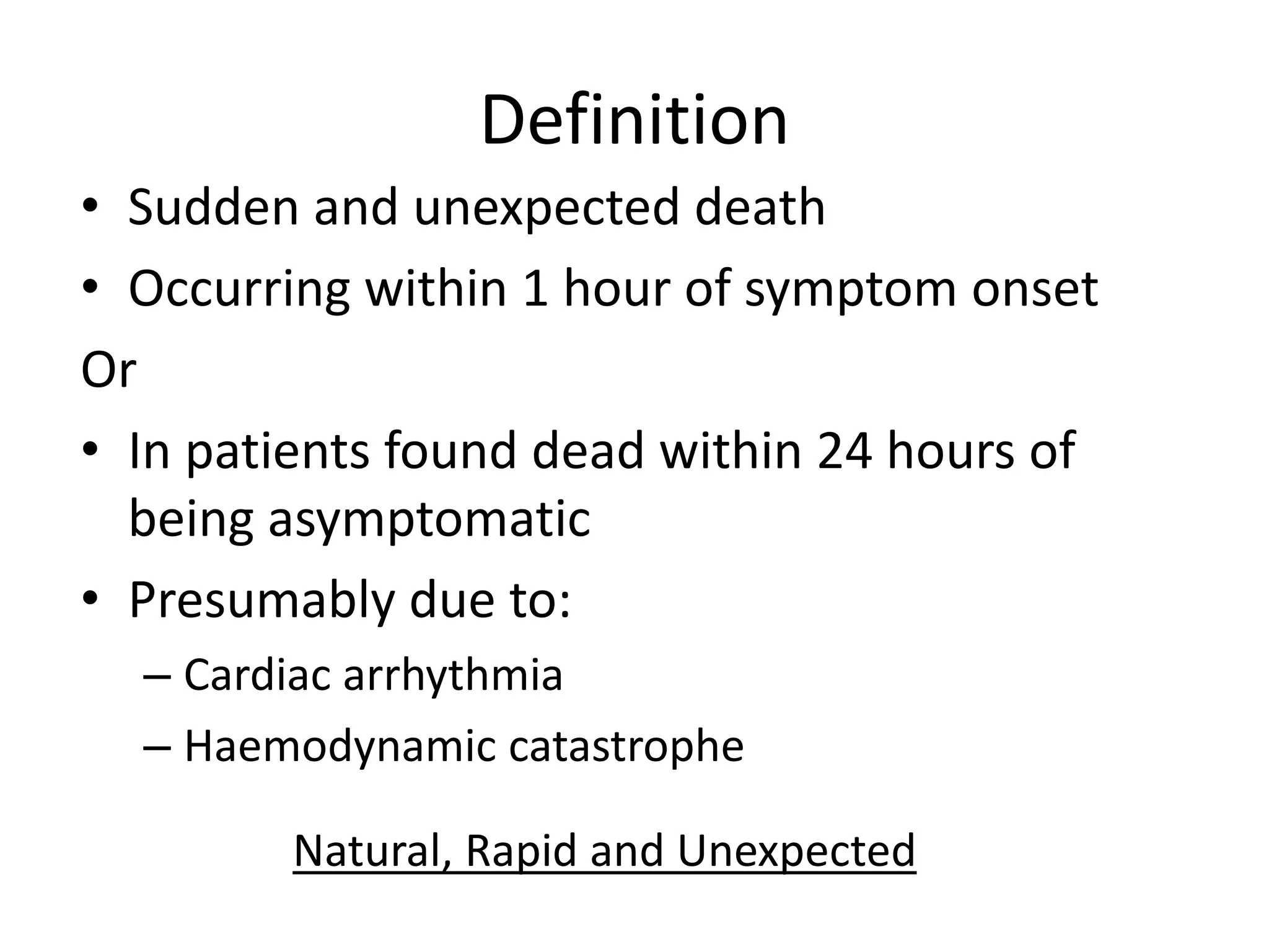
Sudden Cardiac Death
By Dr Niraj
Definition
• Sudden and unexpected death
• Occurring within 1 hour of symptom onset
Or
• In patients found dead within 24 hours of
being asymptomatic
• Presumably due to:
– Cardiac arrhythmia
– Haemodynamic catastrophe
Natural, Rapid and Unexpected
Incidence of SCD
• Almost 50% of all CV deaths
• At least 25% -first symptomatic event
• M>F
• 70%- out-of-hospital*
• Out-of-hospital SR- 6%
• In-hospital SR-24%
• Rapid emergency response, public knowledge
• Prognosis better in shockable > PEA/asystole
Incidence of SCD
• India- >7,00,000 deaths/year and approx 10.3% of mortality (Rao et
al.)
• Peak age – 45 to 75 yrs, M>F (3:1)
• SR <1%
• Vital data esp. from rural areas – unaccounted for
• Rural-urban gradient in CAD (7.6-12% v 3.1-7.4%) - different risk
factors
• Urban India and Western SCD trends similar- Gupta et al. (2007)
• Prevailing RFs- HTN, DM, smoking
Four temporal elements
1. Prodrome- Predictors of an impending
cardiac event
2. Onset of terminal event- from onset of
1st symptom to SCA
3. Cardiac arrest
4. Biologic death- upto 1 hour from
terminal event
diagram
Etiology of SCD
Structural HD Cardiomyopathy
HCM, NIDCM, ARVC
Myocardial scarring and fibrosis
Inherited Arrhythmia syndromes BrS, LQTS, WPW, CPVT
Channelopathies
Ischaemic Heart Disease** - X 2.8-5.3 risk (Framingham)
LVEF < 30-35% *
Valvular HD AS, MVP, Prosthetic valve dysfunction
Mechanical pathologies Acute cardiac tamponade, Massive PE, Acute
intracardiac thrombus, Commotio cordis
Congenital HD Congenital AS, PS, VSD+Eisenmenger
physiology, Anomalous origin of coronary
arteries, post-TOF repair
SIDS- Sudden infant death syndrome - <0.5/1000 live births, M>F infants
Contributory Risk factors
1. Hypertension
2. Type 2 DM
3. Dyslipidaemia
4. CKD
5. Obesity
6. OSA
7. Seizures
8. Smoking
9. Type A personality
10. Nutritional deficiencies
Risk analysis
1. Age
– 2 peaks- <1 year (SIDS) and 45-75 yrs
– Strong predictor, non-linear, >35 yrs, 1 in 1000/yr
– Risk decreases in later years >75 yrs, Framingham
– Steep increase- adolescent to middle-age (IHD)
– HCM, BrS, LQTS, ARVC- important considerations
2. IHD
– MC substrate a/w SCD
– Transition from mid-20s to mid-30s (>40% of cases)
diagram
Risk analysis
3. Diseases presenting in young-
– Myocardial fibrosis and remodelling
– Channelopathies
– CHD
– Myocarditis
– Genetic structural disorders
4. Race-
SCA and SCD risk higher in black >
white/asian > hispanic
Risk analysis
5. Gender
– <65 years- M>F by x 4-7
– 65 + years- M>F – 2:1
– Protection against CAD before menopause
– Severe LV dysfunction- ↓ 50%
– CAD- ↓66% before SCD
6. Heredity
– Inherited arrhythmia syndromes
– Gene loci-encoding ion channel protein mutations
– CAD: Atherogenesis, plaque destabilisation, thrombosis, arrhythmia
diagram
Risk analysis
7. LVEF
– <30%- single most powerful independent predictor of SCD
– MR rate changes the most btw 30-40%
– Infarct size, fibrosis- better predictor of cardiac events
– LV dysfunction- modulates risk in Post-MI pts with VPCs
8. Ventricular arrhythmias
– Benign- most VPCs, NSVT (short runs)- in absence of structural HD
– If present during TMT (peak/recovery)- higher risk
– VPCs in Post-ACS pts- increased risk (>10/hour)
– Polymorphic NSVT- drug/electrolyte/functional
Mechanisms of SCD
1. Automaticity
– Enhanced normal
– Abnormal
2. Afterdepolarisations  Triggered activity
– Early
– Late
3. Re-entry phenomenon
Automaticity
• Phase 4- spontaneous depolarisation of
transmembrane AP
• In MI/ischaemia:↑ EC K+ causes partial
depolarisation of RMP
• Injury currents develop btw healthy and
infarcted/ischaemic myocardium
• This initiates spontaneous activity
• Purkinje fibres and Ventricular myocytes
Triggered Activity
• AP prolongation may occur due to
– ↑ inward currents (Na+/Ca2+/Na+-Ca2+ exchange)
– ↓ repolarising K curents
• Early afterdepolarisations- late phase 3/early
phase 3 of AP
• TdP VT associated with QT-Prolongation
(medications/hereditary)
• Delayed afterdepolarisations- after complete
membrane repolarisation, due to Ca2+ overload
• CPVT, Digoxin toxicity, idiopathic outflow tract VA
Re-entry phenomenon
• Most sustained VA in the presence of
structural HD
• Ischaemia, Brugada syndrome
• Re-entry requires a trigger to initiate the
arrhythmia and a substrate to sustain it
• Trigger- VPC (automaticity)
• Substrate-
1. Fixed anatomical obstacle- patchy fibrosis, scar
(post-MI or post-surgery, CM, hypertrophy)
2. Functional block- functional re-entry. Rotor vs.
Multiple wavelet re-entry
Approach in high-risk patients
• Symptoms of VA:
– Palpitations
– Skipped/extra beats
– Breathlessness (at
rest/on exertion)
– Chest pain
– Syncope/near-syncope
– Lightheadedness
•Symptoms related to
underlying heart disease:
•Exertional dyspnoea
•Orthopnoea/PND
•Chest pain
•Pedal oedema
Precipitating factors: Exercise, emotional stress,
concurrent illness
Approach in high-risk patients
• Medications:
– Anti-arrhythmics
– QTc and TdP
– Alcohol
– Stimulants-
cocaine,
amphetamines
– Anabolic steroids
• Past medical history:
• Thyroid disease
• AKI/CKD
• CVA/embolic events
• Epilepsy
• Unexplained RTA
Clinical examination
• Bigeminy/Trigeminy-
– Effective bradycrdia
– Apico-Radial deficit
– Relative HTN, wide PP
• Carotid bruits
• Peripheral pulses
• JVP
• Peripheral oedema
• S3 gallop
• Cardiac murmurs
• Crepitations
Approach in high-risk patients
• Family history:
– Cardiac channelopathies-LQTS, CPVT, BrS
– Arrhythmias
– Conduction disorders, pacemakers/ICDs
– N-M junction disorders
– Muscular dystrophy
– Epilepsy
Evaluation
• Non-invasive
1. ECG-
– VT, high-grade AV block, sinus brady + pauses, SVT, PPM
malfunction
– Chamber enlargement, previous MI, inherited arrhythmia
disorders
– QRS duration- prognostic
– Microvolt T-wave alternans - predictive
2. Exercise testing-
– Abnormal recovery
– External/implantable recorders
– CPVT
Emerging markers of risk for SCD
• Cardiac MRI-
– Infarct area
– Fibrosis patterns (delayed hyperenhancement)
• Sympathetic imaging-
– 11C-hydroxyephedrine
– MIBG
• Genetic/Familial testing
– Risk profiling
– Familial clustering- CAD

Sudden cardiac death

  • 1.
  • 2.
    Definition • Sudden andunexpected death • Occurring within 1 hour of symptom onset Or • In patients found dead within 24 hours of being asymptomatic • Presumably due to: – Cardiac arrhythmia – Haemodynamic catastrophe Natural, Rapid and Unexpected
  • 3.
    Incidence of SCD •Almost 50% of all CV deaths • At least 25% -first symptomatic event • M>F • 70%- out-of-hospital* • Out-of-hospital SR- 6% • In-hospital SR-24% • Rapid emergency response, public knowledge • Prognosis better in shockable > PEA/asystole
  • 4.
    Incidence of SCD •India- >7,00,000 deaths/year and approx 10.3% of mortality (Rao et al.) • Peak age – 45 to 75 yrs, M>F (3:1) • SR <1% • Vital data esp. from rural areas – unaccounted for • Rural-urban gradient in CAD (7.6-12% v 3.1-7.4%) - different risk factors • Urban India and Western SCD trends similar- Gupta et al. (2007) • Prevailing RFs- HTN, DM, smoking
  • 5.
    Four temporal elements 1.Prodrome- Predictors of an impending cardiac event 2. Onset of terminal event- from onset of 1st symptom to SCA 3. Cardiac arrest 4. Biologic death- upto 1 hour from terminal event
  • 6.
  • 7.
    Etiology of SCD StructuralHD Cardiomyopathy HCM, NIDCM, ARVC Myocardial scarring and fibrosis Inherited Arrhythmia syndromes BrS, LQTS, WPW, CPVT Channelopathies Ischaemic Heart Disease** - X 2.8-5.3 risk (Framingham) LVEF < 30-35% * Valvular HD AS, MVP, Prosthetic valve dysfunction Mechanical pathologies Acute cardiac tamponade, Massive PE, Acute intracardiac thrombus, Commotio cordis Congenital HD Congenital AS, PS, VSD+Eisenmenger physiology, Anomalous origin of coronary arteries, post-TOF repair SIDS- Sudden infant death syndrome - <0.5/1000 live births, M>F infants
  • 8.
    Contributory Risk factors 1.Hypertension 2. Type 2 DM 3. Dyslipidaemia 4. CKD 5. Obesity 6. OSA 7. Seizures 8. Smoking 9. Type A personality 10. Nutritional deficiencies
  • 9.
    Risk analysis 1. Age –2 peaks- <1 year (SIDS) and 45-75 yrs – Strong predictor, non-linear, >35 yrs, 1 in 1000/yr – Risk decreases in later years >75 yrs, Framingham – Steep increase- adolescent to middle-age (IHD) – HCM, BrS, LQTS, ARVC- important considerations 2. IHD – MC substrate a/w SCD – Transition from mid-20s to mid-30s (>40% of cases)
  • 10.
  • 11.
    Risk analysis 3. Diseasespresenting in young- – Myocardial fibrosis and remodelling – Channelopathies – CHD – Myocarditis – Genetic structural disorders 4. Race- SCA and SCD risk higher in black > white/asian > hispanic
  • 12.
    Risk analysis 5. Gender –<65 years- M>F by x 4-7 – 65 + years- M>F – 2:1 – Protection against CAD before menopause – Severe LV dysfunction- ↓ 50% – CAD- ↓66% before SCD 6. Heredity – Inherited arrhythmia syndromes – Gene loci-encoding ion channel protein mutations – CAD: Atherogenesis, plaque destabilisation, thrombosis, arrhythmia
  • 13.
  • 14.
    Risk analysis 7. LVEF –<30%- single most powerful independent predictor of SCD – MR rate changes the most btw 30-40% – Infarct size, fibrosis- better predictor of cardiac events – LV dysfunction- modulates risk in Post-MI pts with VPCs 8. Ventricular arrhythmias – Benign- most VPCs, NSVT (short runs)- in absence of structural HD – If present during TMT (peak/recovery)- higher risk – VPCs in Post-ACS pts- increased risk (>10/hour) – Polymorphic NSVT- drug/electrolyte/functional
  • 15.
    Mechanisms of SCD 1.Automaticity – Enhanced normal – Abnormal 2. Afterdepolarisations  Triggered activity – Early – Late 3. Re-entry phenomenon
  • 16.
    Automaticity • Phase 4-spontaneous depolarisation of transmembrane AP • In MI/ischaemia:↑ EC K+ causes partial depolarisation of RMP • Injury currents develop btw healthy and infarcted/ischaemic myocardium • This initiates spontaneous activity • Purkinje fibres and Ventricular myocytes
  • 17.
    Triggered Activity • APprolongation may occur due to – ↑ inward currents (Na+/Ca2+/Na+-Ca2+ exchange) – ↓ repolarising K curents • Early afterdepolarisations- late phase 3/early phase 3 of AP • TdP VT associated with QT-Prolongation (medications/hereditary) • Delayed afterdepolarisations- after complete membrane repolarisation, due to Ca2+ overload • CPVT, Digoxin toxicity, idiopathic outflow tract VA
  • 18.
    Re-entry phenomenon • Mostsustained VA in the presence of structural HD • Ischaemia, Brugada syndrome • Re-entry requires a trigger to initiate the arrhythmia and a substrate to sustain it • Trigger- VPC (automaticity) • Substrate- 1. Fixed anatomical obstacle- patchy fibrosis, scar (post-MI or post-surgery, CM, hypertrophy) 2. Functional block- functional re-entry. Rotor vs. Multiple wavelet re-entry
  • 19.
    Approach in high-riskpatients • Symptoms of VA: – Palpitations – Skipped/extra beats – Breathlessness (at rest/on exertion) – Chest pain – Syncope/near-syncope – Lightheadedness •Symptoms related to underlying heart disease: •Exertional dyspnoea •Orthopnoea/PND •Chest pain •Pedal oedema Precipitating factors: Exercise, emotional stress, concurrent illness
  • 20.
    Approach in high-riskpatients • Medications: – Anti-arrhythmics – QTc and TdP – Alcohol – Stimulants- cocaine, amphetamines – Anabolic steroids • Past medical history: • Thyroid disease • AKI/CKD • CVA/embolic events • Epilepsy • Unexplained RTA
  • 21.
    Clinical examination • Bigeminy/Trigeminy- –Effective bradycrdia – Apico-Radial deficit – Relative HTN, wide PP • Carotid bruits • Peripheral pulses • JVP • Peripheral oedema • S3 gallop • Cardiac murmurs • Crepitations
  • 22.
    Approach in high-riskpatients • Family history: – Cardiac channelopathies-LQTS, CPVT, BrS – Arrhythmias – Conduction disorders, pacemakers/ICDs – N-M junction disorders – Muscular dystrophy – Epilepsy
  • 23.
    Evaluation • Non-invasive 1. ECG- –VT, high-grade AV block, sinus brady + pauses, SVT, PPM malfunction – Chamber enlargement, previous MI, inherited arrhythmia disorders – QRS duration- prognostic – Microvolt T-wave alternans - predictive 2. Exercise testing- – Abnormal recovery – External/implantable recorders – CPVT
  • 24.
    Emerging markers ofrisk for SCD • Cardiac MRI- – Infarct area – Fibrosis patterns (delayed hyperenhancement) • Sympathetic imaging- – 11C-hydroxyephedrine – MIBG • Genetic/Familial testing – Risk profiling – Familial clustering- CAD