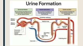
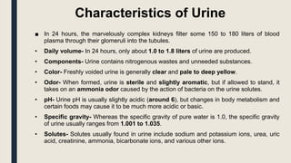
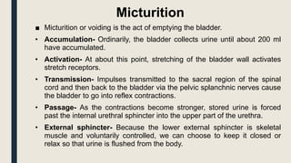
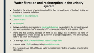
URINE FORMATION AND
MICTURITION
Characteristics of Urine
■ In 24 hours, the marvelously complex kidneys filter some 150 to 180 liters of blood
plasma through their glomeruli into the tubules.
• Daily volume- In 24 hours, only about 1.0 to 1.8 liters of urine are produced.
• Components- Urine contains nitrogenous wastes and unneeded substances.
• Color- Freshly voided urine is generally clear and pale to deep yellow.
• Odor- When formed, urine is sterile and slightly aromatic, but if allowed to stand, it
takes on an ammonia odor caused by the action of bacteria on the urine solutes.
• pH- Urine pH is usually slightly acidic (around 6), but changes in body metabolism and
certain foods may cause it to be much more acidic or basic.
• Specific gravity- Whereas the specific gravity of pure water is 1.0, the specific gravity
of urine usually ranges from 1.001 to 1.035.
• Solutes- Solutes usually found in urine include sodium and potassium ions, urea, uric
acid, creatinine, ammonia, bicarbonate ions, and various other ions.
Micturition
■ Micturition or voiding is the act of emptying the bladder.
• Accumulation- Ordinarily, the bladder collects urine until about 200 ml
have accumulated.
• Activation- At about this point, stretching of the bladder wall activates
stretch receptors.
• Transmission- Impulses transmitted to the sacral region of the spinal
cord and then back to the bladder via the pelvic splanchnic nerves cause
the bladder to go into reflex contractions.
• Passage- As the contractions become stronger, stored urine is forced
past the internal urethral sphincter into the upper part of the urethra.
• External sphincter- Because the lower external sphincter is skeletal
muscle and voluntarily controlled, we can choose to keep it closed or
relax so that urine is flushed from the body.
Water filtration and reabsorption in the urinary
system
■ Regulating the volume of water in each of the fluid compartments of the body is key for
a variety of reasons, including
 Regulation of blood pressure,
 Cardiac output
 Ion transport
 It plays a vital role in maintaining electrolyte balance by regulating the concentration of
ions such as sodium (Na+), potassium (K+), magnesium (Mg2+), and calcium (Ca2+).
 There are two primary sources of H2O in the body: the food/drink we take in
and endogenous water created as a product of aerobic respiration. This endogenous
water is so small it is negligible.
 On average, 180L of water is filtered by the kidneys daily.
 However, only 1.5-2L ends up being excreted as urine.
 This means almost 99% of filtered water is reabsorbed into the circulation or enters the
interstitium.
Storage Phase of Micturition
■ Micturition is the process of eliminating water and electrolytes from the urinary system,
commonly known as urinating.
■ It has two discrete phases: the storage/continence phase, when urine is stored in
the bladder, and the voiding phase, where urine is released through the urethra.
■ These phases require coordinated contraction/relaxation of the bladder and urethral
sphincters, controlled by the sympathetic, parasympathetic, and somatic nervous systems.
■ The storage phase of micturition is controlled at the highest level by the continence
centers of the brain. These, in turn, control the continence centers of the spinal cord.
■ Storage of urine requires relaxation of the detrusor muscle of the bladder
and simultaneous contraction of both the internal urethral sphincters
(IUS) and external urethral sphincters (EUS).
■ The bladder and IUS are under the control of the autonomic nervous system.
■ The EUS is under the control of the somatic nervous system.
■ This means only the EUS can be voluntarily opened or closed to control micturition; the
others are controlled automatically.
Sympathetic Innervation
■ To stimulate storage, impulses from the cerebral cortex travel to the pons. The pons is
responsible for coordinating the actions of the urinary sphincters and the bladder, and the area
involved in the storage phase is the pontine continence center (on the left-hand side of the
pons).
■ From here, signals are sent to sympathetic nuclei in the spinal cord (nerve roots T10-L2) and
finally to the detrusor muscle and internal urethral sphincter (IUS) of the bladder.
■ The impulses travel from the spinal cord to the bladder via the hypogastric nerve (nerve roots
T10-L2). At the bladder, this stimulates:
• Relaxation of the detrusor muscle in the bladder wall – via stimulation of β3-
adrenoreceptors in the fundus and the body of the bladder.
• Contraction of the IUS – via stimulation of α1-adrenoreceptors at the neck of the bladder.
Somatic Innervation
• The EUS is under voluntary somatic control.
• In the storage phase, impulses travel to the EUS via the pudendal nerve (nerve roots S2-S4) to
nicotinic (cholinergic) receptors on the striated muscle, resulting in the contraction of the EUS.
• This prevents any urine from leaking out.
Co-ordinated Effect
■ The coordinated relaxation of the detrusor muscle and contraction of the urethral sphincters allows the bladder to
fill and store urine for many hours.
■ As the bladder fills, the folds in the bladder walls (rugae) flatten, and the walls distend, increasing the bladder’s
capacity.
■ This means that, as the bladder fills, it expands, allowing the inner (intra-vesical) pressure to remain constant and
lower than urethral pressure.
■ This process, known as receptive relaxation, is vital to storing urine and prevents leakage during this phase.
Urinary Incontinence
■ Urinary incontinence is the inability to maintain a storage phase of micturition.
■ Incontinence can arise from multiple pathological processes or be caused by everyday events, such as pregnancy
or excessive urine.
■ Different causes of urinary incontinence include:
■ Stress incontinence – urine leakage when pressure is exerted on the bladder. This is common in pregnancy and
can sometimes happen when laughing or sneezing due to increased intra-abdominal pressure.
■ Urge incontinence – urine leakage as soon as the urge to urinate arises. This is seen in urinary tract infections
(UTIs) and can also be caused by medications, alcohol, or caffeine.
■ Overflow incontinence – urine leakage due to the bladder being overfilled. Causes of this include bladder stones
and chronic urinary retention.
■ Neurological incontinence – urine leakage caused by nerve lesions or neurological conditions, such as multiple
sclerosis or spinal cord compression.
Spinal Cord Lesion above T12 (Reflex Bladder)
■ In an upper motor neuron lesion, sympathetic input to the bladder is lost, leading to an
inability for the detrusor muscle to relax or the IUS to contract.
■ Afferent signals via the pelvic sensory nerve can also not reach the brain, so the EUS
remains constantly relaxed.
■ The result is decreased bladder capacity and detrusor overactivity.
■ The parasympathetic system initiates detrusor wall contraction in response to bladder wall
stretch, automatically emptying the bladder as it fills. This is known as a reflex bladder.
■ The causes of such spinal cord injuries include trauma and multiple sclerosis.
Pontine Continence Centre (PCC) lesions
■ Lesions in the pons can lead to a complete loss of voiding control and the inability to store
urine.
■ In damage to the PCC, sympathetic input to the bladder is lost. This results in the same
symptoms as a reflex bladder, although the damage is in a different location.
■ Typical causes of such brain lesions are strokes, brain tumors and Parkinson’s disease.
Voiding Phase of Micturition
■ Urination is the process of excreting urine from the urinary bladder. This is also known as
the voiding phase of micturition.
■ Most of the time, the bladder (detrusor muscle) stores urine.
■ As it fills, the folded bladder walls (rugae) distend, and a constant pressure in the bladder
(intra-vesicular pressure) is maintained. This is known as the stress-
relaxation phenomenon.
■ The ability to voluntarily control micturition develops for two years as the CNS develops.
Micturition
■ Micturition is also the voiding phase of bladder control and is typically a short-lasting event.
■ The urinary flow rate in a full bladder is:
• 20-25ml/s in men
• 25-30ml/s in women
■ While the bladder’s capacity varies from roughly 300-550ml, afferent nerves in the bladder
wall signal the need to void the bladder at around 400ml of filling.
Contd….
■ The passing of urine is under parasympathetic control.
■ Bladder afferent signals ascend through the spinal cord and then project to the pontine
micturition center and cerebrum.
■ Upon the voluntary decision to urinate, neurons of the pontine micturition center fire to
excite the sacral preganglionic neurons.
■ There is subsequent parasympathetic stimulation to the pelvic nerve (nerve roots S2-4),
causing a release of acetylcholine (ACh), which works on muscarinic ACh receptors
(M3 receptors) on the detrusor muscle, causing it to contract and increase intra-vesicular
pressure.
■ The pontine micturition center also inhibits Onuf’s nucleus, with a resultant reduction in
sympathetic stimulation to the internal urethral sphincter causing relaxation.
■ Finally, a conscious reduction in the voluntary contraction of the external urethral
sphincter from the cerebral cortex allows for distention of the urethra and the passing of
urine.
■ In the female, urination is assisted by gravity, while in the male, bulbospongiosus muscle
contractions along the length of the penis help to expel all of the urine.
Urinary Retention
■ Urinary retention is the inability to void the bladder, i.e., being unable to urinate. It can be
caused by a variety of conditions, such as:
• Benign prostatic hyperplasia (BPH) is the most common cause of urinary
incontinence. BPH causes
• Nerve dysfunction
• Infection – e.g., UTI
• Constipation
• Drugs such as anticholinergics, antidepressants, and opioids can cause incontinence.
■ Patients with urinary incontinence typically present with the following:
• Intermittent flow
• Straining
• Vesical tenesmus (the feeling of incomplete emptying of the bladder following urination)
• Hesitancy (a delay between trying to urinate and the urine stream beginning)
Contd….
■ Complications include:
• Urinary incontinence
• Nocturia (the need to urinate at night)
• Hydronephrosis – high pressure in the bladder can push urine back up ureters into
the kidneys. This causes the renal pelvises to expand.
• Kidney failure
• Sepsis
• Bladder rupture – retention can lead to anuria (inability to pass urine). This can
cause the bladder to stretch and possibly tear.

Urine formation and micturition.pptx

  • 1.
  • 4.
    Characteristics of Urine ■In 24 hours, the marvelously complex kidneys filter some 150 to 180 liters of blood plasma through their glomeruli into the tubules. • Daily volume- In 24 hours, only about 1.0 to 1.8 liters of urine are produced. • Components- Urine contains nitrogenous wastes and unneeded substances. • Color- Freshly voided urine is generally clear and pale to deep yellow. • Odor- When formed, urine is sterile and slightly aromatic, but if allowed to stand, it takes on an ammonia odor caused by the action of bacteria on the urine solutes. • pH- Urine pH is usually slightly acidic (around 6), but changes in body metabolism and certain foods may cause it to be much more acidic or basic. • Specific gravity- Whereas the specific gravity of pure water is 1.0, the specific gravity of urine usually ranges from 1.001 to 1.035. • Solutes- Solutes usually found in urine include sodium and potassium ions, urea, uric acid, creatinine, ammonia, bicarbonate ions, and various other ions.
  • 5.
    Micturition ■ Micturition orvoiding is the act of emptying the bladder. • Accumulation- Ordinarily, the bladder collects urine until about 200 ml have accumulated. • Activation- At about this point, stretching of the bladder wall activates stretch receptors. • Transmission- Impulses transmitted to the sacral region of the spinal cord and then back to the bladder via the pelvic splanchnic nerves cause the bladder to go into reflex contractions. • Passage- As the contractions become stronger, stored urine is forced past the internal urethral sphincter into the upper part of the urethra. • External sphincter- Because the lower external sphincter is skeletal muscle and voluntarily controlled, we can choose to keep it closed or relax so that urine is flushed from the body.
  • 6.
    Water filtration andreabsorption in the urinary system ■ Regulating the volume of water in each of the fluid compartments of the body is key for a variety of reasons, including  Regulation of blood pressure,  Cardiac output  Ion transport  It plays a vital role in maintaining electrolyte balance by regulating the concentration of ions such as sodium (Na+), potassium (K+), magnesium (Mg2+), and calcium (Ca2+).  There are two primary sources of H2O in the body: the food/drink we take in and endogenous water created as a product of aerobic respiration. This endogenous water is so small it is negligible.  On average, 180L of water is filtered by the kidneys daily.  However, only 1.5-2L ends up being excreted as urine.  This means almost 99% of filtered water is reabsorbed into the circulation or enters the interstitium.
  • 7.
    Storage Phase ofMicturition ■ Micturition is the process of eliminating water and electrolytes from the urinary system, commonly known as urinating. ■ It has two discrete phases: the storage/continence phase, when urine is stored in the bladder, and the voiding phase, where urine is released through the urethra. ■ These phases require coordinated contraction/relaxation of the bladder and urethral sphincters, controlled by the sympathetic, parasympathetic, and somatic nervous systems. ■ The storage phase of micturition is controlled at the highest level by the continence centers of the brain. These, in turn, control the continence centers of the spinal cord. ■ Storage of urine requires relaxation of the detrusor muscle of the bladder and simultaneous contraction of both the internal urethral sphincters (IUS) and external urethral sphincters (EUS). ■ The bladder and IUS are under the control of the autonomic nervous system. ■ The EUS is under the control of the somatic nervous system. ■ This means only the EUS can be voluntarily opened or closed to control micturition; the others are controlled automatically.
  • 8.
    Sympathetic Innervation ■ Tostimulate storage, impulses from the cerebral cortex travel to the pons. The pons is responsible for coordinating the actions of the urinary sphincters and the bladder, and the area involved in the storage phase is the pontine continence center (on the left-hand side of the pons). ■ From here, signals are sent to sympathetic nuclei in the spinal cord (nerve roots T10-L2) and finally to the detrusor muscle and internal urethral sphincter (IUS) of the bladder. ■ The impulses travel from the spinal cord to the bladder via the hypogastric nerve (nerve roots T10-L2). At the bladder, this stimulates: • Relaxation of the detrusor muscle in the bladder wall – via stimulation of β3- adrenoreceptors in the fundus and the body of the bladder. • Contraction of the IUS – via stimulation of α1-adrenoreceptors at the neck of the bladder. Somatic Innervation • The EUS is under voluntary somatic control. • In the storage phase, impulses travel to the EUS via the pudendal nerve (nerve roots S2-S4) to nicotinic (cholinergic) receptors on the striated muscle, resulting in the contraction of the EUS. • This prevents any urine from leaking out.
  • 9.
    Co-ordinated Effect ■ Thecoordinated relaxation of the detrusor muscle and contraction of the urethral sphincters allows the bladder to fill and store urine for many hours. ■ As the bladder fills, the folds in the bladder walls (rugae) flatten, and the walls distend, increasing the bladder’s capacity. ■ This means that, as the bladder fills, it expands, allowing the inner (intra-vesical) pressure to remain constant and lower than urethral pressure. ■ This process, known as receptive relaxation, is vital to storing urine and prevents leakage during this phase. Urinary Incontinence ■ Urinary incontinence is the inability to maintain a storage phase of micturition. ■ Incontinence can arise from multiple pathological processes or be caused by everyday events, such as pregnancy or excessive urine. ■ Different causes of urinary incontinence include: ■ Stress incontinence – urine leakage when pressure is exerted on the bladder. This is common in pregnancy and can sometimes happen when laughing or sneezing due to increased intra-abdominal pressure. ■ Urge incontinence – urine leakage as soon as the urge to urinate arises. This is seen in urinary tract infections (UTIs) and can also be caused by medications, alcohol, or caffeine. ■ Overflow incontinence – urine leakage due to the bladder being overfilled. Causes of this include bladder stones and chronic urinary retention. ■ Neurological incontinence – urine leakage caused by nerve lesions or neurological conditions, such as multiple sclerosis or spinal cord compression.
  • 10.
    Spinal Cord Lesionabove T12 (Reflex Bladder) ■ In an upper motor neuron lesion, sympathetic input to the bladder is lost, leading to an inability for the detrusor muscle to relax or the IUS to contract. ■ Afferent signals via the pelvic sensory nerve can also not reach the brain, so the EUS remains constantly relaxed. ■ The result is decreased bladder capacity and detrusor overactivity. ■ The parasympathetic system initiates detrusor wall contraction in response to bladder wall stretch, automatically emptying the bladder as it fills. This is known as a reflex bladder. ■ The causes of such spinal cord injuries include trauma and multiple sclerosis. Pontine Continence Centre (PCC) lesions ■ Lesions in the pons can lead to a complete loss of voiding control and the inability to store urine. ■ In damage to the PCC, sympathetic input to the bladder is lost. This results in the same symptoms as a reflex bladder, although the damage is in a different location. ■ Typical causes of such brain lesions are strokes, brain tumors and Parkinson’s disease.
  • 11.
    Voiding Phase ofMicturition ■ Urination is the process of excreting urine from the urinary bladder. This is also known as the voiding phase of micturition. ■ Most of the time, the bladder (detrusor muscle) stores urine. ■ As it fills, the folded bladder walls (rugae) distend, and a constant pressure in the bladder (intra-vesicular pressure) is maintained. This is known as the stress- relaxation phenomenon. ■ The ability to voluntarily control micturition develops for two years as the CNS develops. Micturition ■ Micturition is also the voiding phase of bladder control and is typically a short-lasting event. ■ The urinary flow rate in a full bladder is: • 20-25ml/s in men • 25-30ml/s in women ■ While the bladder’s capacity varies from roughly 300-550ml, afferent nerves in the bladder wall signal the need to void the bladder at around 400ml of filling.
  • 12.
    Contd…. ■ The passingof urine is under parasympathetic control. ■ Bladder afferent signals ascend through the spinal cord and then project to the pontine micturition center and cerebrum. ■ Upon the voluntary decision to urinate, neurons of the pontine micturition center fire to excite the sacral preganglionic neurons. ■ There is subsequent parasympathetic stimulation to the pelvic nerve (nerve roots S2-4), causing a release of acetylcholine (ACh), which works on muscarinic ACh receptors (M3 receptors) on the detrusor muscle, causing it to contract and increase intra-vesicular pressure. ■ The pontine micturition center also inhibits Onuf’s nucleus, with a resultant reduction in sympathetic stimulation to the internal urethral sphincter causing relaxation. ■ Finally, a conscious reduction in the voluntary contraction of the external urethral sphincter from the cerebral cortex allows for distention of the urethra and the passing of urine. ■ In the female, urination is assisted by gravity, while in the male, bulbospongiosus muscle contractions along the length of the penis help to expel all of the urine.
  • 13.
    Urinary Retention ■ Urinaryretention is the inability to void the bladder, i.e., being unable to urinate. It can be caused by a variety of conditions, such as: • Benign prostatic hyperplasia (BPH) is the most common cause of urinary incontinence. BPH causes • Nerve dysfunction • Infection – e.g., UTI • Constipation • Drugs such as anticholinergics, antidepressants, and opioids can cause incontinence. ■ Patients with urinary incontinence typically present with the following: • Intermittent flow • Straining • Vesical tenesmus (the feeling of incomplete emptying of the bladder following urination) • Hesitancy (a delay between trying to urinate and the urine stream beginning)
  • 14.
    Contd…. ■ Complications include: •Urinary incontinence • Nocturia (the need to urinate at night) • Hydronephrosis – high pressure in the bladder can push urine back up ureters into the kidneys. This causes the renal pelvises to expand. • Kidney failure • Sepsis • Bladder rupture – retention can lead to anuria (inability to pass urine). This can cause the bladder to stretch and possibly tear.