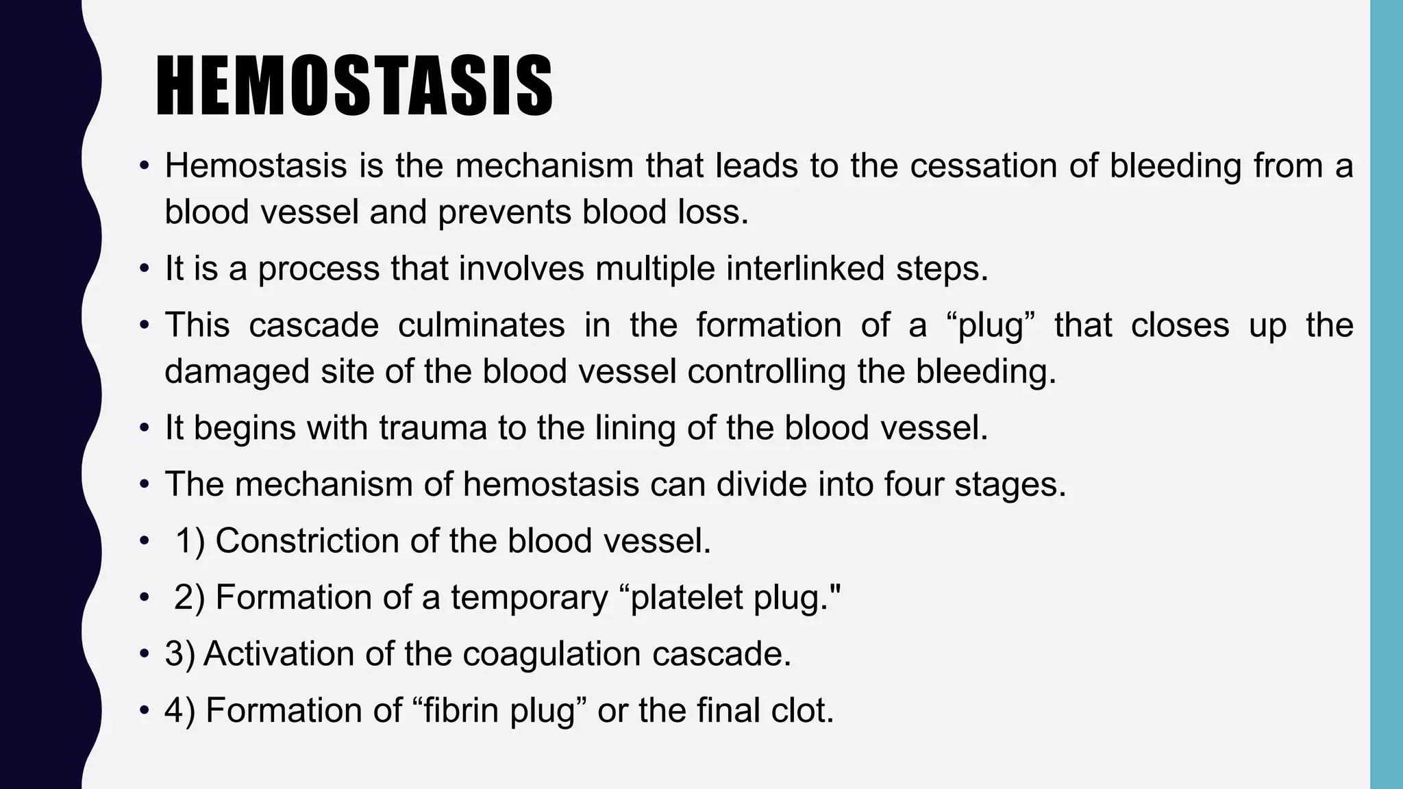
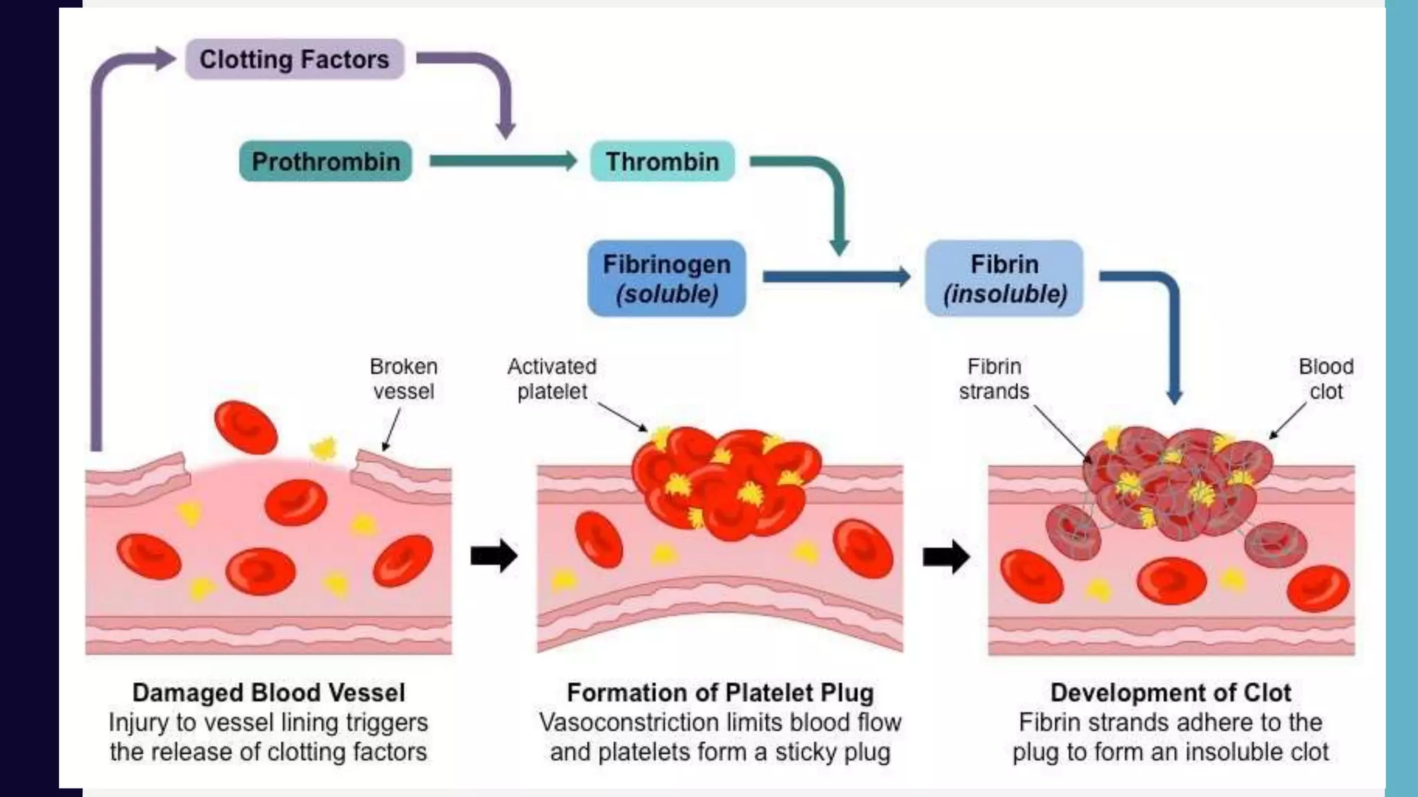
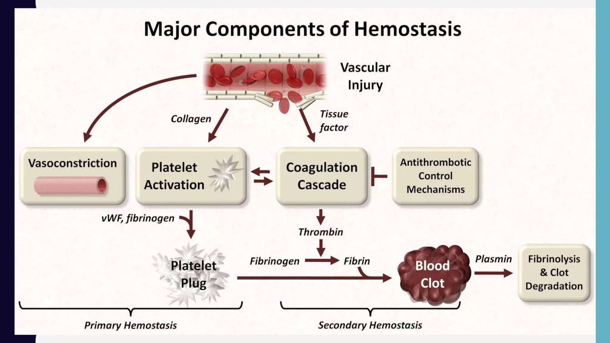
Hemostasis is the process by which bleeding is stopped through a complex cascade of interlinked steps, culminating in the formation of a blood clot. This involves initial platelet aggregation and vasoconstriction to form a temporary plug, followed by activation of coagulation factors that trigger the production of fibrin strands surrounding the platelet plug to form a stable clot. Precise control of coagulation prevents blood loss from injury while maintaining blood fluidity throughout the uninjured circulatory system.
2. HEMOSTASIS
• Hemostasis is the mechanism that leads to the cessation of bleeding from a
blood vessel and prevents blood loss.
• It is a process that involves multiple interlinked steps.
• This cascade culminates in the formation of a “plug” that closes up the
damaged site of the blood vessel controlling the bleeding.
• It begins with trauma to the lining of the blood vessel.
• The mechanism of hemostasis can divide into four stages.
• 1) Constriction of the blood vessel.
• 2) Formation of a temporary “platelet plug."
• 3) Activation of the coagulation cascade.
• 4) Formation of “fibrin plug” or the final clot.
3. BLOOD CLOTTING (COAGULATION)
• The blood clotting process is a multistep activity known as coagulation.
• When the entire coagulation process works appropriately, blood holds firmly together at the site
of an injury, and the bleeding stops.
• When you have a bleeding disorder, you cannot make strong clots quickly or at all.
• The coagulation cascade is a complex chemical process that uses as many as 10 different
proteins (called blood clotting factors or coagulation factors) that are found in plasma.
• The process involves-
1.Injury- A cut on the skin or an internal injury creates a small tear in a blood vessel wall, which
causes blood flow.
2.Vessel constriction- To control blood loss, the blood vessel immediately narrows (called
constriction), which limits blood flow through the vessel.
3.Platelet plug- In response to the injury, tiny cells in the blood called platelets are activated. The
platelets stick to one another and to the wound site to form a plug.
• The protein von Willebrand factor (VWF) helps the platelets stick to each other and to the blood
vessel wall.
4. CONTD….
4.Fibrin clot- Next, blood clotting factors trigger the production of fibrin, a robust and
strand-like substance that surrounds the platelet plug and forms a fibrin clot, a mesh-like
net that keeps the plug firm and stable.
• Over the next several days to weeks, the clot strengthens and then dissolves as the
wounded blood vessel wall heals.
• The clotting factors are-
Factor I (fibrinogen)
Factor IV (ionized
calcium)
Factor VIII
(antihemophilic factor)
Factor XI (plasma
thromboplastin
antecedent)
Factor II (prothrombin)
Factor V (labile factor or
proaccelerin)
Factor IX (plasma
thromboplastin
component or the
Christmas factor)
Factor XII (Hageman
factor)
Factor III (tissue
thromboplastin or tissue
factor)
Factor VII (stable factor
or proconvertin)
Factor X (Stuart-Prower
factor)
Factor XIII (fibrin-
stabilizing factor)
5. CONTD….
• Factor VI was discovered to be part of another factor.
• The liver uses vitamin K to produce some of the factors such as Factors II, VII, IX, and X.
• Normally, vitamin K can be consumed through the diet from plant and animal sources.
The normal flora of the intestine also produces vitamin K.
• Clotting factors III and VIII originate from the endothelial cells while the clotting factor IV
comes from the plasma.
6. SNAPSHOT OF COAGULATION CASCADE
• This cascade is stimulated by clotting factors released from damaged cells (extrinsic
pathway) and platelets (intrinsic pathway).
• Principal events are as follows:
Clotting factors cause platelets to become sticky and adhere to the damaged region to
form a solid plug
These factors also initiate localised vasoconstriction to reduce blood flow through the
damaged region
Additionally, clotting factors trigger the conversion of the inactive zymogen prothrombin into
the activated enzyme thrombin
Thrombin, in turn catalyzes the conversion of the soluble plasma protein fibrinogen into an
insoluble fibrous form called fibrin
The fibrin strands form a mesh of fibres around the platelet plug and traps blood cells to
form a temporary clot
When the damaged region is completely repaired, an enzyme (plasmin) is activated to
dissolve the clot
7.
8. (I) VASCULAR SPASM
A large vessel such as an
artery or vein is severed
Smooth muscle in its
wall contracts in response
to the damage called the
MYOGENIC RESPONSE
Platelets in the ruptured
area release serotonin,
which also brings about
vasoconstriction.
The diameter of the
vessel is
thereby made smaller,
and is blocked by a
blood clot.
If the vessel did
not constrict first, the clot
that forms would quickly
be washed out by the
force of the blood
pressure.
9. (II) PLATELETS PLUGS
When capillaries rupture,
damage is too slight to initiate
the formation of a blood clot.
The rough surface, however,
causes
platelets to change shape
(become spiky and sticky).
These activated platelets stick
to the
edges of the break and to
each other.
The platelets
form a mechanical barrier or
wall to close off the
break in the capillary.
10. (III) CHEMICAL CLOTTING
The stimulus for clotting
is a
rough surface or a break
in the vessel.
Clotting begins, usually
within 15 to 120
seconds.
Stage 1
Platelet factors + tissue
thromboplastin + clotting
factors + calcium ions
form prothrombin
activator
Stage 2
Prothrombin activator
converts prothrombin
to thrombin
Stage 3
Thrombin converts
fibrinogen to fibrin
11. CONTD….
Would platelet plugs be effective for breaks in larger vessels?
• No, they are too small, and though they do form, they are washed away (until a clot
begins to form that can contain them).
Would vascular spasms be effective for capillaries?
• No, because capillaries have no smooth muscle and cannot constrict.