Structure and Functions of the Kidney
•Download as PPTX, PDF•
21 likes•29,548 views

This document summarizes the structure and function of the kidney. It describes that the kidney contains approximately 1-2 million functional units called nephrons. Each nephron contains a glomerular capsule with capillaries that filters blood to form urine. The urine passes through different segments of the nephron and collecting ducts before exiting the kidney. In addition to filtering wastes, the kidneys regulate water, electrolyte and acid-base balance and produce hormones like erythropoietin and renin.

Recommended
More Related Content
What's hot
What's hot (20)
Similar to Structure and Functions of the Kidney
Similar to Structure and Functions of the Kidney (20)
More from MAHESWARI JAIKUMAR
More from MAHESWARI JAIKUMAR (20)
Recently uploaded
Recently uploaded (20)
Structure and Functions of the Kidney
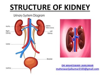
- 1. STRUCTURE OF KIDNEY DR.MAHESWARI JAIKUMAR maheswarijaikumar2103@gmail.com
- 2. • The urinary system is the main excretory system and consists of the following structures: 2 kidneys (secrete urine) 2 ureters (convey urine from the kidney to the urinary bladder) Urinary bladder(collects and stores urine) Urethra (Through which urine leaves the body)
- 4. • The urinary system plays a vital part in maintaining homeostasis of water and electrolytes within the body. • The kidneys produce urine that contains metabolic waste products, including nitrogenous compounds (urea and uric acid, excess ions and some drugs)
- 5. MAIN FUNCTIONS OF KIDNEY • The main functions of kidney are: • Formation of urine, maintaining water and electrolyte balance and acid-base balance • Excretion of waste products
- 6. • Production and secretion of ERYTHROPOETIN (hormone that stimulates formation of RBC) • Production and secretion of RENIN (enzyme to control blood pressure) • Urine is stored in the urinary bladder and excreted by the process of MICTURATION
- 7. • The kidneys lie on the posterior abdominal wall, one on each side of the vertebral column, behind the peritoneum and below the diaphragm • They extend from the level of 12 thoracic vertebra to the 3rd lumbar vertebra
- 8. • The right side kidney is usually slightly lower than the left probably due to considerable space occupied by the liver • Kidneys are bean shaped organs, about 11 cm long, 6 cm wide thick and weigh 150gm
- 9. • They are embedded in and held in position by a mass of fat • A sheath of fibrous connective tissue, the renal fascia, encloses the kidney and the renal fat
- 10. ORGANS ASSOCIATED WITH KIDNEYS • As the kidney lie on either side of the vertebral column each is associated with different structures
- 11. RIGHT KIDNEY • SUPERIORLY- The right Adrenal gland • ANTERIORLY- Right lobe of liver, the duodenum, the hepatic flexure or the colon • POSTERIORLY-diaphragm and muscles of the posterior abdominal wall
- 12. LEFT KIDNEY • SUPERIORLY-Left Adrenal gland • ANTERIORLY-Spleen, stomach, pancreas, jejenum and splenic flexure of the colon • POSTERIORLY- Diaphragm and muscles of the posterior abdominal wall
- 13. GROSS STRUCTURE OF KIDNEY • Three areas of tissue can be distinguished when a longitudinal section of the kidney is viewed with the naked eye • An outer fibrous capsule • The cortex • The medulla
- 17. CORTEX • A reddish brown layer of tissue, immediately below the capsule and outside the renal pyramids
- 19. MEDULLA • The innermost layer, consisting of pale conical shaped striations, the renal pyramids
- 20. HILUM • Is the concave medial border of the kidney where the renal blood and lymph vessels, the ureter and nerves enter
- 22. • Urine formed within the kidney passes through a renal papilla at the apex of a pyramid into a major calyx • Several minor calyx merge into a major calyx and two or three major calyces combine forming the renal pelvis, a funnel shaped structure that narrows when it leaves the kidney as the ureter
- 23. • The walls of the calyces and renal pelvis are lined with transitional epithelium and contain smooth muscle • Peristalsis, intrinsic contractions of smooth muscle, propels the urine through the calyces, renal pelvis and ureters to the bladder
- 25. MICROSCOPIC STRUCTURE • The kidney contains about 1-2 million functional units, the NEPHRONS and smaller number of collecting ducts • The collecting ducts transport urine the pyramids into the calyces, giving the pyramid their striped appearance
- 26. • The collecting ducts are supported by connective tissue, blood vessels, nerves and lymph vessels
- 28. NEPHRON • Is a tubule closed at one end that joins a collecting duct at the other end. • The closed or blind end is indented to form cup-shaped GLOMERULAR CAPSULE (BOWMAN’S CAPSULE) enclosing network of tiny arterial capillaries the glomerulus
- 29. • Continuing from the capsule the remainder of the nephron is about 3 cm long and described in three parts • 1. The proximal convoluted tubule • 2.The medullary loop (Henle’s loop) • 3.The distal convoluted tubule leading into a collecting duct
- 30. • The collecting ducts unite, forming larger ducts that empty into the minor calyces • The kidneys receive about 20% of the cardiac output.
- 31. NEPHRON
- 32. • After entering the kidney at hilum, the renal artery divides into smaller arteries and arterioles. • In the cortex an arteriole (afferent arteriole) enters the glomerular capsule and then subdivides into a cluster of tiny arterial capillaries forming the glomerulus
- 33. • Between these capillary loops are connective tissue phagocytic mesangial cells. • The blood vessel leading away from the glomerulus is the efferent arteriole.
- 34. • The afferent arteriole has a larger diameter than the efferent arteriole, which increases pressure inside the glomerulus and drives filtration across the glomerular capillary walls
- 36. • The efferent arteriole divides into second pertubular (around tubules) capillary network, which wraps around the reminder of the tubule, allowing exchange between the fluid in the tubule and the blood stream
- 38. • This maintains the local supply of oxygen and nutrients and removes waste products • Venous blood drained from this capillary bed eventually leaves the kidney in the renal vein, which empties into the inferior vena cava
- 39. • The walls of the glomerulus and the glomerular capsule consist of a single layer of flattened epithelial cells • The glomerular walls are more permeable than those of other capillaries
- 40. • The remainder of the nephron and the collecting duct are formed by a single layer of simple squamous epithelium • Renal blood vessels are supplied by both sympathetic and parasympathetic nerves
- 41. • The presence of both divisions of the autonomic nervous system controls the renal blood vessel diameter and renal blood flow independently of autoregulation
- 43. THANK YOU