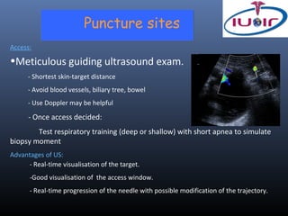
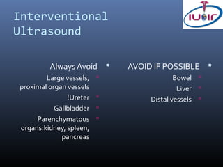
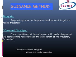
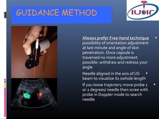
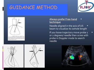
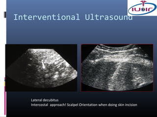
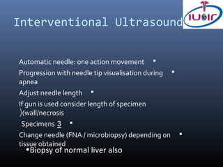
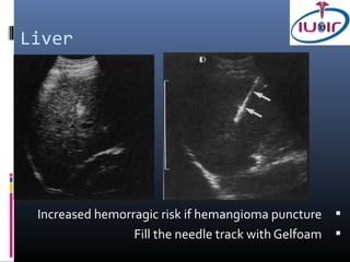
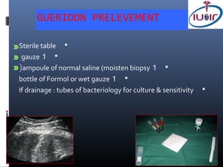
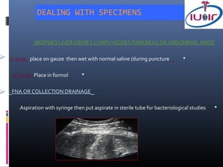
ULTRASOUND & CT
Puncture sites
Access:

•Meticulous guiding ultrasound exam.
      - Shortest skin-target distance
      - Avoid blood vessels, biliary tree, bowel
      - Use Doppler may be helpful

      - Once access decided:
        Test respiratory training (deep or shallow) with short apnea to simulate
biopsy moment
Advantages of US:
     - Real-time visualisation of the target.
      -Good visualisation of the access window.
      - Real-time progression of the needle with possible modification of the trajectory.
Interventional
Ultrasound

         Always Avoid       AVOID IF POSSIBLE 
        Large vessels,                   Bowel 
proximal organ vessels                     Liver 
               !Ureter           Distal vessels 
           Gallbladder   
     Parenchymatous      
organs:kidney, spleen,
             pancreas
CONDITIONS OF REALISATION

One day Hospitalization
(Outpqtient (if cooperative, family at home, classical technique
Fasting
Clinical and imaging data
platelets >150 000             -
PT >70 %s           -
TCA < 2x Normal               -
.Stop anticoagulant TTT 8 days before

Sedation - anxiolytic 2 hours before
!!No sleeping patient
ULTRASOUND

   (.Cleaning of probe, keyboard and cable (protocol                 


Select probe and application depending on procedure                  
            (Try to choose sectorial view if linear probe is used)   
                                                                     
INTERVENTIONAL PROCEDURE


 Skin antispetic measures by technician according to
 protocol
 Patient covered with sterile field
 Sterile material on sterile table

 Cover the US probe with sterile protection
INTERVENTIONAL PROCEDURE


              (Local Anesthesia (10 à 20 cc Lidocaïne 1%                     
                          IM or LP needle according to depth of the target
                                        Evaluation of the trajectory-
                                !Take care of air in the syringe-
                           If liver biopsy go to capsule               -

                                      (Needle guide (US/TDM                  
                                                               anesthesia


  (Skin deep incision with scalpel axis //ribs (intercostal artery           
GUIDANCE METHOD

Biopsy Kit :
        Adaptable systeme on the probe: visualization of target and
needle trajectory


“ Free-hand” Technique :
       Probe is positioned at the entry point with needle along axis of
US beam allowing visualisation of the whole length of the trajectory
(abdominal).


               Always visualize your entry path
                          with real-time needle progression
GUIDANCE METHOD


            Always prefer Free-hand technique 
            possibility of orientation adjustment
            at last minute and angle of skin
            penetration. Once capsule is
            traversed no more adjustment
            possible: withdraw and redress your
            angle
            Needle aligned in the axis of US 
            beam to visualize its swhole length
            If you loose trajectory move probe 1 
            or 2 degrees/ needle then scree with
            probe in Doppler mode to search
            needle
GUIDANCE METHOD

            Always prefer Free-hand 
            technique
            Needle aligned in the axis of US 
            beam to visualize its swhole length
            If you loose trajectory move probe 1   
            or 2 degrees/ needle then scree with
            probe in Doppler mode to search
            needle
Interventional Ultrasound


   Lateral decubitus
   Intercostal approach! Scalpel Orientation when doing skin incision
Needles


Many varieties


Different sizes, calibers, form, shape and nature of the procedure
• Cytology : Chiba needle, Franseen
• Histology : Bard needle
Needles

Many varieties
Interventional Ultrasound

  ’If solid mass : biopsy 18/16 G ‘True cut 
  If cystic mass : initial Fine Needle Aspiration 
  (FNA)-Don’t empty-wall biopsy
  If possible do microbiopsy (histology) of the 
  wall
Co-axial’ Technique‘

types : co-axial - tandem 2 

Coaxial : 1 large bore needle (19G) in contact with the lesion ; 
multiple samples taken with smaller and longer needle inserted
(within it (20G
Advantage : One puncture with multiple samples (<hemorragic
( risk but only one direction

Tandem : 1 needle in the lesion ; biopsy needle parallel 
Advantage : trajectory already done and multiple directions of
biopsy
Interventional Ultrasound


Automatic needle: one action movement 
Progression with needle tip visualisation during   
apnea
Adjust needle length 
If gun is used consider length of specimen
((wall/necrosis
 Specimens 3 
Change needle (FNA / microbiopsy) depending on         
tissue obtained
  •Biopsy of normal liver also
Liver


 Increased hemorragic risk if hemangioma puncture 
                 Fill the needle track with Gelfoam 
Interventional Ultrasound


Possible puncture of distal portal or hepatic branches   


.Biopsy subcapsular lesion by penetration through normal liver   


Use respiration to move the diaphragm and keep away the pleuram      
recess from the needle to get below it


 Coaxial Technique        
GUERIDON PRELEVEMENT

Sterile table 
 gauze 1 
(ampoule of normal saline (moisten biopsy 1 
bottle of Formol or wet gauze 1 
If drainage : tubes of bacteriology for culture & sensitivity   
DEALING WITH SPECIMENS


            :BIOPSIES LIVER KIDNEY LYMPH NODES PANCREAS OR ABDOMINAL MASS

(1st time : place on gauze then wet with normal saline (during puncture         


. 2nd time : Place in formol      


: FNA OR COLLECTION DRAINAGE

     .Aspiration with syringe then put aspirate in sterile tube for bacteriological studies   
COMPLICATIONS


Complications are rare (0,008% à 0,03 %)
-Vasovagal attack


Severe complications
- hemorrhage, arterio-veinous fistula, hematoma and
pneumoperitoneum (liver)
- Acute pancreatitis if normal pancreatic tissue
- Metastatic seeding of the needle track.

Interventional radiology1

  • 1.
  • 2.
    Puncture sites Access: •Meticulous guidingultrasound exam. - Shortest skin-target distance - Avoid blood vessels, biliary tree, bowel - Use Doppler may be helpful - Once access decided: Test respiratory training (deep or shallow) with short apnea to simulate biopsy moment Advantages of US: - Real-time visualisation of the target. -Good visualisation of the access window. - Real-time progression of the needle with possible modification of the trajectory.
  • 3.
    Interventional Ultrasound Always Avoid  AVOID IF POSSIBLE  Large vessels,  Bowel  proximal organ vessels Liver  !Ureter  Distal vessels  Gallbladder  Parenchymatous  organs:kidney, spleen, pancreas
  • 4.
    CONDITIONS OF REALISATION Oneday Hospitalization (Outpqtient (if cooperative, family at home, classical technique Fasting Clinical and imaging data platelets >150 000 - PT >70 %s - TCA < 2x Normal - .Stop anticoagulant TTT 8 days before Sedation - anxiolytic 2 hours before !!No sleeping patient
  • 5.
    ULTRASOUND (.Cleaning of probe, keyboard and cable (protocol  Select probe and application depending on procedure  (Try to choose sectorial view if linear probe is used)  
  • 6.
    INTERVENTIONAL PROCEDURE Skinantispetic measures by technician according to protocol Patient covered with sterile field Sterile material on sterile table Cover the US probe with sterile protection
  • 7.
    INTERVENTIONAL PROCEDURE (Local Anesthesia (10 à 20 cc Lidocaïne 1%  IM or LP needle according to depth of the target Evaluation of the trajectory- !Take care of air in the syringe- If liver biopsy go to capsule - (Needle guide (US/TDM  anesthesia (Skin deep incision with scalpel axis //ribs (intercostal artery 
  • 8.
    GUIDANCE METHOD Biopsy Kit: Adaptable systeme on the probe: visualization of target and needle trajectory “ Free-hand” Technique : Probe is positioned at the entry point with needle along axis of US beam allowing visualisation of the whole length of the trajectory (abdominal). Always visualize your entry path with real-time needle progression
  • 9.
    GUIDANCE METHOD Always prefer Free-hand technique  possibility of orientation adjustment at last minute and angle of skin penetration. Once capsule is traversed no more adjustment possible: withdraw and redress your angle Needle aligned in the axis of US  beam to visualize its swhole length If you loose trajectory move probe 1  or 2 degrees/ needle then scree with probe in Doppler mode to search needle
  • 10.
    GUIDANCE METHOD Always prefer Free-hand  technique Needle aligned in the axis of US  beam to visualize its swhole length If you loose trajectory move probe 1  or 2 degrees/ needle then scree with probe in Doppler mode to search needle
  • 11.
    Interventional Ultrasound Lateral decubitus Intercostal approach! Scalpel Orientation when doing skin incision
  • 12.
    Needles Many varieties Different sizes,calibers, form, shape and nature of the procedure • Cytology : Chiba needle, Franseen • Histology : Bard needle
  • 13.
  • 14.
    Interventional Ultrasound ’If solid mass : biopsy 18/16 G ‘True cut  If cystic mass : initial Fine Needle Aspiration  (FNA)-Don’t empty-wall biopsy If possible do microbiopsy (histology) of the  wall
  • 15.
    Co-axial’ Technique‘ types :co-axial - tandem 2  Coaxial : 1 large bore needle (19G) in contact with the lesion ;  multiple samples taken with smaller and longer needle inserted (within it (20G Advantage : One puncture with multiple samples (<hemorragic ( risk but only one direction Tandem : 1 needle in the lesion ; biopsy needle parallel  Advantage : trajectory already done and multiple directions of biopsy
  • 16.
    Interventional Ultrasound Automatic needle:one action movement  Progression with needle tip visualisation during  apnea Adjust needle length  If gun is used consider length of specimen ((wall/necrosis Specimens 3  Change needle (FNA / microbiopsy) depending on  tissue obtained •Biopsy of normal liver also
  • 17.
    Liver Increased hemorragicrisk if hemangioma puncture  Fill the needle track with Gelfoam 
  • 18.
    Interventional Ultrasound Possible punctureof distal portal or hepatic branches  .Biopsy subcapsular lesion by penetration through normal liver  Use respiration to move the diaphragm and keep away the pleuram  recess from the needle to get below it Coaxial Technique 
  • 19.
    GUERIDON PRELEVEMENT Sterile table gauze 1  (ampoule of normal saline (moisten biopsy 1  bottle of Formol or wet gauze 1  If drainage : tubes of bacteriology for culture & sensitivity 
  • 20.
    DEALING WITH SPECIMENS :BIOPSIES LIVER KIDNEY LYMPH NODES PANCREAS OR ABDOMINAL MASS (1st time : place on gauze then wet with normal saline (during puncture  . 2nd time : Place in formol  : FNA OR COLLECTION DRAINAGE .Aspiration with syringe then put aspirate in sterile tube for bacteriological studies 
  • 21.
    COMPLICATIONS Complications are rare(0,008% à 0,03 %) -Vasovagal attack Severe complications - hemorrhage, arterio-veinous fistula, hematoma and pneumoperitoneum (liver) - Acute pancreatitis if normal pancreatic tissue - Metastatic seeding of the needle track.