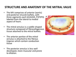
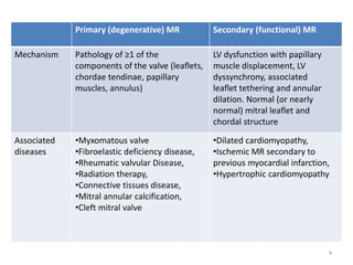
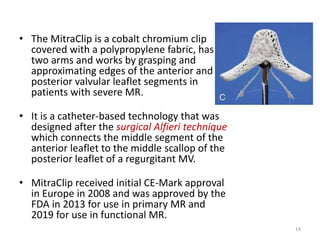
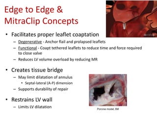
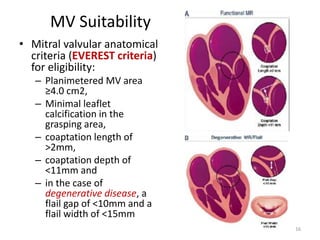
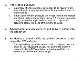
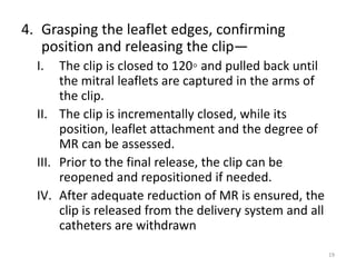
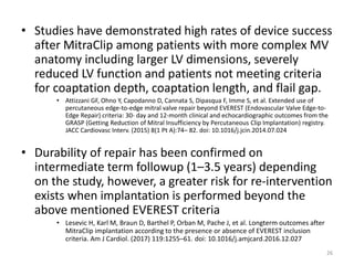
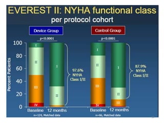
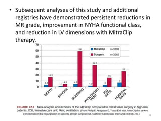
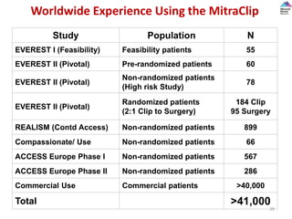
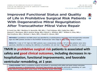
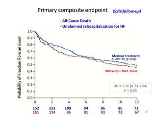
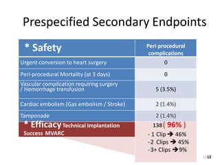
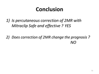
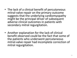
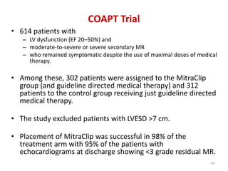
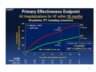
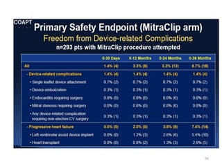
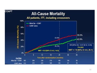
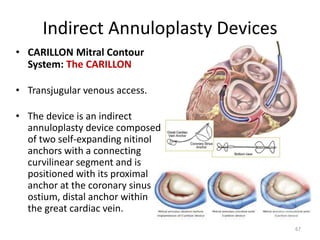
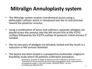
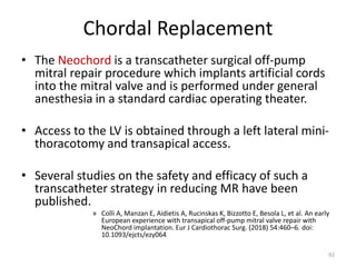
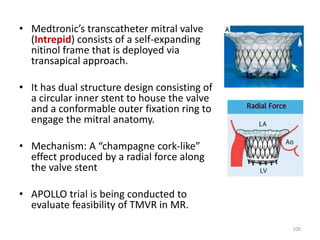
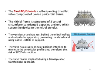
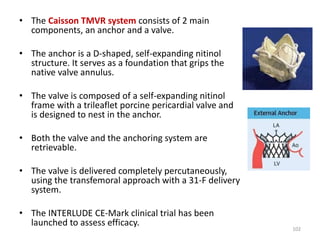
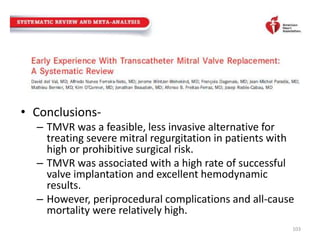
This document discusses percutaneous mitral valve interventions for mitral regurgitation. It begins by describing the anatomy of the mitral valve and causes of mitral regurgitation. It then discusses the natural history of mitral regurgitation and indications for surgery. Current percutaneous options are described including the MitraClip device, which is the only FDA approved one. The MitraClip procedure involves grasping the leaflets edges to reduce regurgitation. Early results show high rates of procedural success for MitraClip in patients at high risk for surgery. Complications are usually low at 15-19% and include bleeding, partial clip detachment, and stroke.
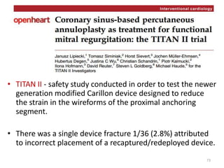
1. The document describes the MitraClip procedure for percutaneous mitral valve repair.
2. The MitraClip procedure involves inserting a clip via the femoral vein to grasp and repair the mitral valve leaflets in a technique similar to the surgical Alfieri repair.
3. The key steps involve transseptal puncture, advancing the clip delivery system into the left atrium, positioning the clip below the mitral valve, grasping the leaflets with the clip, and releasing the clip to create a double orifice mitral valve.
This document discusses development of percutaneous mitral valve repair techniques and clinical trials. It provides background on chronic mitral regurgitation (MR) and the limitations of medical and surgical treatment. Percutaneous mitral valve repair offers benefits over surgery like reduced morbidity and shorter recovery. The document describes the four main percutaneous repair methods and focuses on the MitraClip edge-to-edge leaflet repair system, including patient selection criteria, procedure steps, and clinical trial results demonstrating safety and effectiveness for treating MR.
percutaneous therapies for mitral regurgitationRavi Kanth
This document discusses mitral regurgitation (MR) and percutaneous approaches to mitral valve repair. It provides background on the anatomy of the mitral valve and the types of MR. Surgical repair or replacement is the standard treatment for MR but percutaneous options are being developed to provide less invasive alternatives for high-risk patients. The MitraClip procedure has been used successfully in humans and involves deploying a clip to grasp and oppose the mitral valve leaflets, reducing regurgitation. Indications, outcomes, limitations, and components of the MitraClip system are described.
COAPT was a randomized controlled trial that compared transcatheter mitral valve repair using the MitraClip device plus guideline-directed medical therapy (GDMT) versus GDMT alone in patients with heart failure and secondary mitral regurgitation. The trial found that MitraClip reduced heart failure hospitalizations at 24 months compared to GDMT alone. MitraClip also reduced mortality and improved quality of life. The benefits were seen across different patient subgroups including age and renal function. However, MitraClip was associated with higher costs than GDMT alone.
Percutaneous Balloon Mitral Valvuloplasty (PBMV) is a procedure to dilated the mitral valve in the setting of rheumatic mitral valve stenosis. A catheter is inserted into the femoral vein, advanced to the right atrium and across the interatrial septum. Then the mitral valve is crossed with a balloon and it is inflated to relieve the fusion of the mitral valve commissures effectively acting to increase the mitral valve area and reduce the degree of mitral stenosis. Mitral regurgitation is a potential complication and thus PBMV is contraindicated if moderate or severe regurgitation is present. The Wilkins score examines mitral valve morphology and is determined via echocardiography to assess the likelihood of using PBMV based on certain echocardiographic criteria.
No reflow and slow flow phenomenon during pcirahul arora
This document discusses strategies and prevention of slow flow and no-reflow phenomenon during percutaneous coronary intervention (PCI). It defines no-reflow as inadequate myocardial perfusion through a coronary artery without mechanical obstruction. No-reflow occurs in 8-11% of primary PCIs and is associated with worse clinical outcomes. The pathophysiology involves distal embolization, ischemic injury, reperfusion injury, and individual patient susceptibility. Preventing no-reflow requires reducing thrombus burden, ischemia time, reperfusion injury through anti-inflammatory drugs, and addressing risk factors like diabetes.
Assessment of prosthetic valve functionSwapnil Garde
This document discusses the assessment of prosthetic valve function through various imaging modalities. It begins with an introduction to prosthetic valves and outlines topics to be covered, including classification of valve types. Evaluation methods like chest x-ray, fluoroscopy, echocardiography, and CT are described. Parameters assessed on each modality and guidelines for evaluation are provided. Complications of prosthetic valves and 3D imaging advances are also mentioned.
This document provides an overview of trans-catheter aortic valve implantation (TAVI). It discusses the indications for TAVI including symptomatic severe aortic stenosis in high-risk surgical patients. The pre-procedural workup involves imaging to assess anatomy and risk. The procedure involves accessing the femoral or other arteries and deploying a balloon-expandable or self-expanding bioprosthetic valve. Complications include conduction abnormalities, paravalvular regurgitation, and hypotension. Two clinical cases are presented of high-risk patients undergoing TAVI.
There are many interventional cardiac procedure those need a trans septal puncture of the interatrial septum. This presentation clearly elaborates everything you need to know about the TSP.
DETAILS OF EVIDENCE TAVI FROM ITS EXISTENCE IN INTERVENTIONAL CARDIOLOGY TO THE SURTAVI REGISTRY ..AS AN OPTION FROM HIGH RISK UNOPERABLE PATIENTS TO INTERMEDIATE AND LOW RISK PATIENTS
The document discusses techniques for transseptal puncture (TP). It provides a brief history of septal puncture dating back to the 1950s. It describes the embryology and anatomy of the interatrial septum. The common landmarks and techniques used for fluoroscopy-guided TP are described, including Inoue's angiographic and Hung's modified fluoroscopic methods. Indications for TP include percutaneous mitral commissurotomy and electrophysiology studies. The basic steps of the TP procedure and potential complications are summarized.
This document discusses various echocardiographic scoring systems used to assess mitral valve anatomy and predict outcomes of percutaneous balloon mitral valvuloplasty (PBMV). The Wilkins score and Commissural Calcification score are described in detail. The Wilkins score grades leaflet thickening, mobility, calcification and subvalvular involvement on a scale of 4-16. A score ≤8 indicates favorable anatomy for PBMV. The Commissural Calcification score quantifies calcification at each commissure. Other discussed scores include the Cormier score, RT-3DE score, Chen score, Reid score and Nobuyoshi score. Limitations of the scoring systems and ideas for an ideal future scoring
Ventricular septal defect after myocardial infarctionRamachandra Barik
This document discusses ventricular septal defects (VSDs) that occur after a myocardial infarction. It provides several key points:
1. Surgical repair is the gold standard treatment for post-infarction VSD, but patients are at high risk for complications and mortality rates remain poor.
2. Transcatheter device closure of VSDs is an emerging alternative to surgery, with studies showing high rates of technical success but also risks of major complications and death within 30 days.
3. Factors associated with increased mortality from both surgical repair and device closure include older age, cardiogenic shock, renal dysfunction, and larger defect size. Overall, post-infarction VSD continues to carry
Echocardiography plays an essential role in MitraClip procedures by guiding patient selection, procedural guidance, assessment of results, and follow-up. Three-dimensional TEE provides detailed mitral valve anatomy and allows visualization of catheters and devices in a single view. Key steps of MitraClip include transseptal puncture, clip delivery system advancement, clip positioning and grasping of leaflets, and assessment of residual regurgitation. Residual MR is best assessed using multiple parameters like Doppler, pressures, volumes rather than a single method due to limitations with multiple jets. Three-dimensional TEE may improve quantification of residual MR.
The document discusses transcatheter therapy for mitral regurgitation. It describes the anatomy of the mitral valve and causes of mitral regurgitation. Surgical repair or replacement is the standard treatment but some high-risk patients are not candidates. The MitraClip procedure is described which uses a percutaneous clip to grasp the leaflets and reduce regurgitation as a less invasive alternative for selected patients. Effectiveness has been demonstrated in clinical trials but long-term outcomes are still being evaluated.
This document discusses left main coronary artery (LMCA) percutaneous coronary intervention (PCI). Key points:
1. LMCA disease can be treated with balloon angioplasty or stenting. Drug-eluting stents have reduced restenosis rates compared to bare-metal stents.
2. Assessment of LMCA lesions includes angiography as well as intravascular ultrasound (IVUS) and fractional flow reserve (FFR) to determine significance. An IVUS minimum luminal area <4.5mm2 or FFR<0.80 indicate a significant stenosis.
3. Techniques for stenting LMCA lesions depend on location and involve strategies like single crossover stenting or provisional two
1. Guide catheters provide support for advancing devices into coronary arteries and injecting contrast for visualization. Their selection depends on factors like coronary anatomy, aortic root size, and desired level of support.
2. Common guide catheters include the Judkins, Amplatz, and extra-backup guides. The Judkins provides balanced support while the Amplatz offers firm passive support. Long tip catheters provide coaxial support and manipulation.
3. Achieving proper coaxial alignment and maintaining backup support are important for device delivery and preventing complications. Catheter size, curves, and deep seating techniques impact the level of passive versus active support provided.
Mitral valve morphology assessed via echocardiography is the strongest predictor of outcomes for percutaneous mitral balloon dilation (PTMC). A combined echocardiographic scoring system that includes assessment of leaflet thickness, calcification, mobility and subvalvular thickening can help identify patients likely to have optimal versus suboptimal results. A total score above 11 generally indicates PTMC will be suboptimal, while a score below 9 suggests an optimal outcome. Pre-procedural transesophageal echocardiography is also important to rule out left atrial thrombus, especially in patients with atrial fibrillation. Careful patient selection based on echocardiographic scoring and screening for thrombus can help maximize benefits and
The document discusses mitral regurgitation (MR), including the anatomy of the mitral valve, mechanisms and etiologies of MR, assessment of MR severity using echocardiography techniques like Doppler imaging, and consequences and management of MR. It provides details on evaluating MR severity based on vena contracta width, proximal isovelocity surface area, mitral-aortic time velocity integral ratios, and pulmonary venous flow. Primary causes of MR include degenerative diseases of the valve like Barlow's syndrome, while secondary MR is typically functional and due to left ventricular remodeling without structural valve abnormalities.
This document discusses bifurcation lesions and various techniques for treating them. It begins by defining a bifurcation lesion and different types. It then discusses several classifications of bifurcation lesions including the Medina classification. It provides details on techniques such as T-stenting, crush stenting, culotte stenting and kissing balloon inflation. It summarizes several clinical trials that have compared outcomes of provisional side branch stenting versus systematic two-stent approaches. The document emphasizes that a provisional approach is generally preferred with side branch stenting only if needed. It provides guidance on wire and catheter selection, optimal techniques and the role of kissing balloon inflation.
- The SYNTAX trial compared outcomes of percutaneous coronary intervention (PCI) using drug-eluting stents versus coronary artery bypass grafting (CABG) for treating three-vessel or left main coronary artery disease.
- For the primary endpoint of major adverse cardiac and cerebrovascular events at 12 months, CABG was superior to PCI. However, outcomes were similar between treatments for patients with less complex disease as measured by low or intermediate SYNTAX scores.
- For patients with more complex disease and high SYNTAX scores, CABG had significantly fewer primary events than PCI at 12 months.
Tavi,Transcatheter Aortic Valve Replacement, TAVI,TAVR,Dr.Hasan Mahmud
Transcatheter aortic valve implantation (TAVI) has been developed as an alternative to surgical aortic valve replacement for high-risk patients. TAVI involves threading a collapsible valve through blood vessels and implanting it to replace the diseased valve. Over 30,000 high-risk patients with severe aortic stenosis have undergone TAVI, based on evidence from studies showing it is safer than surgery for this group. TAVI indications may expand as longer-term data on outcomes becomes available and the procedure requires a multidisciplinary team approach and dedicated training.
A lecture on the echocardiographic evaluation of hypertrophic cardiomyopathy. Starts with an overview of the topic then a systematic approach to diagnosis and then a differential diagnosis followed by take-home messages and conclusion.
- The document discusses the Fontan procedure for univentricular heart defects. It covers the evolution of the Fontan concept from the original atriopulmonary connection to lateral tunnel and extracardiac conduit techniques. It also discusses indications for Fontan, complications such as arrhythmias and ventricular dysfunction, and strategies to optimize outcomes like fenestration.
This document describes the step-by-step procedure for transseptal valve-in-valve transcatheter mitral valve replacement (ViV-TMVR) using an Edwards SAPIEN 3 valve. Key steps include patient selection and imaging to assess risk of left ventricular outflow tract obstruction, transseptal puncture to access the left atrium, valve deployment under rapid pacing, and closure of the septal defect if needed. ViV-TMVR provides an alternative to repeat surgery for high-risk patients with a failing bioprosthetic mitral valve. The major risk is left ventricular outflow tract obstruction, which techniques like alcohol septal ablation aim to prevent.
This document discusses the echocardiographic evaluation of mitral valve prolapse (MVP). It describes the use of M-mode, 2D, and 3D echocardiography to diagnose MVP and assess mitral regurgitation severity. Measurement of the vena contracta and use of the proximal isovelocity surface area method are emphasized for accurate regurgitant quantification. Surgical indications and repair techniques are also summarized.
This document discusses trans-septal puncture, which involves puncturing the septum between the right and left atria to access the left side of the heart. It outlines the evolving indications for trans-septal puncture including interventions for mitral valve disease, closure of defects, left atrial procedures, and arrhythmia ablation. The key steps are reviewed - having the proper anatomical landmarks, hardware including sheaths and needles, and imaging guidance. Complications are discussed and how to successfully perform the puncture is summarized as being familiar with the anatomy, hardware, and vigilance for potential complications.
This document discusses various percutaneous mitral valve interventions that are being developed, including those targeting the leaflets (e.g. percutaneous leaflet plication), annulus (e.g. indirect approaches using the coronary sinus), chordae, and left ventricle. It summarizes major clinical trials on the MitraClip device and the benefits seen with percutaneous mitral valve repair compared to medical management alone. Challenges with percutaneous mitral valve replacement are also outlined.
This document discusses the history and techniques of mitral valve surgery. It begins with a brief history of mitral valve repair surgery from 1902 to present. It then describes various techniques for mitral valve repair including leaflet resection, sliding plasty, chordal replacement, and annuloplasty. Indications for mitral valve surgery include symptomatic patients with severe mitral regurgitation or asymptomatic patients with reduced left ventricular function. Mitral valve repair is generally preferred over replacement when possible. Surgical outcomes are improved with repair compared to replacement.
Percutaneous Transcatheter Mitral Valve ReplacementShadab Ahmad
Symptomatic mitral regurgitation (MR) conveys significant morbidity and mortality. However, many patients with severe MR are not treated with surgery due to advanced age, left ventricular (LV) dysfunction, or other comorbidities. This unmet clinical need has driven the development of safer, catheter-based treatments for mitral valve disease.
Transcatheter mitral valve repair can be safe and effective in patients with suitable anatomy.
Chordal preservation DR NIKUNJ R SHEKHADA (MBBS,MS GEN SURG ,DNB CTS SR)DR NIKUNJ SHEKHADA
This document discusses different techniques for chordal preservation during mitral valve replacement surgery. It begins by explaining the importance of preserving left ventricular function during replacement when repair is not possible. Several historical techniques are described, including Lillehei and David's, which aimed to maintain chordal attachments to support left ventricular geometry and function. The advantages of complete chordal preservation are discussed, such as reducing mortality and improving survival by preserving left ventricular structure and function. The document provides an overview of the physiological rationale and technical considerations for different chordal preservation approaches.
Trans catheter intervention is emerging field in cardiac intervention. due to complex anatomy of mitral valve understanding of anatomy and three dimensional imaging is most important aspect of successful intervention and could be life saving in high risk surgical candidate
Surgical Treatment of Ischemic Mitral RegurgitationNora Albogami
This document discusses surgical treatment options for ischemic mitral regurgitation (IMR). It begins by describing the anatomy and physiology behind IMR, as well as medical therapies. Revascularization alone often does not sufficiently reduce IMR. The document then reviews evidence on mitral valve repair versus replacement. While repair traditionally had higher recurrence rates, newer techniques like leaflet augmentation and edge-to-edge repair are showing promise. Overall, the optimal surgical approach for treating IMR remains unclear due to heterogeneity in patient populations and outcomes data.
Successful Valve in Ring Abolishing the Stenosis and Regurgitation with Robus...Premier Publishers
Transcatheter mitral valve-in ring implantation (TMViRI), is a novel alternative treatment strategy and promising technique for patients at high risk of repeat open-heart surgery. In this report we demonstrate a case of 61 years old male with multiple co morbidities who underwent mitral valve repair long time ago who successfully treated and dramatically improved through trans-septal approach, under trans oesophageal echocardiography and fluoroscopic guidance in Hybrid catheterization laboratory.
The document describes the history and evolution of minimally invasive cardiac surgery, including early procedures using smaller incisions rather than full sternotomy, and the development of techniques like port-access bypass which use peripheral cannulation and an endoaortic balloon to occlude the aorta and allow procedures like CABG or mitral valve surgery to be done through smaller incisions. It also covers the various approaches and techniques used for minimally invasive procedures, as well as patient selection considerations and how to harvest vessels like the LIMA through smaller incisions.
The document describes various surgical approaches for acquired mitral valve disease. It discusses considerations for mitral valve repair versus replacement and emphasizes the importance of surgeon experience for complex repairs. Standard median sternotomy is described as the most common approach, while minimally invasive approaches through smaller incisions are also discussed. Specific techniques for exposing the mitral valve through the right atrium, interatrial groove, or left atrial dome are outlined. Factors such as concomitant procedures, myocardial protection, and closure are addressed.
1) The document discusses various surgical procedures for treating aortic root pathologies. It describes the anatomy of the aortic root and various conditions that can affect it like aneurysms and dissections.
2) Surgical techniques discussed include different types of composite graft replacements, valve sparing procedures, and re-do operations. Specific procedures mentioned are the Bentall procedure and the Ross procedure.
3) Factors that determine whether the aortic valve should be replaced or repaired are discussed. Guidelines for intervention based on aortic root size are also provided.
This document discusses the use of echocardiography to guide percutaneous interventions involving the mitral valve, including:
1. Percutaneous balloon mitral valvuloplasty to treat mitral stenosis, assessing valve anatomy and suitability.
2. Edge-to-edge repair with the Mitraclip device to treat mitral regurgitation, including patient selection, valve assessment, and guidance during the procedure.
3. Closure of prosthetic paravalvular mitral leaks, with echocardiography used for assessment, guidance, and monitoring complications.
Journal Club - Extra axial Endoscopic Third Ventriculostomy.pptxDr. Rahul Jain
This document summarizes a presentation on recent advances in endoscopic third ventriculostomy (ETV). It describes the surgical procedure for an extra-axial approach to ETV through the lamina terminalis (EAETV) and provides results from initial studies. EAETV was found to significantly reduce ventricular size in acute hydrocephalus cases and decrease size in chronic cases, with minimal complications. Advantages of EAETV include avoiding restenosis risks and providing an alternative fenestration site. The presentation concludes that while preliminary EAETV results are encouraging, further studies are needed to evaluate its long-term efficacy.
Carotid artery stenting is an alternative to carotid endarterectomy for treating carotid artery stenosis. The document discusses the indications for and procedures involved in carotid revascularization. It summarizes several key studies comparing stenting to endarterectomy. For symptomatic patients, stenting was found to be non-inferior to surgery with the risk of stroke or death below 6%. Recent advances discussed include new embolic protection devices, stent designs like double layer mesh stents, and the transradial approach to reduce manipulation of complex aortic arches. Overall the document provides an overview of carotid stenting procedures and updates on recent technology improvements aimed at reducing risks.
Transeptal access is an integral skill for interventional cardiologists for a multitude of cardiac interventions including,
balloon mitral valvotomy a commonly performed procedure
in India and south Asia. The procedure was first performed by
Braunwald, Ross and Morrow and later refined by Brockenbrough
and Mullins, whose names have been intricately
linked with this procedure.1e3 The procedure, however,
evokes considerable trepidation in many young interventionalists due its steep learning curve and potential catastrophic complications. However, the procedure is relatively
simple in most patients, barring patients with extremely distorted
anatomy like aneursymally dilated left/right atria
where the anatomy of the interatrial septum is often grossly
altered.
The document discusses complications and limitations of the elephant trunk procedure for treating complex aortic aneurysms. Some key points:
- The elephant trunk procedure has acceptable short and long-term outcomes but carries risks of left recurrent nerve injury, aortic rupture early after surgery, and rupture before the second stage.
- Only 61% of patients underwent the planned second-stage repair, and some refused the second procedure.
- Alternatives like a single-stage approach or performing the distal anastomosis more distally can reduce risks but were not always favored in the past.
- Emerging techniques like using the elephant trunk graft to enable endovascular stent grafting of the descending aorta may address some limitations
This document provides guidelines for obtaining optimal views and measurements during a transesophageal echocardiogram (TEE) for mitral valve repair. It recommends obtaining 0, 60-90, 110-130 degree views of the mitral valve as well as additional views such as of the pulmonary veins and 3D imaging. It describes how to obtain measurements of flail gap, flail width, and vertical coaptation length for degenerative and functional mitral regurgitation. The document also provides indications for use and safety information for the MitraClip device intended for reducing mitral regurgitation in high-risk surgical patients.
This document discusses post-myocardial infarction ventricular septal rupture (VSR). It notes that VSR incidence has decreased with improved reperfusion therapies. Surgical repair is the definitive treatment but is high risk, while percutaneous closure and mechanical support have improved outcomes. The timing and presentation of VSR depends on its pathophysiology, which can include acute or delayed rupture. Diagnosis is via echocardiography. Management involves surgical closure if stable, while unstable patients may be supported with devices or surgery delayed. Percutaneous closure is an option for inoperable cases.
Dr. Abhishek presented on coronary artery perforation during PCI. Key points included:
- Incidence ranges from 0.19-3% with increased mortality risk. Risk factors include complex lesions and older age.
- Perforations are classified anatomically and by severity (Ellis classification). Large vessel perforations are highest risk.
- Management involves balloon inflation, covered stents, or catheter techniques to seal the perforation. Distal perforations can be managed with balloon occlusion or embolization.
- Outcomes depend on severity but type III perforations have high mortality. Monitoring for delayed tamponade is important.
Optical coherence tomography (OCT) uses near-infrared light to generate high-resolution images of coronary arteries in vivo.
The near-infrared light with a wavelength of about 1.3 μm is invisible to the human eye.
To generate cross-sectional images, OCT uses low-coherence interferometry by measuring the echo time delay and intensity of the light reflected from internal structures in tissue.
Radiographic Contrast Agents And Contrast-induced Nephropathy
All contrast agents have a basic structure of a benzene ring, which is composed of 6 joined carbon atoms, each of which has an attached hydrogen atom.
Contrast media consist of triiodinated benzene rings, whereby 3 hydrogen atoms are replaced with attached iodine atoms.
Monomers contain 1 triiodinated benzene ring, and dimers contain 2 triiodinated benzene rings
Tetralogy of Fallot (TOF) is a congenital heart defect characterized by four anatomical features - ventricular septal defect, right ventricular outflow tract obstruction, overriding aorta, and right ventricular hypertrophy. It is the most common cyanotic heart defect in children beyond infancy. Patients typically present with cyanosis in infancy which worsens with activity due to right-to-left shunting through the ventricular septal defect. On examination, a systolic murmur may be heard due to the outflow tract obstruction. Without surgical repair, long-term outcomes are poor.
Brugada Syndrome is a genetic cardiac condition characterized by an abnormal ECG pattern and increased risk of sudden cardiac death. It is caused by mutations that result in loss of function of cardiac ion channels, most commonly sodium channels encoded by SCN5A. The ECG typically shows ST segment elevation in leads V1-V3. Brugada Syndrome presents variably from asymptomatic to sudden cardiac death, usually during sleep. Drug challenges may help diagnose when the ECG is unclear. An ICD is recommended for those with symptoms or inducible arrhythmias on electrophysiological study to prevent sudden death.
The funny current, also known as the If current or pacemaker current, underlies the generation of the diastolic depolarization phase of the cardiac action potential and is responsible for repetitive cardiac activity. It is a mixed sodium/potassium current that is activated during membrane hyperpolarization in the diastolic range. The If current is modulated by intracellular cyclic AMP levels and its main channel subunits are HCN channels. Ivabradine is a specific inhibitor of the If current that reduces heart rate by decreasing the slope of diastolic depolarization without substantially changing action potential duration.
- Koch's triangle delineates the location of the atrioventricular node. It is bounded posteriorly by the tendon of Todaro, anteriorly by the tricuspid valve septal leaflet, and inferiorly by the coronary sinus ostium.
- The atrioventricular node and His bundle are located near the apex of the triangle where the His bundle penetrates the central fibrous body. Catheter ablation for atrioventrial nodal reentrant tachycardia often targets the slow pathway region within the triangle.
- The dimensions and structures within Koch's triangle can vary between individuals, which is clinically relevant for catheter ablation procedures guided by anatomic landmarks in this region.
This document discusses bifurcation lesions and interventions. It begins by defining bifurcation lesions as coronary artery narrowings adjacent to or involving the origin of a significant side branch. True bifurcations involve narrowings in both the main branch and side branch. Classification systems like the Medina classification are used to describe bifurcation lesions. The preferred approach for most lesions is provisional stenting of the main branch first, followed by stenting the side branch only if needed. Techniques like wiring both branches, main branch stenting, proximal optimization, and kissing balloon inflations are described. Clinical trials have shown provisional stenting to have similar outcomes to two-stent approaches.
- T-wave alternans (TWA) refers to beat-to-beat variability in the T-wave morphology and amplitude on ECGs and reflects temporal heterogeneity in ventricular repolarization, an important mechanism for arrhythmias.
- TWA can be measured using frequency or time domain analyses and has been associated with increased risk of sudden cardiac death and mortality in various populations, though limitations exist.
- TWA testing can yield positive, negative, or indeterminate results but its use in guiding therapy such as ICD implantation requires further evidence.
Coronary intravascular lithotripsy and lasers/ IVLYogesh Shilimkar
1. The document discusses coronary intravascular lithotripsy (IVL), a new technique for treating severely calcified coronary lesions by using low-pressure shockwaves to fracture calcium deposits.
2. Early studies found IVL to be safe and effective for facilitating stent delivery and expansion in calcified lesions, with low rates of complications.
3. Larger trials confirmed IVL's safety and ability to modify calcium, with most lesions experiencing calcium fracture and acute gains in lumen size post-IVL.
The left atrial appendage (LAA) is a remnant of the left atrium that can be a source of thrombus and stroke in patients with atrial fibrillation. Several percutaneous devices have been developed to occlude the LAA to prevent thrombus formation and reduce the risk of stroke, including the Watchman device. The Watchman is a nitinol frame covered with PET fabric that is implanted via transseptal puncture and deployed in the LAA orifice. Correct placement is confirmed using TEE and fluoroscopy to ensure the device is properly positioned, anchored, sized, and sealing the LAA opening.
This document summarizes the history and mechanisms of action of SGLT2 inhibitors, a class of diabetes medications. Key points include:
- SGLT2 inhibitors work by blocking glucose reabsorption in the kidney, inducing glucosuria and lowering blood glucose levels.
- Several large clinical trials demonstrated SGLT2 inhibitors effectively lower HbA1c and blood glucose with a low risk of hypoglycemia compared to other diabetes medications.
- Beyond glycemic control, SGLT2 inhibitors provide additional benefits like weight loss, blood pressure reduction, and improved cardiac outcomes.
8 Surprising Reasons To Meditate 40 Minutes A Day That Can Change Your Life.pptxHolistified Wellness
We’re talking about Vedic Meditation, a form of meditation that has been around for at least 5,000 years. Back then, the people who lived in the Indus Valley, now known as India and Pakistan, practised meditation as a fundamental part of daily life. This knowledge that has given us yoga and Ayurveda, was known as Veda, hence the name Vedic. And though there are some written records, the practice has been passed down verbally from generation to generation.
- Video recording of this lecture in English language: https://youtu.be/Pt1nA32sdHQ
- Video recording of this lecture in Arabic language: https://youtu.be/uFdc9F0rlP0
- Link to download the book free: https://nephrotube.blogspot.com/p/nephrotube-nephrology-books.html
- Link to NephroTube website: www.NephroTube.com
- Link to NephroTube social media accounts: https://nephrotube.blogspot.com/p/join-nephrotube-on-social-media.html
Our backs are like superheroes, holding us up and helping us move around. But sometimes, even superheroes can get hurt. That’s where slip discs come in.
Cell Therapy Expansion and Challenges in Autoimmune DiseaseHealth Advances
There is increasing confidence that cell therapies will soon play a role in the treatment of autoimmune disorders, but the extent of this impact remains to be seen. Early readouts on autologous CAR-Ts in lupus are encouraging, but manufacturing and cost limitations are likely to restrict access to highly refractory patients. Allogeneic CAR-Ts have the potential to broaden access to earlier lines of treatment due to their inherent cost benefits, however they will need to demonstrate comparable or improved efficacy to established modalities.
In addition to infrastructure and capacity constraints, CAR-Ts face a very different risk-benefit dynamic in autoimmune compared to oncology, highlighting the need for tolerable therapies with low adverse event risk. CAR-NK and Treg-based therapies are also being developed in certain autoimmune disorders and may demonstrate favorable safety profiles. Several novel non-cell therapies such as bispecific antibodies, nanobodies, and RNAi drugs, may also offer future alternative competitive solutions with variable value propositions.
Widespread adoption of cell therapies will not only require strong efficacy and safety data, but also adapted pricing and access strategies. At oncology-based price points, CAR-Ts are unlikely to achieve broad market access in autoimmune disorders, with eligible patient populations that are potentially orders of magnitude greater than the number of currently addressable cancer patients. Developers have made strides towards reducing cell therapy COGS while improving manufacturing efficiency, but payors will inevitably restrict access until more sustainable pricing is achieved.
Despite these headwinds, industry leaders and investors remain confident that cell therapies are poised to address significant unmet need in patients suffering from autoimmune disorders. However, the extent of this impact on the treatment landscape remains to be seen, as the industry rapidly approaches an inflection point.
Muktapishti is a traditional Ayurvedic preparation made from Shoditha Mukta (Purified Pearl), is believed to help regulate thyroid function and reduce symptoms of hyperthyroidism due to its cooling and balancing properties. Clinical evidence on its efficacy remains limited, necessitating further research to validate its therapeutic benefits.
Rasamanikya is a excellent preparation in the field of Rasashastra, it is used in various Kushtha Roga, Shwasa, Vicharchika, Bhagandara, Vatarakta, and Phiranga Roga. In this article Preparation& Comparative analytical profile for both Formulationon i.e Rasamanikya prepared by Kushmanda swarasa & Churnodhaka Shodita Haratala. The study aims to provide insights into the comparative efficacy and analytical aspects of these formulations for enhanced therapeutic outcomes.
Histololgy of Female Reproductive System.pptxAyeshaZaid1
Dive into an in-depth exploration of the histological structure of female reproductive system with this comprehensive lecture. Presented by Dr. Ayesha Irfan, Assistant Professor of Anatomy, this presentation covers the Gross anatomy and functional histology of the female reproductive organs. Ideal for students, educators, and anyone interested in medical science, this lecture provides clear explanations, detailed diagrams, and valuable insights into female reproductive system. Enhance your knowledge and understanding of this essential aspect of human biology.
Osteoporosis - Definition , Evaluation and Management .pdfJim Jacob Roy
Osteoporosis is an increasing cause of morbidity among the elderly.
In this document , a brief outline of osteoporosis is given , including the risk factors of osteoporosis fractures , the indications for testing bone mineral density and the management of osteoporosis
These lecture slides, by Dr Sidra Arshad, offer a quick overview of the physiological basis of a normal electrocardiogram.
Learning objectives:
1. Define an electrocardiogram (ECG) and electrocardiography
2. Describe how dipoles generated by the heart produce the waveforms of the ECG
3. Describe the components of a normal electrocardiogram of a typical bipolar lead (limb II)
4. Differentiate between intervals and segments
5. Enlist some common indications for obtaining an ECG
6. Describe the flow of current around the heart during the cardiac cycle
7. Discuss the placement and polarity of the leads of electrocardiograph
8. Describe the normal electrocardiograms recorded from the limb leads and explain the physiological basis of the different records that are obtained
9. Define mean electrical vector (axis) of the heart and give the normal range
10. Define the mean QRS vector
11. Describe the axes of leads (hexagonal reference system)
12. Comprehend the vectorial analysis of the normal ECG
13. Determine the mean electrical axis of the ventricular QRS and appreciate the mean axis deviation
14. Explain the concepts of current of injury, J point, and their significance
Study Resources:
1. Chapter 11, Guyton and Hall Textbook of Medical Physiology, 14th edition
2. Chapter 9, Human Physiology - From Cells to Systems, Lauralee Sherwood, 9th edition
3. Chapter 29, Ganong’s Review of Medical Physiology, 26th edition
4. Electrocardiogram, StatPearls - https://www.ncbi.nlm.nih.gov/books/NBK549803/
5. ECG in Medical Practice by ABM Abdullah, 4th edition
6. Chapter 3, Cardiology Explained, https://www.ncbi.nlm.nih.gov/books/NBK2214/
7. ECG Basics, http://www.nataliescasebook.com/tag/e-c-g-basics
2. STRUCTURE AND ANATOMY OF THE MITRAL VALVE
• The MV comprises of anterior (aortic)
and posterior (mural) leaflets, with
three segments each (A1A2A3, P1P2P3)
labeled from the lateral to medial
aspect of heart.
• The mitral annulus is a saddle shaped
structure composed of fibrocollagenous
tissue attached to the mitral leaflets.
• The anterior portion of the mitral
annulus is attached to the fibrous
trigones which contain conduction
tissue
• The posterior annulus is less well
developed, more muscular and prone
to dilation. 2
6. Natural History of Mitral Regurgitation
• The natural history of chronic primary MR is highly variable
and depends on a combination of the volume of regurgitation,
state of the myocardium, and cause of the underlying disorder.
• Asymptomatic patients with mild primary MR usually remain in
a stable state for many years.
• Acute rheumatic fever is a frequent cause of isolated severe
MR among adolescents in developing nations, and these
patients often have a rapidly progressive course.
• In asymptomatic patients with severe MR, the rate of
progression to symptoms, LV dysfunction, pulmonary
hypertension, or AF is 30% to 40% at 5 years.
» Bonow RO. Chronic mitral regurgitation and aortic regurgitation: have
indications for surgery changed? J Am Coll Cardiol. 2013;61:693–701
6
7. • Patients with severe MR have decreased survival,
whether symptomatic or not, and surgery is often
recommended.
• In asymptomatic patients with primary MR and
preserved LV function, a “watchful waiting” or “active
surveillance” approach can be considered until the
development of symptoms, LV dysfunction, pulmonary
hypertension, or atrial fibrillation, and current
guidelines recommend surgery in patients who have
reached these endpoints.
• Surgery may also be considered for asymptomatic
patients with normal LV function in whom there is a
high likelihood of successful mitral valve repair.
7
8. • Conclusion
– Surgery was denied in 49% of patients with severe
symptomatic MR.
– Impaired LVEF, older age, and comorbidity were the
most striking characteristics of patients who were
denied surgery.
8
9. • Further, the role of surgery in patients with functional MR
(FMR) remains unproven.
• Surgery improves survival in observational studies but is
associated with mortality rates of 1% to 5% and additional
morbidity rates of 10% to 20%, including stroke,
reoperation, renal failure, and prolonged ventilation.
• As a result, percutaneous technology is poised to
significantly alter the treatment paradigm for chronic MR.
• Percutaneous MVRe offers the potential benefits of
decreased morbidity, improved recovery time, and shorter
hospital stay compared with open heart surgery.
9
10. • Current percutaneous options are loosely based
on surgical repair techniques, with four primary
methods to accomplish a reduction in MR:
– (1) edge-to-edge (E2E) leaflet repair,
– (2) indirect coronary sinus annuloplasty,
– (3) direct annuloplasty, and
– (4) septal-lateral annular cinching.
• Percutaneous MV replacement is also being
developed.
10
11. Percutaneous MV Repair
• Minimally invasive approach to treatment of
certain patients with symptomatic chronic
significant MR.
• Aim - to provide relief from severe MR in patients
who would otherwise not be candidates for
surgical correction.
• Currently, the only US Food and Drug
Administration (FDA) approved device for
percutaneous MV repair in primary and
secondary MR is the MitraClip.
11
13. History
• Dr. Fred St. Goar
– sketched the first
drawings of this catheter-
based device.
• The first human implant
of Mitraclip was
performed in June 2003
by Dr. Jose Condado on
55-year old Octalina
Mendoza from,
Venezuela.
13
14. • The MitraClip is a cobalt chromium clip
covered with a polypropylene fabric, has
two arms and works by grasping and
approximating edges of the anterior and
posterior valvular leaflet segments in
patients with severe MR.
• It is a catheter-based technology that was
designed after the surgical Alfieri technique
which connects the middle segment of the
anterior leaflet to the middle scallop of the
posterior leaflet of a regurgitant MV.
• MitraClip received initial CE-Mark approval
in Europe in 2008 and was approved by the
FDA in 2013 for use in primary MR and
2019 for use in functional MR.
14
15. Edge to Edge &
MitraClip Concepts
• Facilitates proper leaflet coaptation
– Degenerative - Anchor flail and prolapsed leaflets
– Functional - Coapt tethered leaflets to reduce time and force required
to close valve
– Reduces LV volume overload by reducing MR
• Creates tissue bridge
– May limit dilatation of annulus
• Septal-lateral (A-P) dimension
– Supports durability of repair
• Restrains LV wall
– Limits LV dilatation Porcine model, 6M
16. MV Suitability
• Mitral valvular anatomical
criteria (EVEREST criteria)
for eligibility:
– Planimetered MV area
≥4.0 cm2,
– Minimal leaflet
calcification in the
grasping area,
– coaptation length of
>2mm,
– coaptation depth of
<11mm and
– in the case of
degenerative disease, a
flail gap of <10mm and a
flail width of <15mm
16
17. Procedure Technique
• The percutaneous procedure is performed
with the patient under general anesthesia
using transthoracic, transesophageal
echocardiography and fluoroscopic guidance
in the cardiac catheterization laboratory.
• The MitraClip procedure consists of several
steps following femoral venous access:
17
18. 1. Trans-septal puncture:
i. In primary MR, the puncture site needs to be roughly 5 cm
above the mitral annulus to allow sufficient catheter and clip
maneuvering.
ii. In functional MR the puncture site needs to be more inferior
and closer to the annular plane (about 3.5 cm above annular
plane) since tethering of leaflets results in coaptation
occurring below the plane of the mitral annulus.
2. Advancement of guide catheter and delivery system into
the left atrium
3. Positioning of the MitraClip into the left ventricle to just
below the MV leaflets—
I. The clip delivery system is steered until it is aligned over the
origin of the regurgitant jet, its arms opened to orient it
perpendicular to MV coaptation and advanced into the
ventricle just below the leaflet edges.
18
19. 4. Grasping the leaflet edges, confirming
position and releasing the clip—
I. The clip is closed to 120◦ and pulled back until
the mitral leaflets are captured in the arms of
the clip.
II. The clip is incrementally closed, while its
position, leaflet attachment and the degree of
MR can be assessed.
III. Prior to the final release, the clip can be
reopened and repositioned if needed.
IV. After adequate reduction of MR is ensured, the
clip is released from the delivery system and all
catheters are withdrawn
19
20. • In cases with residual MR, additional clips can
similarly be placed in the way of regurgitant jets
while ensuring no evidence of significant de novo
mitral stenosis.
• The entire procedure is performed on intravenous
heparin while serially checking activated coagulation
time (goal >250 s).
• After the clip is placed, patients are treated with
aspirin 325mg daily for 6–12 months and clopidogrel
75mg daily for 30 days.
• These recommendations are based on estimated
time to device endothelialization.
20
22. Contraindications to MitraClip
• Inability to tolerate procedural anticoagulation
or antiplatelet agents postprocedure,
• Active MV endocarditis,
• Rheumatic MV disease,
• Mitral stenosis from any cause and
• Thrombosis of femoral access vein, inferior
vena cava or left sided intracardiac structures
22
23. Complications of MitraClip
• In the EVEREST II clinical trial, major adverse events of
death & major stroke were similar patients receiving
MitraClip and those undergoing MV surgery .
• Patients undergoing surgery needed more blood
transfusions and longer mechanical ventilation while
MitraClip implantation was associated with greater
onset of new AF and acute renal failure.
• Rate of 30-day complications is usually in the range of
15–19% following such transcatheter MV repair.
23
24. • Bleeding is largely peri-procedural from the
vascular access site for MitraClip due to its large
sheath size.
• Partial clip detachment is most common in the
first year post-procedure but occurs in <5% cases.
• Clip embolization and complete detachment or
hemodynamically significant mitral stenosis are
rare.
• Risk for endocarditis involving the MitraClip is
unclear since most data comes from case reports
and use of periprocedural antibiotic prophylaxis
among recipients of MitraClip is controversial
24
25. Early results
• Included 564 patients with high surgical risk with
predominantly primary MR
• Procedural success – 90.6%
• In hospital mortality – 2.3%
• 30 day complication rates – death in 5.8%, stroke
in 1.8%, bleeding in 2.6%, device related
complications – 1.4%
25
26. • Studies have demonstrated high rates of device success
after MitraClip among patients with more complex MV
anatomy including larger LV dimensions, severely
reduced LV function and patients not meeting criteria
for coaptation depth, coaptation length, and flail gap.
• Attizzani GF, Ohno Y, Capodanno D, Cannata S, Dipasqua F, Imme S, et al. Extended use of
percutaneous edge-to-edge mitral valve repair beyond EVEREST (Endovascular Valve Edge-to-
Edge Repair) criteria: 30- day and 12-month clinical and echocardiographic outcomes from the
GRASP (Getting Reduction of Mitral Insufficiency by Percutaneous Clip Implantation) registry.
JACC Cardiovasc Interv. (2015) 8(1 Pt A):74– 82. doi: 10.1016/j.jcin.2014.07.024
• Durability of repair has been confirmed on
intermediate term followup (1–3.5 years) depending
on the study, however, a greater risk for re-intervention
exists when implantation is performed beyond the
above mentioned EVEREST criteria
• Lesevic H, Karl M, Braun D, Barthel P, Orban M, Pache J, et al. Longterm outcomes after
MitraClip implantation according to the presence or absence of EVEREST inclusion
criteria. Am J Cardiol. (2017) 119:1255–61. doi: 10.1016/j.amjcard.2016.12.027
26
28. EVEREST II: randomized clinical trial
• Compared outcomes of TMVRe with surgical
mitral repair among 279 patients with moderate
to severe (grade 3+ or 4+) MR
• Most of the patients have heart failure at
baseline (91% in TMVRe group & 78% in surgical
group)
• Most of the patients (73%) had primary
(degenerative) MR
28
30. • Major adverse events at 30 days were significantly
less frequent with MitraClip therapy (9.6% versus
57% with surgery; P < 0.0001), although much of the
difference could be attributed to the greater need for
blood transfusions with surgery.
30
35. EVEREST II Conclusion
• Although percutaneous repair was less effective
at reducing mitral regurgitation than surgery
before hospital discharge, at 12 and 24 months
the rates of reduction in mitral regurgitation were
similar.
• Percutaneous treatment was associated with
increased safety, improved left ventricular
dimensions, and clinical improvements in NYHA
class and quality of life.
35
37. • The final 5-year results of the EVEREST II trial supported the
superiority of surgery in reducing MR but clearly supported
the long-term safety of the MitraClip and the durability of
MR reduction after percutaneous repair.
• Patients treated with percutaneous repair more commonly
required surgery for residual MR during the first year after
treatment, but between 1- and 5-year follow-up, comparably
low rates of surgery for MV dysfunction with either
percutaneous or surgical therapy endorse the durability of
MR reduction with both repair techniques.
• Similarly, improvements in heart failure symptoms and in LV
dimensions remained stable through 5-year follow-up,
mitigating concerns that residual MR after device placement.
37
38. • Subsequent analyses of this study and additional
registries have demonstrated persistent reductions in
MR grade, improvement in NYHA functional class,
and reduction in LV dimensions with MitraClip
therapy.
38
39. Worldwide Experience Using the MitraClip
Study Population N
EVEREST I (Feasibility) Feasibility patients 55
EVEREST II (Pivotal) Pre-randomized patients 60
EVEREST II (Pivotal)
Non-randomized patients
(High risk Study)
78
EVEREST II (Pivotal)
Randomized patients
(2:1 Clip to Surgery)
184 Clip
95 Surgery
REALISM (Contd Access) Non-randomized patients 899
Compassionate/ Use Non-randomized patients 66
ACCESS Europe Phase I Non-randomized patients 567
ACCESS Europe Phase II Non-randomized patients 286
Commercial Use Commercial patients >40,000
Total >41,000
39
40. • Given the excellent results of MVRe in patients
with primary MR who had acceptable surgical
risk, interest turned to the use of the MitraClip
for those patients at prohibitive surgical risk.
40
41. TMVR in prohibitive surgical risk patients is associated with
safety and good clinical outcomes, including decreases in re-
hospitalization, functional improvements, and favorable
ventricular remodeling, at 1 year.
41
43. Chronic Secondary MR
• Surgery in patients with secondary MR is generally
high risk, has poor durability, and is associated
with worse overall survival than in patients with
DMR.
• As a result, the major professional societies,
including the ACC/AHA, the European Society of
Cardiology (ESC), and the International Society for
Heart and Lung Transplantation (ISHLT) provide a
class IIb indication for MV surgery in patients with
FMR.
43
44. • Among 149 patients with secondary MR included
in the high-risk surgical cohort of EVEREST II and
the continued access registry, 63% had prior
cardiac surgery and 87% had NYHA class III or IV
symptoms.
• Despite their high-risk profile, these patients
demonstrated a low (4.7%) mortality rate at 30
days.
• At 1 year, 82% had 2+ or less MR, and patients
demonstrated significant reduction in LV
volumes.
44
45. • The ACCESS-EU trial (A Two-Phase Observational
Study of the MitraClip System in Europe) involved 567
high-risk patients, including 393 patients (69%) with
FMR
– 66% had an LVEF of less than 40%.
• Despite the high-risk profile, the mortality rate was
only
– 2.8% at 30 days and
– 17% at 1 year.
• At 1 year,
– 78.6% of the FMR patients had 2+ or less MR, and
– 71% of the entire group had NYHA class I or II symptoms.
45
48. • Two separate randomized controlled trials
compared the efficacy of percutaneous MV
repair using MitraClip to medical therapy
among patients with significant secondary MR
and underlying LV abnormality
48
49. MITRA-FR trial
• The trial enrolled
– 304 symptomatic patients with LV dysfunction (EF 15–40%)
and
– moderate to severe secondary MR
• Randomized either to percutaneous mitral-valve repair
plus medical therapy (n = 152) or medical therapy
alone (n = 152).
• Placement of MitraClip was successful in 95.8% of the
treatment arm with 91.9% of the patients showing <3
grade residual MR at discharge
49
50. months
152 123 109 94 86 80 73
151 114 95 91 81 73 67
Primary composite endpoint (99% follow-up)
- All-Cause Death
- Unplanned rehospitalization for HF
Mitraclip + Med. treat.
Medical treatment
OR = 1.16 (0.73-1.84)
P = 0.53
50
52. Conclusion
1) Is percutaneous correction of 2MR with
Mitraclip Safe and effective ? YES
2) Does correction of 2MR change the prognosis ?
NO
52
53. • The lack of a clinical benefit of percutaneous
mitral-valve repair on the primary outcome
suggests that the underlying cardiomyopathy
might be the principal driver of subsequent
adverse clinical outcomes in patients with
secondary mitral regurgitation.
• Another explanation for the lack of clinical
benefit observed could be the fact that some of
the patients who underwent percutaneous
mitral-valve repair had incomplete correction of
mitral regurgitation.
53
54. COAPT Trial
• 614 patients with
– LV dysfunction (EF 20–50%) and
– moderate-to-severe or severe secondary MR
– who remained symptomatic despite the use of maximal doses of medical
therapy.
• Among these, 302 patients were assigned to the MitraClip
group (and guideline directed medical therapy) and 312
patients to the control group receiving just guideline directed
medical therapy.
• The study excluded patients with LVESD >7 cm.
• Placement of MitraClip was successful in 98% of the
treatment arm with 95% of the patients with
echocardiograms at discharge showing <3 grade residual MR.
54
59. Conclusions of COAPT trial
• In pts with HF and moderate-to-severe or severe
secondary MR who remained symptomatic despite
maximally-tolerated GDMT, transcatheter mitral leaflet
approximation with the MitraClip was safe, provided
durable reduction in MR, reduced the rate of HF
hospitalizations, and improved survival, quality-of-life
and functional capacity during 24-month follow-up
• MitraClip shown to improve the prognosis of patients
with HF by reducing secondary MR due to LV
dysfunction
59
60. • At 36 months MitraClip was
– safe,
– provided durable reduction in MR,
– reduced the rate of HF hospitalizations, and
– improved survival, QOL and functional capacity
compared to GDMT alone.
60
61. Why are the COAPT Results so Different from MITRA-FR?
Possible Reasons
MITRA-FR (n=304) COAPT (n=614)
Severe MR entry criteria
EROA >20 mm2 or
RV >30 mL/beat
EROA >30 mm2 or
RV >45 mL/beat
EROA (mean ± SD) 31 ± 10 mm2 41 ± 15 mm2
LVEDV (mean ± SD) 135 ± 35 mL/m2 101 ± 34 mL/m2
GDMT at baseline and FU
Receiving HF meds at baseline
– allowed variable adjustment
in each group during follow-up
per “real-world” practice
CEC confirmed pts were
failing maximally-tolerated
GDMT at baseline – few
major changes during follow-
up
Acute results: No clip / ≥3+
MR
9% / 9% 5% / 5%
Procedural complications* 14.6% 8.5%
12-mo MitraClip ≥3+ MR 17% 5%
63. • The PASCAL TMVr system is
designed to overcome
shortcomings of the MitraClip
system by
– facilitating easy steering within the
left atrium,
– larger implant size,
– broader paddles with central
spacer within device to reduce MR
by maximizing leaflet coaptation,
– ability to grasp individual leaflets
and implant elongation to
promote safe subvalvular
maneuvering.
63
64. • Placement of the device occurs via transvenous access
(femoral) and transseptal approach similar to the
MitraClip.
• In its first human study, the PASCAL TMVr was studied in
23 patients with symptomatic severe degenerative,
functional or mixed etiology MR (NYHA functional class
III to IV) and patients were deemed high to inoperable
surgical risk
• Patients were not considered candidates for MitraClip
repair either due to anatomical complexity (short
posterior leaflet, large malcoaptation area, severe
annular dilatation >61mm) or lack of an approved
indication for use.
64
65. • Procedural success was obtained in 22/23 patients
(96%), residual MR was <3 grade in 96% patients and
reduction in NYHA functional class ≤II grade occurred
in 95% of the cases.
• Direct procedure related complications occurred in
two cases (9%) from a minor bleeding event and
transient ischemic attack, respectively.
• The device expanded patient eligibility for repair
especially in case of short posterior leaflets and larger
flail gaps and needs additional data on durability and
future head-to-head comparisons with newer
generations of the MitraClip device.
65
67. Indirect Annuloplasty Devices
• CARILLON Mitral Contour
System: The CARILLON
• Transjugular venous access.
• The device is an indirect
annuloplasty device composed
of two self-expanding nitinol
anchors with a connecting
curvilinear segment and is
positioned with its proximal
anchor at the coronary sinus
ostium, distal anchor within
the great cardiac vein.
67
68. • Upon deployment the device plicates the tissue next to the
MV annulus reducing mitral annular dilation and degree of
MR by bringing the anterior and posterior leaflets closer.
• Coronary angiography is also performed to evaluate for left
circumflex-obtuse marginal arterial system compression
following deployment due to its close proximity.
• Current technology allows either recapture and
repositioning of device implant during same procedure or
recapture and removal of original device followed by new
device implantation when needed.
• Such manipulation becomes possible due to its benign
design and availability in multiple sizes.
68
69. • Clinical feasibility was evaluated using the next-generation
CARILLON XE device in 48 patients with FMR and LV systolic
dysfunction.
• The device was successfully implanted in 30 patients.
• At the 6-month follow-up, there was a durable and
significant decrease in
– mitral annulus diameter (from 4.2 to 3.78 cm [10%]),
– MR (average reduction, 23%), and
– NYHA class (from 2.9 to 1.8), and
• Improvement in the quality-of-life score and 6-minute walk
testing (from 307 to 403 meters).
AMADEUS trial
(CARILLON Mitral Annuloplasty Device European Union Study)
69
70. • With respect to safety, six patients (13%) experienced a
total of seven complications within 30 days of the
procedure: one patient died of multiorgan failure,
three experienced myocardial infarction (none
requiring percutaneous coronary intervention), and
three experienced coronary sinus dissection or
perforation.
• The complications were clustered early in the
experience and resulted in changes to the implantation
procedure; improvement in safety was observed later
in the study.
• On the basis of this early work, the CARILLON system
was granted the CE mark of approval for use in Europe.
70
71. • The objective of the TITAN trial was to assess the haemodynamic
and clinical significance of treating FMR percutaneously in HF
patients using the Carillon Mitral Contour System.
• Follow-up of patients in whom the device was recaptured during the
index procedure provided a non-randomized, non-blinded
comparison group.
• Device implantation was shown to be safe, with no major adverse
event attributable to the annuloplasty device.
• In contrast to the comparison group, implanted patients displayed a
significant reduction in the degree of FMR that was associated with
reverse remodelling.
• There was sustained benefit in clinical parameters of NYHA class and
6MWD up to 24 months.
71
72. • The earlier generation device had a
reasonable safety profile, but asymptomatic
wire-form fractures were seen at the level of
the high strain proximal anchor locking
mechanism in the Carillon device in 25% of
cases.
72
73. • TITAN II - safety study conducted in order to test the newer
generation modified Carillon device designed to reduce
the strain in the wireforms of the proximal anchoring
segment.
• There was a single device fracture 1/36 (2.8%) attributed
to incorrect placement of a recaptured/redeployed device.
73
74. • The primary end point of 30-day major adverse event
rate was 2.8%.
• The 1-year mortality was 23% (7 of 30 patients) and no
deaths were adjudicated to be device related.
• From the efficacy standpoint, TITAN II showed similar
clinical and echocardiographic benefits as in TITAN with
reduction in MR, mitral annular dimension,
improvement in NYHA functional class and a trend
toward reduction in ventricular size suggestive of
reverse remodeling.
TITAN II Results
74
76. • The simplicity of this approach is supported by
the study being largely done by centers
previously unfamiliar with the technology.
• Other advantages include the right-sided
approach, avoidance of trans-septal puncture,
and the fact that Carillon device placement does
not preclude any future mitral valve treatment if
needed later.
• Studies are now underway to assess the effect of
this approach on clinical outcomes.
76
77. • The MONARC percutaneous
transvenous annuloplasty device
consists of
– a stent-like anchor placed in the great
cardiac vein,
– a connecting bridge, and
– a second anchor located proximally at the
coronary sinus ostium.
• The compressed device can be
introduced from the jugular vein
using a long sheath.
• Device was evaluated in the
EVOLUTION I feasibility trial, which
enrolled 72 symptomatic patients
with grade 2 functional MR. Interim
analysis found a reduction in MR
severity and functional class in the
majority of patients.
77
78. ARTO device
• The MVRx ARTO
transcatheter annular
reduction therapy is an
indirect annuloplasty
system that includes
transvenous delivery of 2
anchors:
– one through the interatrial
septum,
– the other to the coronary
sinus
• It acts by reducing the
anteroposterior diameter
of the mitral annulus.
78
79. MAVERIC trial
• To evaluate safety and feasibility of ARTO system in patients with MR
with congestive heart failure.
• In the first phase of MAVERIC trial, 11 patients underwent successful
device implantation with one device displacement and one pericardial
effusion requiring surgical intervention.
• At 30-day follow up, a decrease in regurgitant volumes from 45.4 ±
15.0 ml to 19.5±10.2 ml was demonstrated with a beneficial effect on
LV volumes.
• LV end-systolic volume index improved from 77.5 ± 24.3 ml/m2 to 68.5
± 21.4 ml/m2, and LV end-diastolic volume index from 118.7 ± 28.6
ml/m2 to 103.9 ± 21.2 ml/m2.
• Mitral annular AP diameter decreased from 45.0 ± 3.3 mm to 38.7 ±
3.0 mm with an associated improvement in New York Heart
Association (NYHA) functional class.
79
80. • Data at 2-year follow up demonstrated a
consistent significant
– improvement in functional MR grade, regurgitant
volumes and
– reduction in mitral annular AP diameter.
• These changes were associated with an
improvement in symptomatic status from
81.8% NHYA functional class III/IV at baseline
to 60.0% NYHA functional class I/II at 2 years.
80
81. Mitral Loop Cerclage Catheter System
• In the mitral loop cerclage
procedure, both the femoral
vein and left subclavian vein
are accessed.
• The cerclage is accomplished
by using a guidewire to enter
the coronary sinus and great
cardiac vein, crossing the
interventricular septum from
the anterior interventricular
vein into the right ventricle,
snaring this wire from the
right ventricular outflow tract
and forming a loop around
mitral annular plane.
81
82. • The procedure is performed either under general
anaesthesia or moderate sedation.
• The first in human study attempted the procedure in
5 patients, was successful in 4/5 patients but
aborted in one due to unfavourable anatomy .
• The device resulted in immediate reduction in MR
that was sustained upto 6-months and reduced left
atrial and LV chamber volumes over time.
• Device related complications were coronary artery
occlusion, new bundle branch block and need for a
repositioning procedure.
82
83. CARDIOBAND
• The Cardioband device
delivers direct sutureless
anchors around the mitral
annulus to connect the
annuloplasty device.
• The cardioband system
enables adjustable septo-
lateral diameter
compression, reducing MV
annulus size and severity of
MR.
83
84. Cardioband Mitral System CE mark trial
• In this largest multicenter study of 60 patients with moderate
to severe secondary MR who underwent Cardioband
implantation early results raised issue with device design
leading to device modification half way through the study.
• Anchor disengagement was observed in 10 patients, resulting
in device inefficacy in five patients but most (9/10 anchor
disengagement) occurred prior to device modification.
• There were no device related deaths and 2-year overall
survival was 79%.
• The Cardioband system gained CE Mark approval for the
treatment of functional MR in 2015.
84
88. Mitralign Annuloplasty system
• The Mitralign system involves transfemoral access using a
deflectable catheter which is introduced into the LV and directed
toward the posterior annulus.
• Using a combination of wires and catheters, polyester pledgets are
placed across the annulus into the left atrium first at the P1/P2
scallops followed by the P2/P3 scallops of posterior mitral annulus if
needed.
• One to two pairs of pledgets are plicated, locked and the result is a
reduction in MV annular diameter.
• The device has been tested in a prospective,multicenter singlearm
feasibility study where 45 patients underwent procedure.
– Nickenig G, Schueler R, Dager A, Martinez Clark P, Abizaid A, Siminiak T, et al.
Treatment of chronic functional mitral valve regurgitation with a percutaneous
annuloplasty system. J Am Coll Cardiol. (2016) 67:2927– 36. doi:
10.1016/j.jacc.2016.03.591 88
89. • There were no intraprocedural deaths or conversion to surgery,
but pericardial tamponade occurred in 4 (8%) patients.
• Exclusion of LVEDD <5 cm and second generation catheter
systems have decreased the risk of tamponade.
• Within 6-months, all-cause mortality was 12.2%, 7 (17%)
patients underwent MitraClip placement and one patients
received non-emergent MV surgery.
• Improvement was noted in MR severity in 50% of the patients
with worsening in 15.4% cases, with greater trend for
improvement in those who received 2 pledgets.
• HF symptoms and 6-min walk test improved at 6-months from
baseline.
• Mitralign system has gained CE mark approval for functional MR
in 2016.
89
90. Newer Devices
• The Millipede IRIS ring
– A semirigid “zigzag” shaped annuloplasty ring, with eight helical stainless steel
anchors that anchor directly into the mitral annulus.
– Device has eight tensioning sliders that can be used to actuate the device and
reduce the annulus size .
• The AccuCinch Ventricular Repair System (Ancora Heart, Santa Clara, CA)
– uses a retrograde arterial mechanism to implant a series of adjustable anchors
within the LV wall tethered by a cable below themitral valve annulus.
– The cable is tightened to cinch the left ventricular wall, reducing ventricular size
and consequently mitral annulus, thus succeeding in lowering regurgitant
volume.
– Unlike other systems within this section, the AccuCinch system represents more
of a ventriculoplasty than direct annuloplasty due to its direct support to and
placement within the left ventricular myocardium; consequently, this device is
current being tested in heart failure patients with dilated left ventricles but
without significant valvular lesions.
• Prospective clinical data is awaited on these devices and studies are
underway.
90
91. Cardiac Chamber Remodeling Devices
• These include
– Coapsys and iCoapsys
– Percutaneous Septal Sinus Shortening (PS3)
system
– Basal Annuloplasty of the Cardia Externally (BACE)
• Financial hardship has led to abandonment of
the Coapsys device, and the PS3 and BACE
devices have been demonstrated only in
limited use.
91
92. Chordal Replacement
• The Neochord is a transcatheter surgical off-pump
mitral repair procedure which implants artificial cords
into the mitral valve and is performed under general
anesthesia in a standard cardiac operating theater.
• Access to the LV is obtained through a left lateral mini-
thoracotomy and transapical access.
• Several studies on the safety and efficacy of such a
transcatheter strategy in reducing MR have been
published.
» Colli A, Manzan E, Aidietis A, Rucinskas K, Bizzotto E, Besola L, et al. An early
European experience with transapical off-pump mitral valve repair with
NeoChord implantation. Eur J Cardiothorac Surg. (2018) 54:460–6. doi:
10.1093/ejcts/ezy064
92
93. • The ReChord Trial
– Randomized Trial of the NeoChord DS1000 System
versus Open Surgical Repair
– The objective of this study is to assess the safety
and effectiveness of the NeoChord DS1000 System
in subjects with degenerative mitral valve disease.
93
94. • Another similar device is the
Harpoon MV Repair System
that anchors artificial cords
on the flaps to take the
place of the natural cords via
transapical off-pump
surgical technique using
transcatheter technology.
• Chordal replacement is
more commonly used in
degenerative MV disease
and no current transvenous
or transarterial systems
mimic either of these
techniques.
94
95. Transcatheter MV Replacement
• Mitral valve disease is a highly heterogenous
disorder and the mitral valve is a remarkably
complicated structure.
• Challenges lie in:
– (I) delivery systems;
– (II) valve sizing;
– (III) prevention of paravalvular leak;
– (IV) prevention of LVOTO;
– (V) valve thrombosis;
– (VI) weighing up the variations between the devices.
95
96. Valve in Valve (Sapien 3)
• Careful preoperative CTA and echocardiographic
assessment should be performed to demonstrate that the
annular calcifications are relatively circular and their size is
sufficient to allow for seating of the valve before the
operation begins.
• Transseptal route is frequently used
• As per VIVID registry data, 88.8% of 349 mitral VIV
procedures were clinically successful.
• In June 2017, Sapien 3 valve received US-FDA approval for
VIV procedures at aortic and mital poitions in high risk
patients.
96
97. • The Edwards FORTIS
transcatheter mitral valve is a
cloth-covered, self-expanding
nitinol stent with three bovine
pericardial leaflets that is
implanted via a transapical
approach.
• It is non-recapturable, tethered
via the subvalvular apparatus
and is circularly oriented.
• A high rate of valve thrombosis
in three patients (19%) led
cessation of implantation of this
valve in 2016.
97
98. • The Tendyne mitral valve system –
– a trileaflet porcine pericardial valve on a
self-expanding nitinol double-frame stent,
an adjustable tether, and an apical
fixation/sealing pad.
• There is an atrial cuff designed to
– provide sealing (prevention of diastolic
paravalvular leak) and
– anchoring (preventing valve from pulling
into the ventricle when force is applied to
the tether).
• A left ventricular apical tethering
system with an apical pad anchors the
device and assists with apical closure.
• The device is implanted using a
transapical approach and can be fully
retrieved or repositioned even after full
deployment
98
99. • The Neovasc Tiara
– non-recapturable, tethered via a fibrous
trigone capture with native leaflet
engagement and is D-shaped.
• Mechanism:
– native leaflet engagement :Ventricular
anchors to grasp the free margins of the
native leaflets
• It comes in a 35 and 40 mm
configuration, with two anterior and one
posterior anchoring structures.
• It is implanted via a transapical
approach.
• Currently TIARA I & TIARA II trials are
undergoing.
99
100. • Medtronic’s transcatheter mitral valve
(Intrepid) consists of a self-expanding
nitinol frame that is deployed via
transapical approach.
• It has dual structure design consisting of
a circular inner stent to house the valve
and a conformable outer fixation ring to
engage the mitral anatomy.
• Mechanism: A “champagne cork-like”
effect produced by a radial force along
the valve stent
• APOLLO trial is being conducted to
evaluate feasibility of TMVR in MR.
100
101. • The CardiAQ-Edwards - self-expanding trileaflet
valve composed of bovine pericardial tissue.
• The nitinol frame is composed of 2 sets of
circumference-oriented opposing anchors which
secure the device to the mitral annulus.
• The ventricular anchors rest behind the mitral leaflets
and subvalvular apparatus, preserving the chords and
using native leaflets as support.
• The valve has a supra-annular position intended to
minimize the ventricular profile and, therefore, the
risk of LVOT obstruction.
• The valve can be implanted using a transapical or
transfemoral approach.
101
102. • The Caisson TMVR system consists of 2 main
components, an anchor and a valve.
• The anchor is a D-shaped, self-expanding nitinol
structure. It serves as a foundation that grips the
native valve annulus.
• The valve is composed of a self-expanding nitinol
frame with a trileaflet porcine pericardial valve and
is designed to nest in the anchor.
• Both the valve and the anchoring system are
retrievable.
• The valve is delivered completely percutaneously,
using the transfemoral approach with a 31-F delivery
system.
• The INTERLUDE CE-Mark clinical trial has been
launched to assess efficacy.
102
103. • Conclusions-
– TMVR was a feasible, less invasive alternative for
treating severe mitral regurgitation in patients with
high or prohibitive surgical risk.
– TMVR was associated with a high rate of successful
valve implantation and excellent hemodynamic
results.
– However, periprocedural complications and all-cause
mortality were relatively high.
103
105. MitraClip is good but there are issues
• Failure to eliminate MR
– Is moderate(≤2+) good enough?
– Residual severe (3-4+) MR in 5% patients
• Late recurrence of MR
– Reduces option for surgical repair
• Mitral stenosis
– MPG <5 mmHg in cath lab can be worse when
patient is ambulatory
105
106. TMVR vs MitraClip for secondary MR
• MitraClip reduces MR
• TMVR eliminates MR
• Study hypothesis need to be tested is that can
elimination of MR with TMVR will be more
effective than reduction of MR with MitraClip
• Problem is safety
– Safety profile of MitraClip is very high
– TMVR must become transfemoral/transseptal!
106