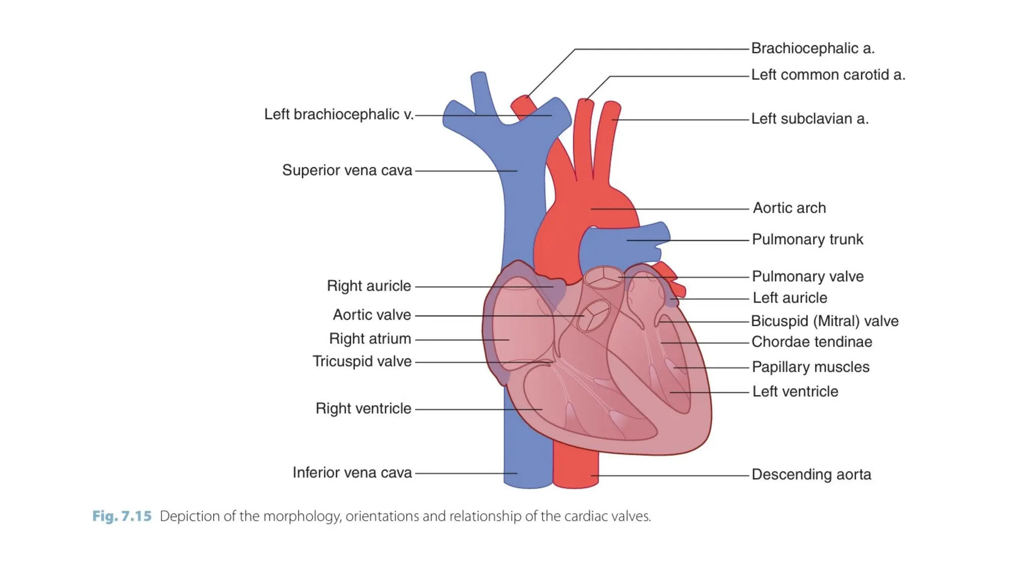
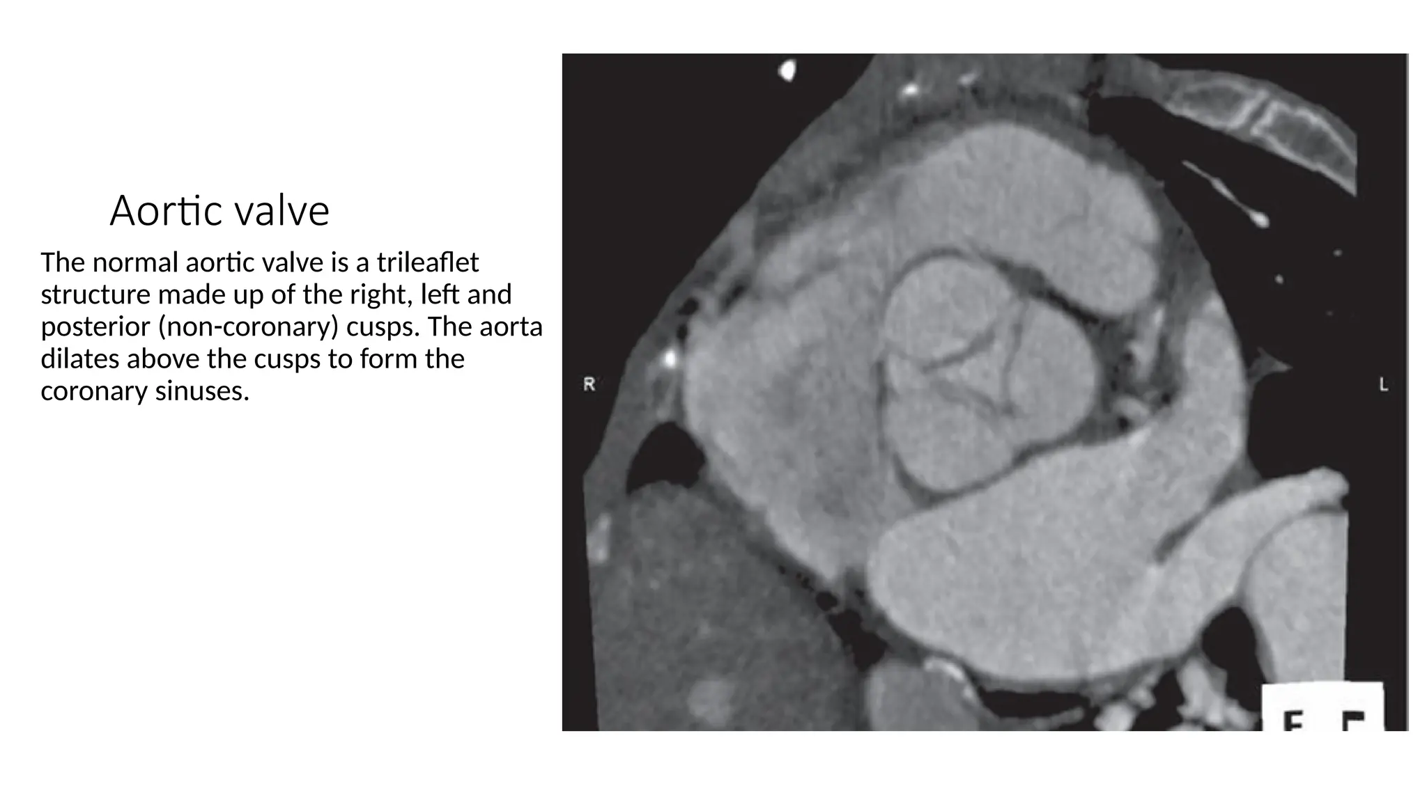
Radiology Anatomy Of The
Heart And Great Vessels
Dr. Abdirahman
Tutor: Prof. Alina
OUTLINE
• Heart gross anatomy and orientation
• Pericardium
• Pericardial recesses
• Heart chambers
• Heart commonly depicted planes
• Cardiac Valves
• Great Vessels
Gross anatomy and orientation
• The heart is pyramidal in shape and lies obliquely in the chest. Its square-shaped base points
posteriorly and the elongated apex to the left and inferiorly.
• Base or posterior part, The left atrium with the superior and inferior pulmonary veins draining
into its four corners.
• Right border The right atrium, with superior and inferior venaecavae draining into its upper and
lower parts.
• The apex and left border are formed by the left ventricle.
• Anterior part The right ventricle.
• The inferior (diaphragmatic) part is formed by both ventricles anteriorly and a small part of right
atrium posteriorly where the IVC enters this chamber.
Cont.
• The interatrial and interventricular septa lie in the left anterior oblique plane
• The tricuspid and mitral valves, are roughly vertically oriented.
• The plane of the valves is also inclined inferiorly and to the left .
• The transverse axis of the pair of valves runs to the right and anteriorly, and they lie in the right
anterior oblique plane.
• The heart is rotated in a clockwise fashion about its axis.
Pericardium
• Is a closed sac consisting of parietal and visceral layers that enclose a potential space which
contains 20 – 25 mL of serous fluid, and It is draped over the heart and great vessels.
• Layers:
• The visceral/epicardium layer adheres to the myocardium.
• The parietal layer is free, except inferiorly, where it is bound to the central tendon of the
diaphragm, and superiorly where it fuses with the covering of the great vessels.
• pericardial reflections: are the boundaries of the closed sac, and are found posteriorly around
the IVC and pulmonary veins where the space between the veins forms the oblique sinus of the
pericardium.
• The serous pericardial layers extend antero-superiorly over the superior vena cava (SVC) and-
separately over the aorta and pulmonary artery.
• The transverse sinus of pericardium: is The space between the aorta and pulmonary artery.
Cont.
• The pericardium extends superiorly for 2 – 3 cm over the ascending aorta and over the
pulmonary artery almost to its bifurcation.
• Fat is present between the epicardium and myocardium, between the pericardium and
mediastinal pleura, and in the anterior and lateral cardio phrenic angles if extensive it is know as
the pericardial fat pad.
Pericardial recesses
• The reflections of the pericardium give rise to a number of sinuses and recesses.
• The fluid-filed recesses and sinuses can be mistaken for enlarged lymph nodes, tumours or cysts.
1. Transverse sinus. This lies just above the left atrium and posterior to the great arteries. The
recesses formed by the transverse sinus are:
2. Superior aortic recess. is triangular in shape. Fluid here can mimic an aortic dissection.
3. The lateral/the superior pericardial recess or superior sinus. Fluid in this portion can be
mistaken for lymph nodes.
• Inferior aortic recess.
• Pulmonic recesses. Fluid collections within the pulmonic recesses can mimic the appearance of
lymph- nodes.
• Oblique sinus.
• Posterior pericardial recess. Fluid in this location can mimic subcarinal lymph node enlargement
Cont.
• The two recesses formed from the pericardial cavity proper are:
• Pulmonic vein recesses. Fluid in these recesses can be mistaken for hilar lymph nodes.
• Postcaval recess
• CT Appearance: As small, well-defined fluid collections.
• MRI: offers high-resolution images distinguishes them from masses or other pathologies due to
its superior soft tissue contrast.
• Echocardiograph: appear as echo-free spaces adjacent to the heart.
Normal cross-sectional anatomy of pericardial sinus and recess. A. Cross-sectional drawing of pericardial
sinus and recess. B. Due to fluid collection in rSAR, right lateral margin of rSAR migrate into right lateral
margin of SVC, wrapping one half of SVC (arrow). rSAR = right lateral portion of superior aortic recess, aSAR =
anterior portion of superior aortic recess, pSAR = posterior portion of superior aortic recess, LPR = left
pulmonic recess, PPR = posterior pericardial recess, AA = ascending aorta, SVC = superior vena cava, RPA =
right pulmonary artery, DA = descending aorta, MPA = main pulmonary artery.
Heart chambers
• Right atrium
• The superior (SVC) and inferior-venacavae (IVC) enter the right atrium at the superior and inferior
extremities of the posterior wall respectively.
• The Eustachian valve is a rudimentary valvular structure at the opening of the IVC that functions
to direct oxygenated blood flow towards the foramenovale in fetal life.
• The opening of the coronary sinus (CS) is located along the posterior wall between the IVC and
the tricuspid valve. The Thebesian valve is situated at the opening of the CS.
• The crista terminalis is a vertically oriented ridge between the openings of the SVC and IVC
which represents the line of fusion between the anterior trabeculated atrial portion and the
posterior smooth-walled sinus venosus portion.
Right ventricle
• is the most anterior cardiac chamber. It is relatively thin-walled and more complex in shape than
the left ventricle.
• It contains anterior, posterior and medial papillary muscles. The anterior papillary muscle has
chordae tendinae that attach to the anterior and posterior cusps of the tricuspid valve, the
posterior papillary muscle has chordae tendinae that attach to the posterior and medial (septal)
cusps, and the medial papillary muscle has chordae tendinae that attach to the anterior and
medial (septal) cusps.
• The moderator band is a ridge of tissue that extends across the right ventricular apex from the
anterior papillary muscle to the interventricular septum.
Left atrium
• The most posterior and cranially situated chamber.
• The left atrioventricular valve is the mitral valve.
• The annulus of the mitral valve is contiguous with the annulus of the aortic valve.
• The chamber has a thin muscular wall and is divided from the right atrium by the inter-atrial
septum.
• Typically four pulmonary veins (left and right superior and inferior veins) drain into the LA.
Additional pulmonary veins (PV) when present occur more commonly on the right.
Left ventricle
• The left ventricular myocardium is considerably thicker than the right ventricular myocardium.
• As a result of this and the pressure gradient between the right and left ventricles, the
interventricular septum bulges slightly into the right ventricle. Flattening or bulging of the
interventricular septum into the left ventricle is an indicator of raised right ventricular pressure.
• The chordae tendinae arising from the anterior and posterior papillary muscles attach to the
anterior and posterior mitral valve leaflets.
The heart in commonly depicted planes
• Significant overlap exists in the imaging planes used to depict the cardiac chambers in nuclear
medicine, MR imaging and echocardiography. MPR of cardiac CT images can also reproduce these
planes. The standard planes include;
• Vertical long axis (left ventricular long axis, apical two chamber)
• Parasaggital plane along the long axis of the left ventricle lumen. In addition to depicting the left
atrium and left ventricle, useful information regarding the structure and function of the mitral
valve can be obtained.
Horizontal long axis
(four chamber)
Provides a horizontal image through
all four chambers of the heart.
Useful in assessing chamber size
and valve position. In particular the
area of the left atrium can be
measured on this view.
Left ventricular outflow
tract (three chamber,
left parasternal)
This is an oblique long-axis view
that is used to assess the LA, LV,
aortic root, mitral valve and
aortic valve.
Left ventricular short axis
• This is an oblique coronal plane perpendicular to the long axis of the LV lumen.
• The 17-segment standardized myocardial segmentation and nomenclature for tomographic
imaging of the heart as recommended by the American Heart Association is derived mainly from
this plane.
• According to this model, the LV is divided into equal thirds/three circular slices. The anatomical
landmarks used to divide the LV into slices are:
• Basal – from the mitral annulus to the tips of the papillary muscles at end-diastole (6)
• Mid cavity – the region that includes the entire length of the papillary muscles.(6)
• Apical – the area beyond the papillary muscles to just before the cavity ends.(4)
• The basal and mid-cavity portions are further sub-divided into six segments of 60° each. The
apical portion is divided into four segments.
• The true apex is the area of myocardium beyond the end of the left ventricular cavity and is
illustrated on the horizontal/vertical long axis images . (1)
Cardiac valves
• Echocardiography is currently the principal imaging modality used for assessment of valve
structure and function. However, with advancement in cardiac CT (MDCT) and MR (CMR)
technology, comparable data can now be achieved with these modalities.
• Four-dimensional MDCT provides detailed depiction of leaflet anatomy during late diastole.
• Valvular calcification is better evaluated and quantified with MDCT.
• The higher temporal resolution of CMR allows visualization of rapid valve movements. In addition,
phase-contrast CMR can be used to quantify flow, allowing estimation of stroke volume and mean
velocity across a cardiac valve.
Aortic valve
The normal aortic valve is a trileaflet
structure made up of the right, left and
posterior (non-coronary) cusps. The aorta
dilates above the cusps to form the
coronary sinuses.
Mitral valve (MV)
The MV apparatus consists of the mitral valve
leaflets, chordae tendinae, papillary muscles
and the mitral valve annulus (MVA). The
normal mitral valve is a bileaflet structure
with an ovoid orifice.
The anterior leaflet tends to be more mobile
and thicker than the posterior leaflet. The
leaflets show complex movements during the
cardiac cycle.
Initially, passive opening is followed by rapid,
maximal opening with atrial contraction.
Then there is partial closure at end-diastole
followed by complete closure
during ventricular contraction.
Tricuspid and pulmonary valves
• The normal tricuspid valve is a trileaflet structure with anterior, septal and posterior leaflets. The-
pulmonary valve is similarly a trileaflet structure .
Cardiac Valves on CXR
• On PA views the valves lie close to a line from the left atrium to the lowest point of the left heart
border.
• On a lateral view the pulmonary and aortic valves lie just above and the mitral and tricuspid
valves just below from a line drawn from T5 to the apex of the heart.
•
Coronary circulation
• Left coronary artery(LCA)
• normally arises from the left coronary sinus. However, the LCA can arise from the right coronary
sinus and follow anomalous courses.
• It gives rise to the left anterior descending (LAD) and left circumflex (LCx) arteries.
• The LAD courses anterolaterally in the anterior interventricular groove and supplies the majority
of the left ventricle. The branches of the LAD are:
• lateral diagonal branches that supply the LV free wall medial septal branches that supply the
interventricular septum.
• The LCx courses in the left atrioventricular groove, giving rise to the obtuse marginal branches.
• In approximately 15% of patients, a third branch, the ramus intermedius (RI), arises at the division
of the LCA, resulting in a trifurcation. The course of the RI is similar to the diagonal branches of
the LAD
Right coronary artery (RCA)
• arises from the right sinus of Valsalva and courses in the right atrioventricular groove towards the
crux of the heart.
• The conus artery is the first branch 50–60% of patients. And In 30–35% of patients the conus
artery arises directly from the aorta.
• In 58% of patients the sinoatrial nodal artery arises from the RCA and in the remaining 42% of
patients it arises from the LCx. The ventricular branches arising from the RCA are the marginal
arteries.
• The RCA terminates in the posterior descending artery in at least 70% of the population, along
the inferior aspect of the interventricular septum, and the posterolateral branch to the
posterolateral wall of the left ventricle. This indicates a right dominant system.
Dominance
• The posterior descending artery (PDA) and the posterolateral branch supply the diaphragmatic
wall of the left ventricle.
• The coronary artery that gives rise to the PDA and posterolateral branch is referred to as the
‘dominant’ artery. The RCA is dominant in 70% of cases and the LCA is dominant in 10% of cases.
When the LCA is dominant, the PDA and posterolateral branch arise from the LCx artery. In the
remaining cases the RCA and LCA are codominant.
Coronary veins
• The venous anatomy of the heart is variable but the most constant structure is the coronary sinus
(CS).
• The CS runs along the posterior atrioventricular groove before emptying into the right atrium.
• The first branch of the CS is the middle cardiac vein, which courses in the posterior
interventricular groove.
• The next branches are the posterior vein of the left ventricle and the left marginal vein.
• The CS then becomes the great cardiac vein, which courses in the left atrioventricular groove with
the LCx artery.
• The anterior cardiac veins empty directly into the right atrium.
Aorta
In the thorax the aorta is divided into:
Aortic root
The first few centimeters of aorta from its
valve to just above the coronary sinuses is
the aortic root and this segment is invested
within the pericardium.
• Ascending aorta
• The ascending aorta courses upwards,
anteriorly and to the right for a distance
of approximately 5 cm, where it becomes
the aortic arch
Aortic arch
• The aortic arch runs posteriorly from right to left . At f i rst it lies anterior to the trachea and
oesophagus, then over the pulmonary trunk and left main bronchus to a position left of the
fourth dorsal vertebral body.
• Beneath the arch the pulmonary trunk bifurcates and the right pulmonary artery passes to the
right under the arch. The left pulmonary artery is attached to the junction of the arch and
descending aorta by the ligamentum arteriosum.
• In 65% the major vessels arising from the arch are: – brachiocephalic trunk, which subsequently
divides into the right common carotid and right subclavian arteries – left common carotid artery –
left subclavian artery. – variations in origin of the major vessels are common.
• Descending aorta · Passes down the posterior mediastinum to the aortic hiatus of the diaphragm
at the level of the 12th dorsal vertebral body. (T12).
Bronchial arteries
• Have variable anatomy in terms of origin, branching pattern and course.
• Originate directly from the descending thoracic
• Aorta, most commonly between the T5 and T6 vertebrae.
• The four typical bronchial artery branching patterns are: two on the left and one on the right
that arises as an intercostobronchial trunk (ICBT) (41%) one on the left and one ICBT on the right
(21%) two on the left and two on the right, one of which is an ICBT (21%) one on the left and two
on the right, one of which is an ICBT (10%).
• The right ICBT is the most consistently seen vessel at angiography (80% of individuals).
• It usually arises from the right posterolateral aspect of the thoracic aorta and the normal right
and left bronchial arteries from the anterolateral aspect of the aorta.
• Right and left bronchial arteries that arise from the aorta as a common trunk are not uncommon
at angiography.
Great veins
• The right and left brachiocephalic veins are located anterior to the arch vessels.
• The right brachiocephalic vein has a vertical course and lies anterior and to the right of the trachea.
• The left brachiocephalic vein follows a more horizontal course.
• The superior vena cava (SVC) is formed by the confluence of the two brachiocephalic veins. The SVC lies to
the right of the aortic arch and drains into the right atrium.
• Double SVC is the commonest variant, occurs as an incidental finding or be associated with cardiac
abnormalities such as an atrial septal defect (ASD), Fallot’s tetralogy and coarctation of the aorta. The
persistent left SVC represents failure of obliteration of the left anterior cardinal vein present in early
embryological development. In 90% of cases the persistent left -sided SVC connects to the right atrium via
the coronary sinus. In 10% the left SVC connects to the left atrium, resulting in a right-to left shunt. The
left SVC situated at the angle between the left pulmonary veins and the left atrial appendage can be
mistaken for lymphadenopathy on CT.
• A single left-sided SVC is a much rarer variant and may be mistaken for partial anomalous pulmonary
venous return.
Cont.
• The azygos vein passes behind the right crus of the diaphragm to lie in the azygo-oesophageal
recess, passing cranially until it arches forward over the tracheobronchial junction on the right to
join the posterior surface of the SVC.
• The hemiazygos vein usually drains into the azygos vein at T8 but the hemiazygos–azygos system
is quite variable in its course and drainage pattern. The hemiazygos vein may drain directly into
the SVC or into the left brachiocephalic vein.
2. Pulmonary artery
The main pulmonary trunk (MPT) is the
continuation of the right ventricular outflow
tract separated by the pulmonary valve. It
bifurcates into right (RPA) and left (LPA)
pulmonary arteries.
The LPA is a direct posterior continuation of
the MPT. It courses anteroinferior to
posterosuperior over the left main bronchus
before dividing into upper and lower lobe
trunks.
The RPA arises almost perpendicular to the
MPT and LPA. The RPA then crosses the
mediastinum from left to right.
almost horizontally under the aortic arch
before dividing into a smaller upper and a
larger lower trunk. Anomalous origin of the
LPA, which may form a pulmonary sling, can be
symptomatic in infancy or adulthood. It could
also represent an incidental finding in.
Cont.
• an asymptomatic adult. Usually the aberrant LPA arises from the RPA and initially passes lateral to the
trachea. It then turns abruptly to the left and passes in between the trachea and oesophagus before
reaching the left hilum.
• Pulmonary veins
• Typically four pulmonary veins drain into the left atrium ( Fig. 7.8 ). Normally the upper lobe veins drain
the upper and middle lobes on the right and the upper lobe and lingula on the left . The lower lobe veins
drain their corresponding lower lobe.
• On the left , it is relatively common to see a common pulmonary vein trunk formed by the confluence of
the upper and lower lobe veins. A common right pulmonary vein trunk is relatively uncommon.
• The site of drainage of the right middle pulmonary vein can be variable ( Fig. 7.34 ). Variants include: –
directly into the left atrium – common ostium with the proximal right superior pulmonary vein – right
inferior pulmonary vein.
THANK YOU

Radiological Anatomy Of the heart and great vessels.pptx

  • 1.
    Radiology Anatomy OfThe Heart And Great Vessels Dr. Abdirahman Tutor: Prof. Alina
  • 2.
    OUTLINE • Heart grossanatomy and orientation • Pericardium • Pericardial recesses • Heart chambers • Heart commonly depicted planes • Cardiac Valves • Great Vessels
  • 3.
    Gross anatomy andorientation • The heart is pyramidal in shape and lies obliquely in the chest. Its square-shaped base points posteriorly and the elongated apex to the left and inferiorly. • Base or posterior part, The left atrium with the superior and inferior pulmonary veins draining into its four corners. • Right border The right atrium, with superior and inferior venaecavae draining into its upper and lower parts. • The apex and left border are formed by the left ventricle. • Anterior part The right ventricle. • The inferior (diaphragmatic) part is formed by both ventricles anteriorly and a small part of right atrium posteriorly where the IVC enters this chamber.
  • 4.
    Cont. • The interatrialand interventricular septa lie in the left anterior oblique plane • The tricuspid and mitral valves, are roughly vertically oriented. • The plane of the valves is also inclined inferiorly and to the left . • The transverse axis of the pair of valves runs to the right and anteriorly, and they lie in the right anterior oblique plane. • The heart is rotated in a clockwise fashion about its axis.
  • 11.
    Pericardium • Is aclosed sac consisting of parietal and visceral layers that enclose a potential space which contains 20 – 25 mL of serous fluid, and It is draped over the heart and great vessels. • Layers: • The visceral/epicardium layer adheres to the myocardium. • The parietal layer is free, except inferiorly, where it is bound to the central tendon of the diaphragm, and superiorly where it fuses with the covering of the great vessels. • pericardial reflections: are the boundaries of the closed sac, and are found posteriorly around the IVC and pulmonary veins where the space between the veins forms the oblique sinus of the pericardium. • The serous pericardial layers extend antero-superiorly over the superior vena cava (SVC) and- separately over the aorta and pulmonary artery. • The transverse sinus of pericardium: is The space between the aorta and pulmonary artery.
  • 12.
    Cont. • The pericardiumextends superiorly for 2 – 3 cm over the ascending aorta and over the pulmonary artery almost to its bifurcation. • Fat is present between the epicardium and myocardium, between the pericardium and mediastinal pleura, and in the anterior and lateral cardio phrenic angles if extensive it is know as the pericardial fat pad.
  • 16.
    Pericardial recesses • Thereflections of the pericardium give rise to a number of sinuses and recesses. • The fluid-filed recesses and sinuses can be mistaken for enlarged lymph nodes, tumours or cysts. 1. Transverse sinus. This lies just above the left atrium and posterior to the great arteries. The recesses formed by the transverse sinus are: 2. Superior aortic recess. is triangular in shape. Fluid here can mimic an aortic dissection. 3. The lateral/the superior pericardial recess or superior sinus. Fluid in this portion can be mistaken for lymph nodes. • Inferior aortic recess. • Pulmonic recesses. Fluid collections within the pulmonic recesses can mimic the appearance of lymph- nodes. • Oblique sinus. • Posterior pericardial recess. Fluid in this location can mimic subcarinal lymph node enlargement
  • 17.
    Cont. • The tworecesses formed from the pericardial cavity proper are: • Pulmonic vein recesses. Fluid in these recesses can be mistaken for hilar lymph nodes. • Postcaval recess • CT Appearance: As small, well-defined fluid collections. • MRI: offers high-resolution images distinguishes them from masses or other pathologies due to its superior soft tissue contrast. • Echocardiograph: appear as echo-free spaces adjacent to the heart.
  • 20.
    Normal cross-sectional anatomyof pericardial sinus and recess. A. Cross-sectional drawing of pericardial sinus and recess. B. Due to fluid collection in rSAR, right lateral margin of rSAR migrate into right lateral margin of SVC, wrapping one half of SVC (arrow). rSAR = right lateral portion of superior aortic recess, aSAR = anterior portion of superior aortic recess, pSAR = posterior portion of superior aortic recess, LPR = left pulmonic recess, PPR = posterior pericardial recess, AA = ascending aorta, SVC = superior vena cava, RPA = right pulmonary artery, DA = descending aorta, MPA = main pulmonary artery.
  • 21.
    Heart chambers • Rightatrium • The superior (SVC) and inferior-venacavae (IVC) enter the right atrium at the superior and inferior extremities of the posterior wall respectively. • The Eustachian valve is a rudimentary valvular structure at the opening of the IVC that functions to direct oxygenated blood flow towards the foramenovale in fetal life. • The opening of the coronary sinus (CS) is located along the posterior wall between the IVC and the tricuspid valve. The Thebesian valve is situated at the opening of the CS. • The crista terminalis is a vertically oriented ridge between the openings of the SVC and IVC which represents the line of fusion between the anterior trabeculated atrial portion and the posterior smooth-walled sinus venosus portion.
  • 23.
    Right ventricle • isthe most anterior cardiac chamber. It is relatively thin-walled and more complex in shape than the left ventricle. • It contains anterior, posterior and medial papillary muscles. The anterior papillary muscle has chordae tendinae that attach to the anterior and posterior cusps of the tricuspid valve, the posterior papillary muscle has chordae tendinae that attach to the posterior and medial (septal) cusps, and the medial papillary muscle has chordae tendinae that attach to the anterior and medial (septal) cusps. • The moderator band is a ridge of tissue that extends across the right ventricular apex from the anterior papillary muscle to the interventricular septum.
  • 25.
    Left atrium • Themost posterior and cranially situated chamber. • The left atrioventricular valve is the mitral valve. • The annulus of the mitral valve is contiguous with the annulus of the aortic valve. • The chamber has a thin muscular wall and is divided from the right atrium by the inter-atrial septum. • Typically four pulmonary veins (left and right superior and inferior veins) drain into the LA. Additional pulmonary veins (PV) when present occur more commonly on the right.
  • 27.
    Left ventricle • Theleft ventricular myocardium is considerably thicker than the right ventricular myocardium. • As a result of this and the pressure gradient between the right and left ventricles, the interventricular septum bulges slightly into the right ventricle. Flattening or bulging of the interventricular septum into the left ventricle is an indicator of raised right ventricular pressure. • The chordae tendinae arising from the anterior and posterior papillary muscles attach to the anterior and posterior mitral valve leaflets.
  • 30.
    The heart incommonly depicted planes • Significant overlap exists in the imaging planes used to depict the cardiac chambers in nuclear medicine, MR imaging and echocardiography. MPR of cardiac CT images can also reproduce these planes. The standard planes include; • Vertical long axis (left ventricular long axis, apical two chamber) • Parasaggital plane along the long axis of the left ventricle lumen. In addition to depicting the left atrium and left ventricle, useful information regarding the structure and function of the mitral valve can be obtained.
  • 32.
    Horizontal long axis (fourchamber) Provides a horizontal image through all four chambers of the heart. Useful in assessing chamber size and valve position. In particular the area of the left atrium can be measured on this view.
  • 33.
    Left ventricular outflow tract(three chamber, left parasternal) This is an oblique long-axis view that is used to assess the LA, LV, aortic root, mitral valve and aortic valve.
  • 35.
    Left ventricular shortaxis • This is an oblique coronal plane perpendicular to the long axis of the LV lumen. • The 17-segment standardized myocardial segmentation and nomenclature for tomographic imaging of the heart as recommended by the American Heart Association is derived mainly from this plane. • According to this model, the LV is divided into equal thirds/three circular slices. The anatomical landmarks used to divide the LV into slices are: • Basal – from the mitral annulus to the tips of the papillary muscles at end-diastole (6) • Mid cavity – the region that includes the entire length of the papillary muscles.(6) • Apical – the area beyond the papillary muscles to just before the cavity ends.(4) • The basal and mid-cavity portions are further sub-divided into six segments of 60° each. The apical portion is divided into four segments. • The true apex is the area of myocardium beyond the end of the left ventricular cavity and is illustrated on the horizontal/vertical long axis images . (1)
  • 38.
    Cardiac valves • Echocardiographyis currently the principal imaging modality used for assessment of valve structure and function. However, with advancement in cardiac CT (MDCT) and MR (CMR) technology, comparable data can now be achieved with these modalities. • Four-dimensional MDCT provides detailed depiction of leaflet anatomy during late diastole. • Valvular calcification is better evaluated and quantified with MDCT. • The higher temporal resolution of CMR allows visualization of rapid valve movements. In addition, phase-contrast CMR can be used to quantify flow, allowing estimation of stroke volume and mean velocity across a cardiac valve.
  • 40.
    Aortic valve The normalaortic valve is a trileaflet structure made up of the right, left and posterior (non-coronary) cusps. The aorta dilates above the cusps to form the coronary sinuses.
  • 43.
    Mitral valve (MV) TheMV apparatus consists of the mitral valve leaflets, chordae tendinae, papillary muscles and the mitral valve annulus (MVA). The normal mitral valve is a bileaflet structure with an ovoid orifice. The anterior leaflet tends to be more mobile and thicker than the posterior leaflet. The leaflets show complex movements during the cardiac cycle. Initially, passive opening is followed by rapid, maximal opening with atrial contraction. Then there is partial closure at end-diastole followed by complete closure during ventricular contraction.
  • 44.
    Tricuspid and pulmonaryvalves • The normal tricuspid valve is a trileaflet structure with anterior, septal and posterior leaflets. The- pulmonary valve is similarly a trileaflet structure .
  • 45.
    Cardiac Valves onCXR • On PA views the valves lie close to a line from the left atrium to the lowest point of the left heart border. • On a lateral view the pulmonary and aortic valves lie just above and the mitral and tricuspid valves just below from a line drawn from T5 to the apex of the heart. •
  • 47.
    Coronary circulation • Leftcoronary artery(LCA) • normally arises from the left coronary sinus. However, the LCA can arise from the right coronary sinus and follow anomalous courses. • It gives rise to the left anterior descending (LAD) and left circumflex (LCx) arteries. • The LAD courses anterolaterally in the anterior interventricular groove and supplies the majority of the left ventricle. The branches of the LAD are: • lateral diagonal branches that supply the LV free wall medial septal branches that supply the interventricular septum. • The LCx courses in the left atrioventricular groove, giving rise to the obtuse marginal branches. • In approximately 15% of patients, a third branch, the ramus intermedius (RI), arises at the division of the LCA, resulting in a trifurcation. The course of the RI is similar to the diagonal branches of the LAD
  • 50.
    Right coronary artery(RCA) • arises from the right sinus of Valsalva and courses in the right atrioventricular groove towards the crux of the heart. • The conus artery is the first branch 50–60% of patients. And In 30–35% of patients the conus artery arises directly from the aorta. • In 58% of patients the sinoatrial nodal artery arises from the RCA and in the remaining 42% of patients it arises from the LCx. The ventricular branches arising from the RCA are the marginal arteries. • The RCA terminates in the posterior descending artery in at least 70% of the population, along the inferior aspect of the interventricular septum, and the posterolateral branch to the posterolateral wall of the left ventricle. This indicates a right dominant system.
  • 52.
    Dominance • The posteriordescending artery (PDA) and the posterolateral branch supply the diaphragmatic wall of the left ventricle. • The coronary artery that gives rise to the PDA and posterolateral branch is referred to as the ‘dominant’ artery. The RCA is dominant in 70% of cases and the LCA is dominant in 10% of cases. When the LCA is dominant, the PDA and posterolateral branch arise from the LCx artery. In the remaining cases the RCA and LCA are codominant.
  • 55.
    Coronary veins • Thevenous anatomy of the heart is variable but the most constant structure is the coronary sinus (CS). • The CS runs along the posterior atrioventricular groove before emptying into the right atrium. • The first branch of the CS is the middle cardiac vein, which courses in the posterior interventricular groove. • The next branches are the posterior vein of the left ventricle and the left marginal vein. • The CS then becomes the great cardiac vein, which courses in the left atrioventricular groove with the LCx artery. • The anterior cardiac veins empty directly into the right atrium.
  • 58.
    Aorta In the thoraxthe aorta is divided into: Aortic root The first few centimeters of aorta from its valve to just above the coronary sinuses is the aortic root and this segment is invested within the pericardium. • Ascending aorta • The ascending aorta courses upwards, anteriorly and to the right for a distance of approximately 5 cm, where it becomes the aortic arch
  • 60.
    Aortic arch • Theaortic arch runs posteriorly from right to left . At f i rst it lies anterior to the trachea and oesophagus, then over the pulmonary trunk and left main bronchus to a position left of the fourth dorsal vertebral body. • Beneath the arch the pulmonary trunk bifurcates and the right pulmonary artery passes to the right under the arch. The left pulmonary artery is attached to the junction of the arch and descending aorta by the ligamentum arteriosum. • In 65% the major vessels arising from the arch are: – brachiocephalic trunk, which subsequently divides into the right common carotid and right subclavian arteries – left common carotid artery – left subclavian artery. – variations in origin of the major vessels are common. • Descending aorta · Passes down the posterior mediastinum to the aortic hiatus of the diaphragm at the level of the 12th dorsal vertebral body. (T12).
  • 63.
    Bronchial arteries • Havevariable anatomy in terms of origin, branching pattern and course. • Originate directly from the descending thoracic • Aorta, most commonly between the T5 and T6 vertebrae. • The four typical bronchial artery branching patterns are: two on the left and one on the right that arises as an intercostobronchial trunk (ICBT) (41%) one on the left and one ICBT on the right (21%) two on the left and two on the right, one of which is an ICBT (21%) one on the left and two on the right, one of which is an ICBT (10%). • The right ICBT is the most consistently seen vessel at angiography (80% of individuals). • It usually arises from the right posterolateral aspect of the thoracic aorta and the normal right and left bronchial arteries from the anterolateral aspect of the aorta. • Right and left bronchial arteries that arise from the aorta as a common trunk are not uncommon at angiography.
  • 65.
    Great veins • Theright and left brachiocephalic veins are located anterior to the arch vessels. • The right brachiocephalic vein has a vertical course and lies anterior and to the right of the trachea. • The left brachiocephalic vein follows a more horizontal course. • The superior vena cava (SVC) is formed by the confluence of the two brachiocephalic veins. The SVC lies to the right of the aortic arch and drains into the right atrium. • Double SVC is the commonest variant, occurs as an incidental finding or be associated with cardiac abnormalities such as an atrial septal defect (ASD), Fallot’s tetralogy and coarctation of the aorta. The persistent left SVC represents failure of obliteration of the left anterior cardinal vein present in early embryological development. In 90% of cases the persistent left -sided SVC connects to the right atrium via the coronary sinus. In 10% the left SVC connects to the left atrium, resulting in a right-to left shunt. The left SVC situated at the angle between the left pulmonary veins and the left atrial appendage can be mistaken for lymphadenopathy on CT. • A single left-sided SVC is a much rarer variant and may be mistaken for partial anomalous pulmonary venous return.
  • 66.
    Cont. • The azygosvein passes behind the right crus of the diaphragm to lie in the azygo-oesophageal recess, passing cranially until it arches forward over the tracheobronchial junction on the right to join the posterior surface of the SVC. • The hemiazygos vein usually drains into the azygos vein at T8 but the hemiazygos–azygos system is quite variable in its course and drainage pattern. The hemiazygos vein may drain directly into the SVC or into the left brachiocephalic vein.
  • 71.
    2. Pulmonary artery Themain pulmonary trunk (MPT) is the continuation of the right ventricular outflow tract separated by the pulmonary valve. It bifurcates into right (RPA) and left (LPA) pulmonary arteries. The LPA is a direct posterior continuation of the MPT. It courses anteroinferior to posterosuperior over the left main bronchus before dividing into upper and lower lobe trunks. The RPA arises almost perpendicular to the MPT and LPA. The RPA then crosses the mediastinum from left to right. almost horizontally under the aortic arch before dividing into a smaller upper and a larger lower trunk. Anomalous origin of the LPA, which may form a pulmonary sling, can be symptomatic in infancy or adulthood. It could also represent an incidental finding in.
  • 72.
    Cont. • an asymptomaticadult. Usually the aberrant LPA arises from the RPA and initially passes lateral to the trachea. It then turns abruptly to the left and passes in between the trachea and oesophagus before reaching the left hilum. • Pulmonary veins • Typically four pulmonary veins drain into the left atrium ( Fig. 7.8 ). Normally the upper lobe veins drain the upper and middle lobes on the right and the upper lobe and lingula on the left . The lower lobe veins drain their corresponding lower lobe. • On the left , it is relatively common to see a common pulmonary vein trunk formed by the confluence of the upper and lower lobe veins. A common right pulmonary vein trunk is relatively uncommon. • The site of drainage of the right middle pulmonary vein can be variable ( Fig. 7.34 ). Variants include: – directly into the left atrium – common ostium with the proximal right superior pulmonary vein – right inferior pulmonary vein.
  • 73.

Editor's Notes

  • #5 LIS-R
  • #6 RA-LIB
  • #7 BIL-AAR
  • #14 1. Pericardium 2. Right ventricle 3. Left ventricle 4. Interventricular septum 5. Aorta 6. Oesophagus
  • #16 Sinus pocket Are extentions or folds of the pericardial sac and are important in radiology because they can be mistaken for pathologies such as lymphadenopathay or cysts.
  • #17 Echo is used less
  • #20 rSAR(right superior aortic recesses )
  • #22 8. Right atrium
  • #24 The right ventricle is separated from the right atrium by the right coronary artery. 1. is right ventricle
  • #28 2. Is left ventricle
  • #29 Left Ventricular Orientation. Lon gitudinal two-chamber (A), three-chamber (B) and four-chamber (C) reconstructions. Left ventricular short-axis reconstructions at the base (D), mid-ventricular (E) and apical level (F). Ao, Aorta; LA, left atrium; LV, left ventricle; RA, right atrium; RV, right ventricle.
  • #30 Echocardioghraphy planes Echocardiography long Axis(PLAX) Parasternal Short Axis (PSAX) Apical Four Champer(A4C) Apical Two Chambers (A2C) CT and MRI 1. Axial planes: horizontal planes divides the body into superior and inferior parts. 2. Coronal plane 3. Sagittal Plane 4. Four Chamber View 5. Short Axis View 6. Long Axis View
  • #34 Left ventricular outflow tract
  • #35 When accurate visualization of the whole LV cannot be made to estimate equal thirds (e.g. echocardiography), 6,6, and 4 segmental
  • #38 MDCT-multi detector computed tomography CMR-cardiovasrcular MR
  • #39 Aortic branch-bcs
  • #43 A short axis view through the left ventricle through the left ventricle demonstrating Mitral valve
  • #46 C) AP chest D) later CXR
  • #50 Left circumflex
  • #52 Refers to the coronary artery that suppies the posterior descending artery PDA and the posteriolateral branches of the heart
  • #53 Coronary Dominance. Three-dimensional volume-rendered computed tomography images with frontal view and view from below showing right dominant coronary artery circulation (A) and left dominant coronary artery circulation (B). In right dominant coronary artery circulation, the posterior descending artery (PD) arises from the right coronary artery (RCA). In left dominant coronary artery circulation, the PD arises from the circumflex artery (Cx) (B). Note the short RCA with empty right atrioventricular groove (RAVG), which is normal in left dominant coronary artery circulation, and should not be confused with RCA occlusion. Side branches visualised: AM, acute marginal branch; D, diagonal branch; LAD, left anterior descending coronary artery; LV, left ventricle; OM, obtuse marginal branch; PL, posterolateral branch.
  • #56 The anterior and posterior of the heart depicting the coronary veins
  • #57 The interrelationship of the great vessels within the superior mediastinum
  • #58 Image derived from enhanced ct
  • #59 1. Aorta 2. Superior aortic recess of the transverse sinus 3. Superior vena cava 4. Right pulmonary artery 5. Aorta 6. Oblique sinus
  • #64 Commonly encountred variations B.
  • #71 The pulmonary arteries