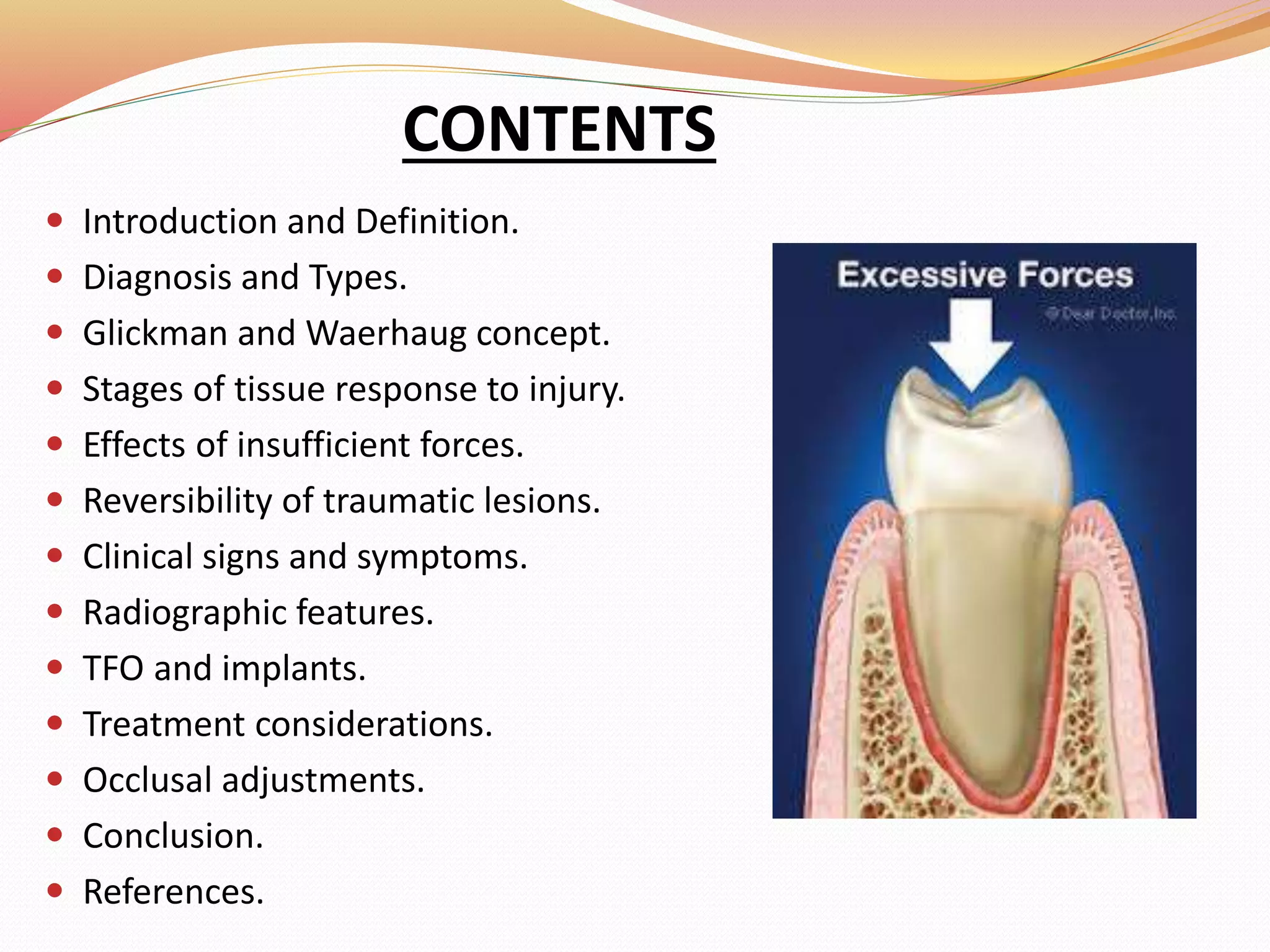
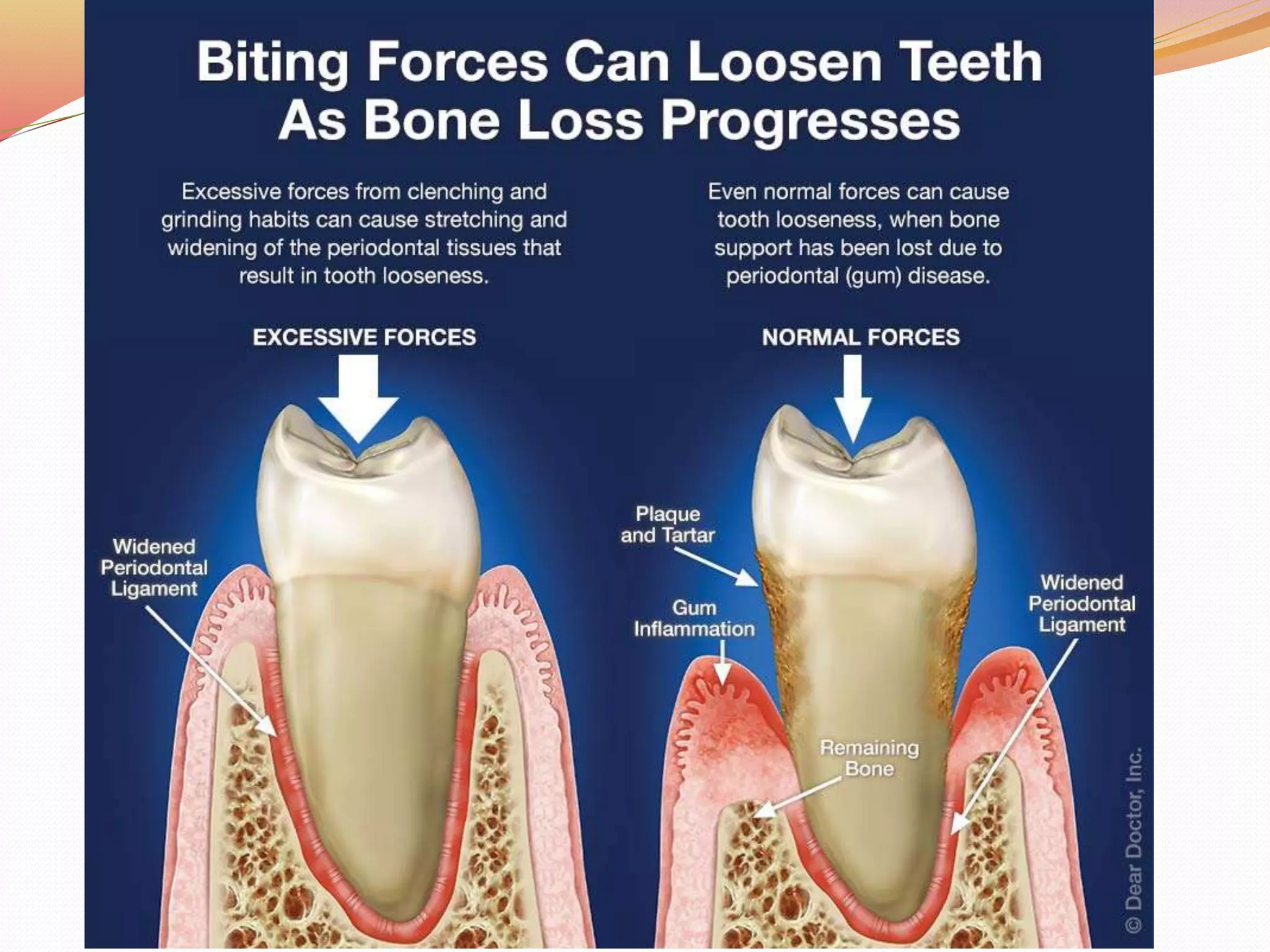
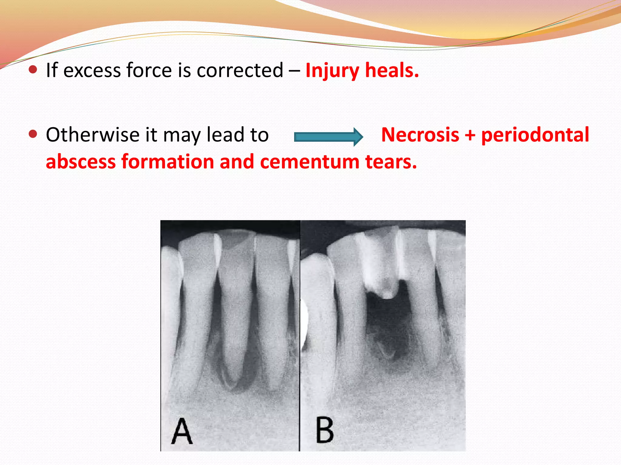
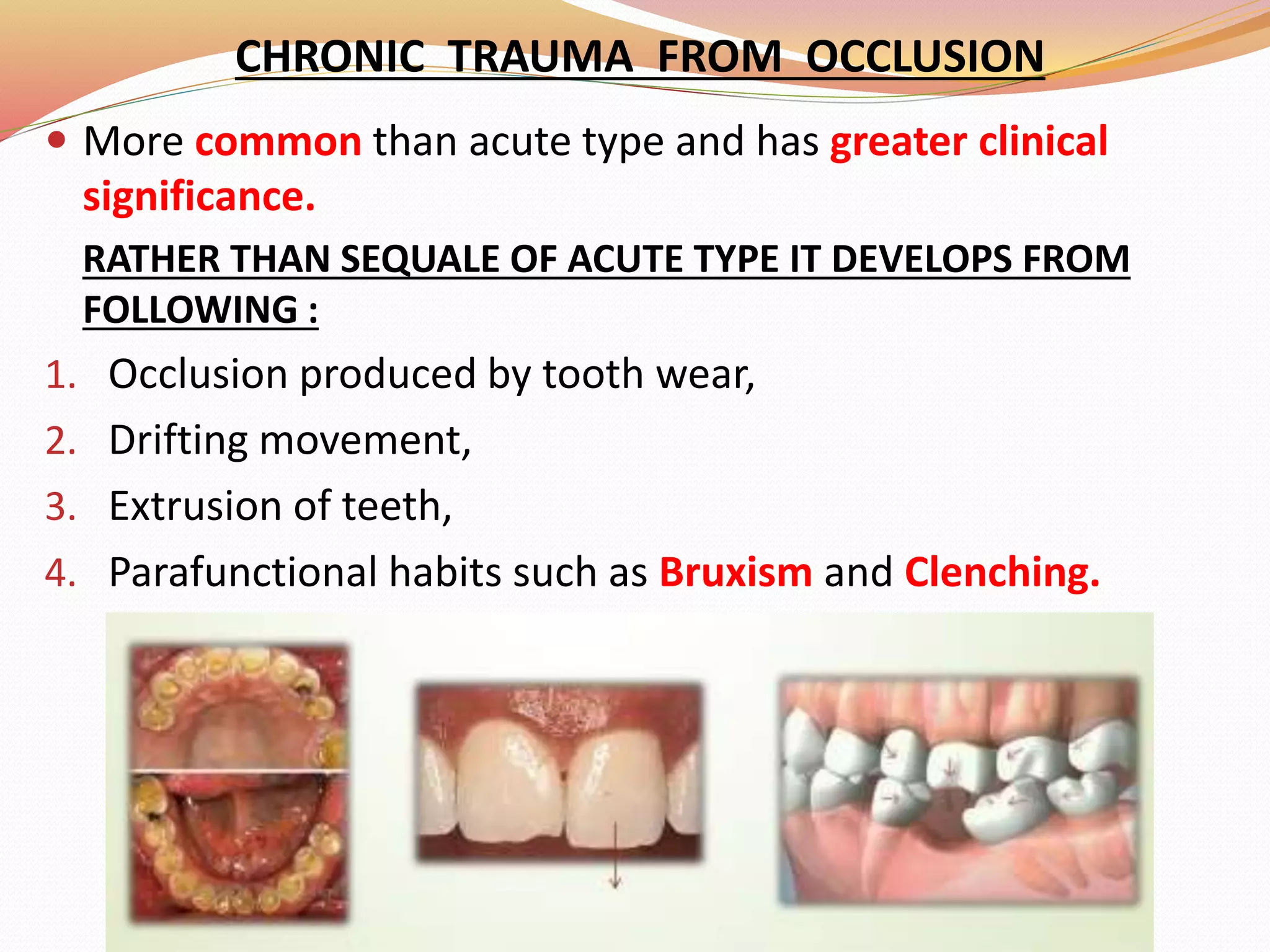
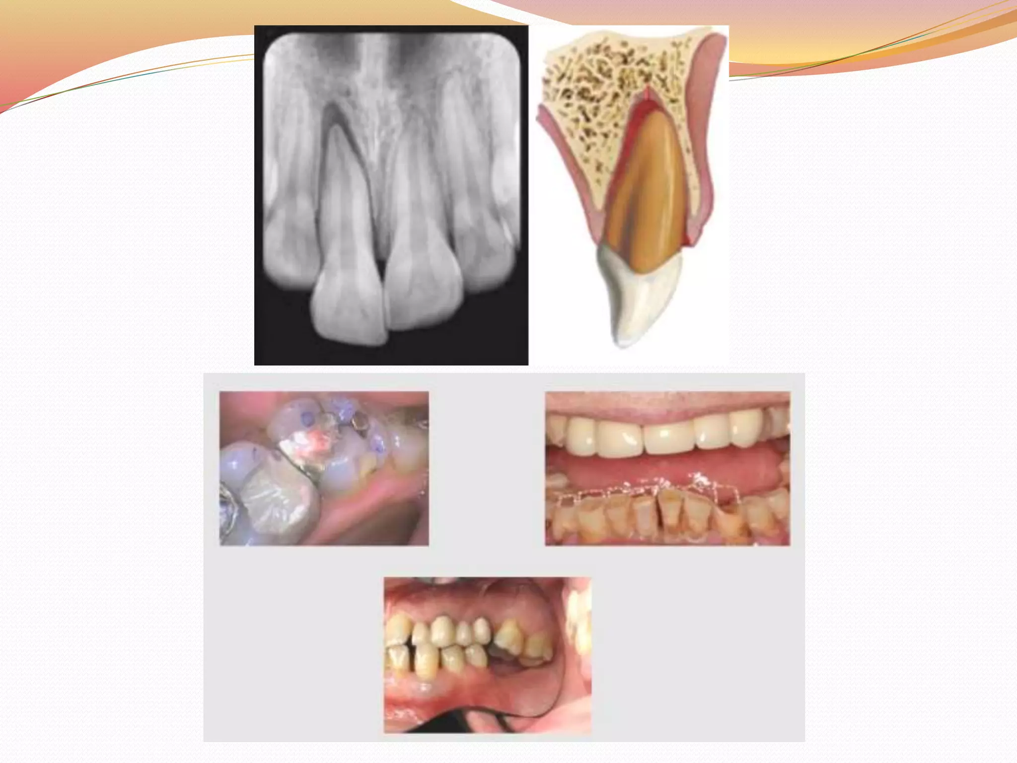
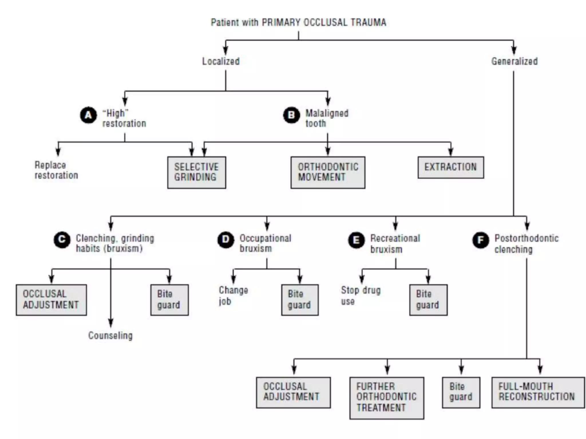
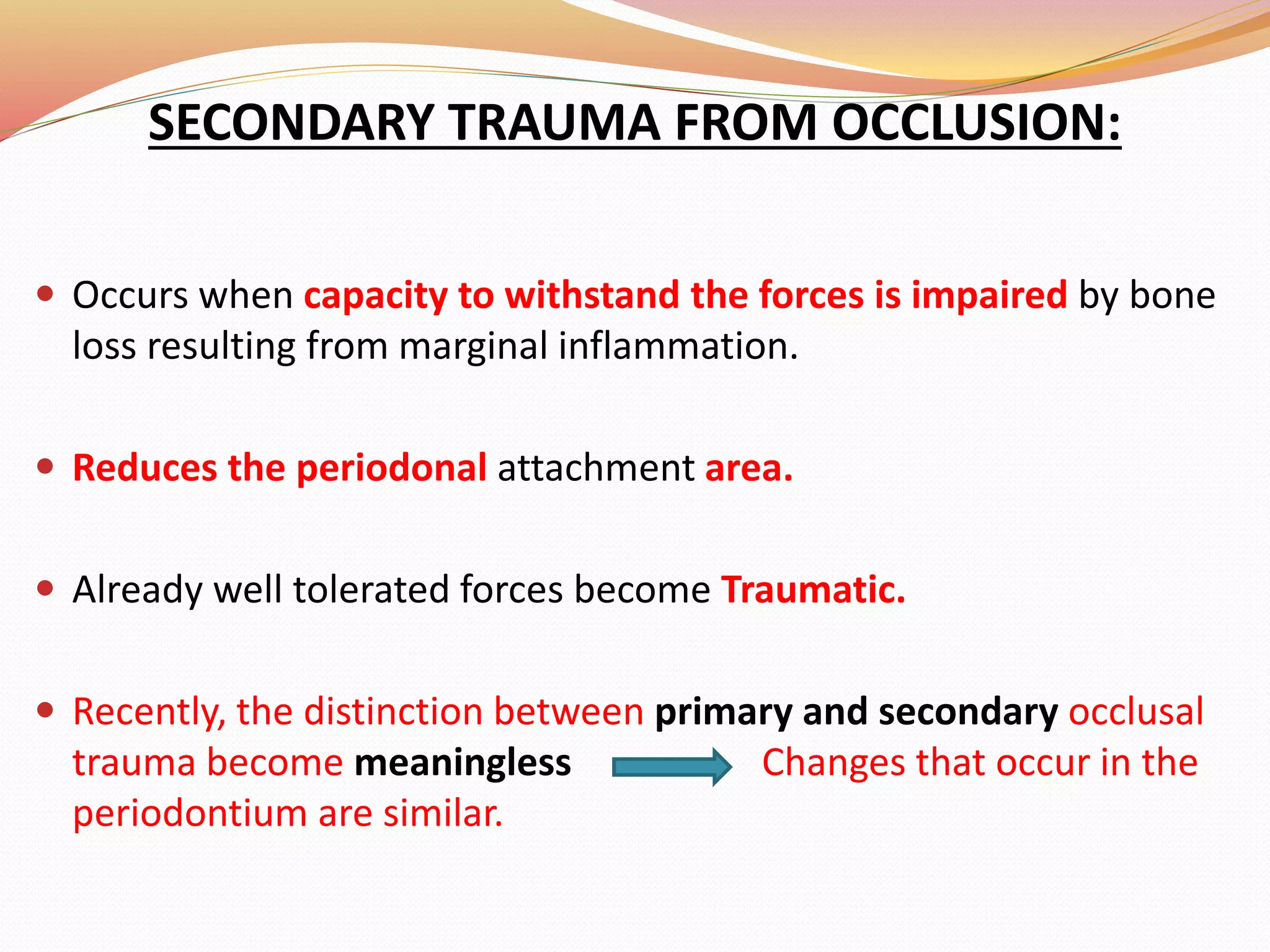
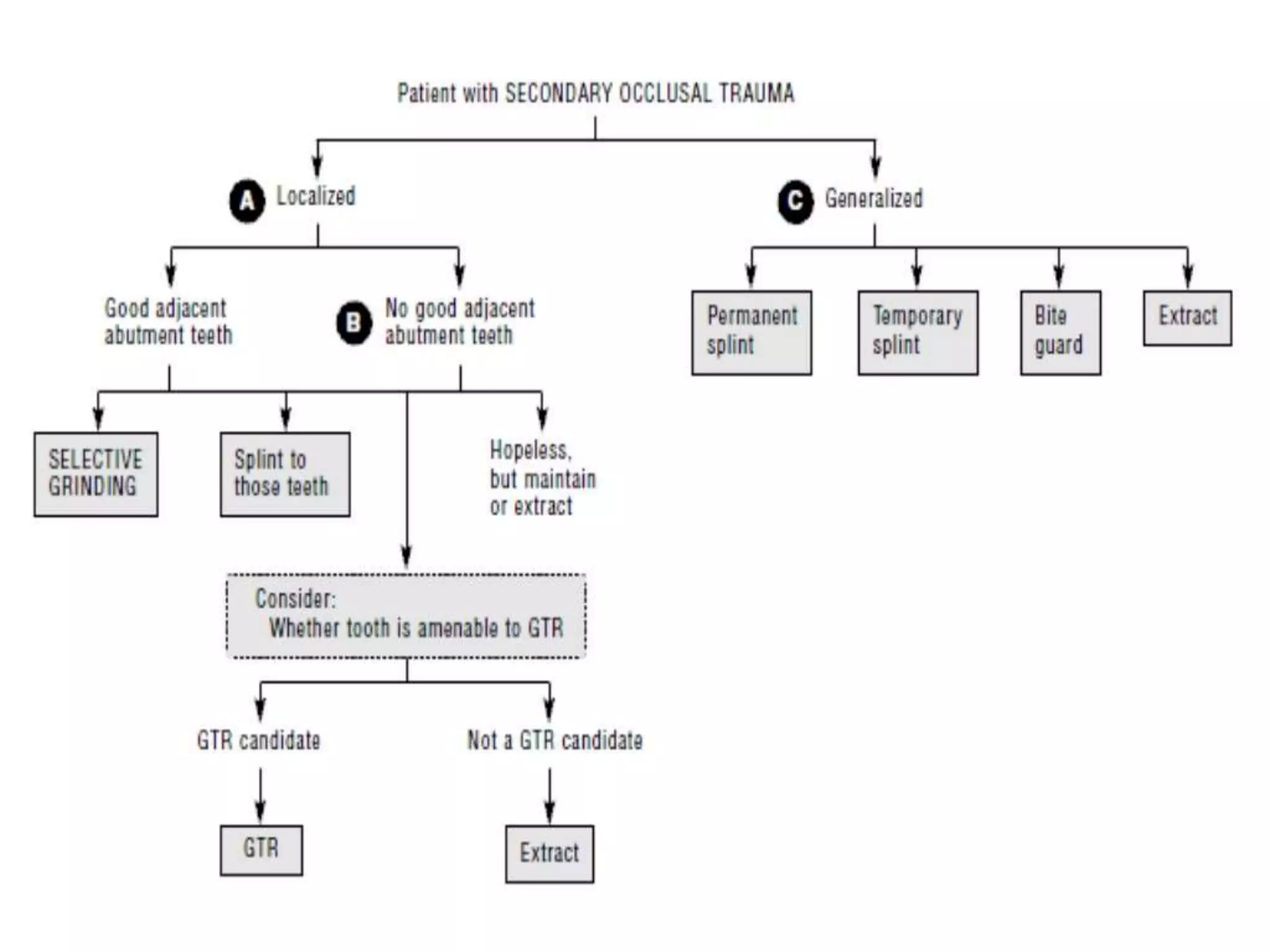
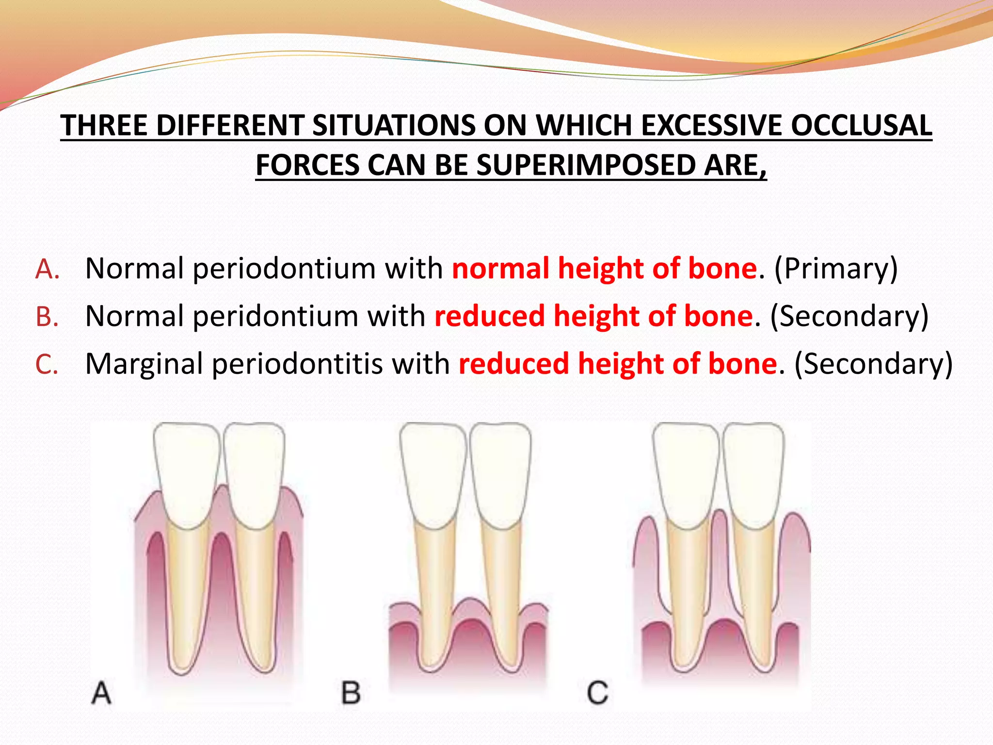
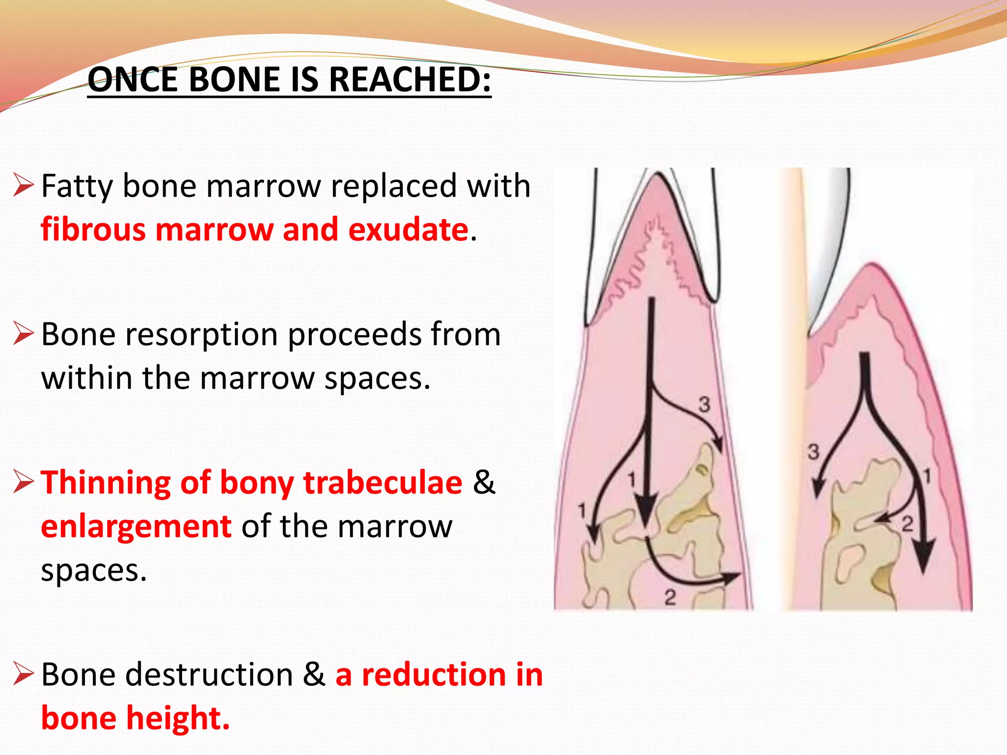
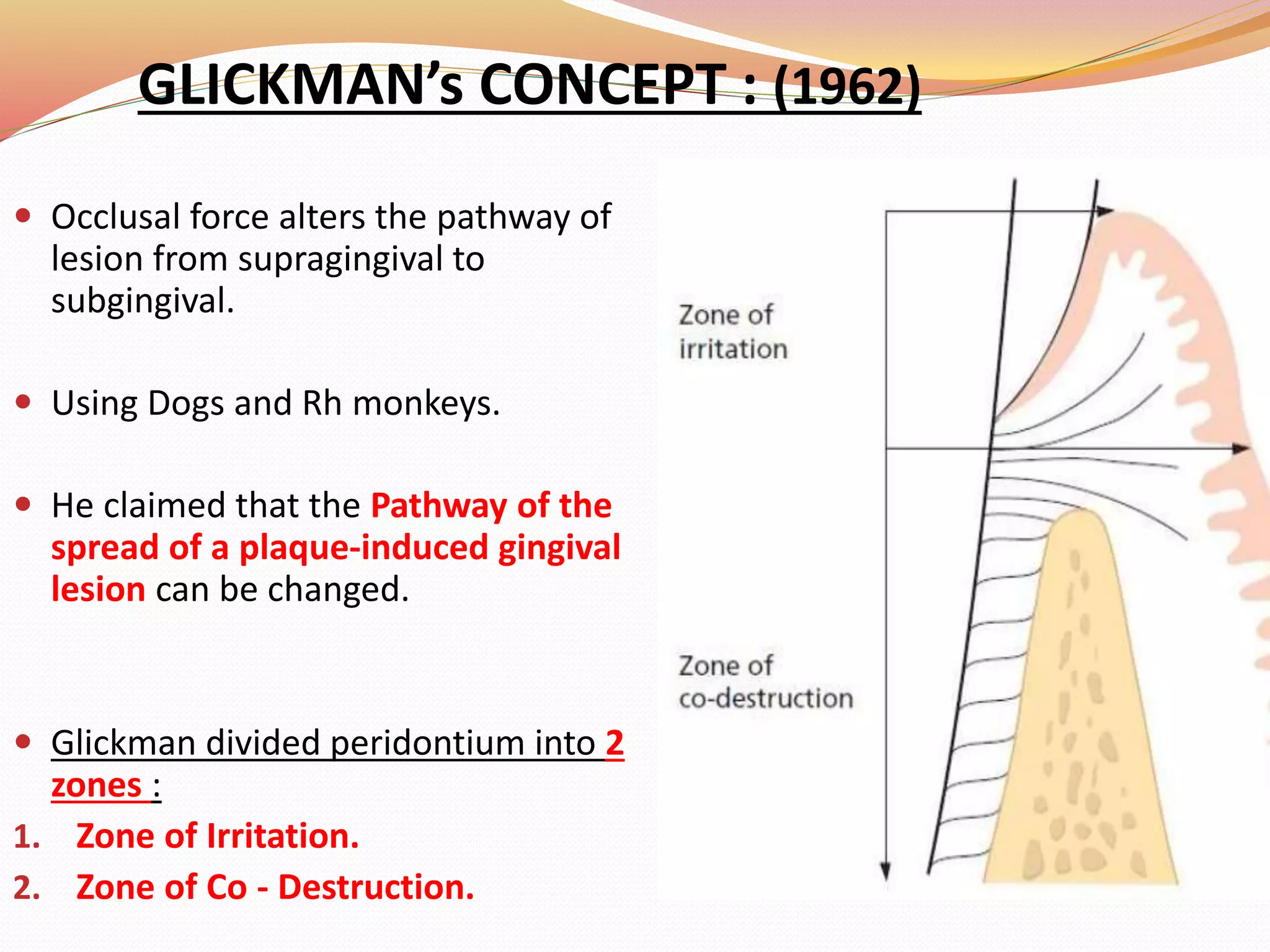
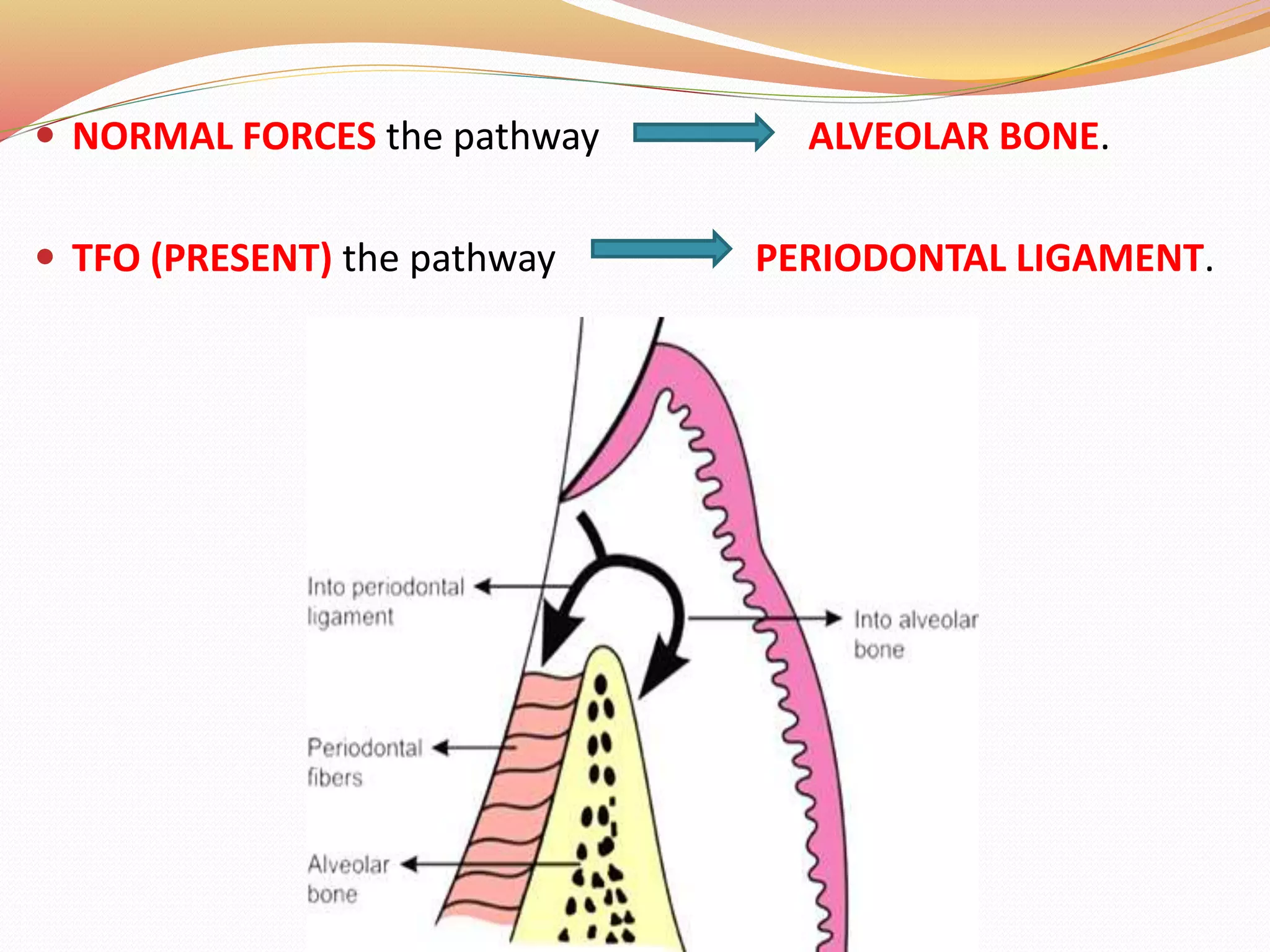
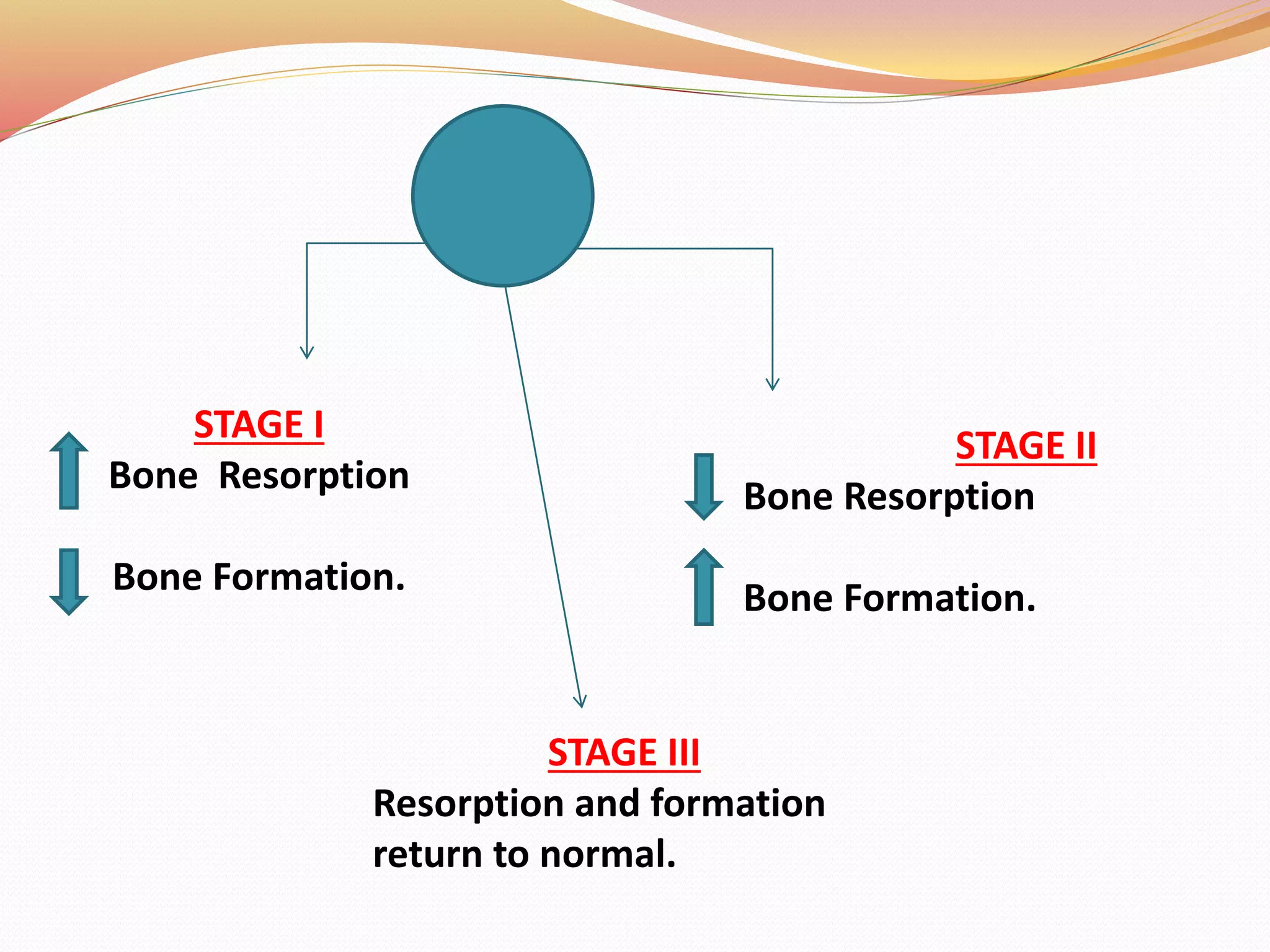
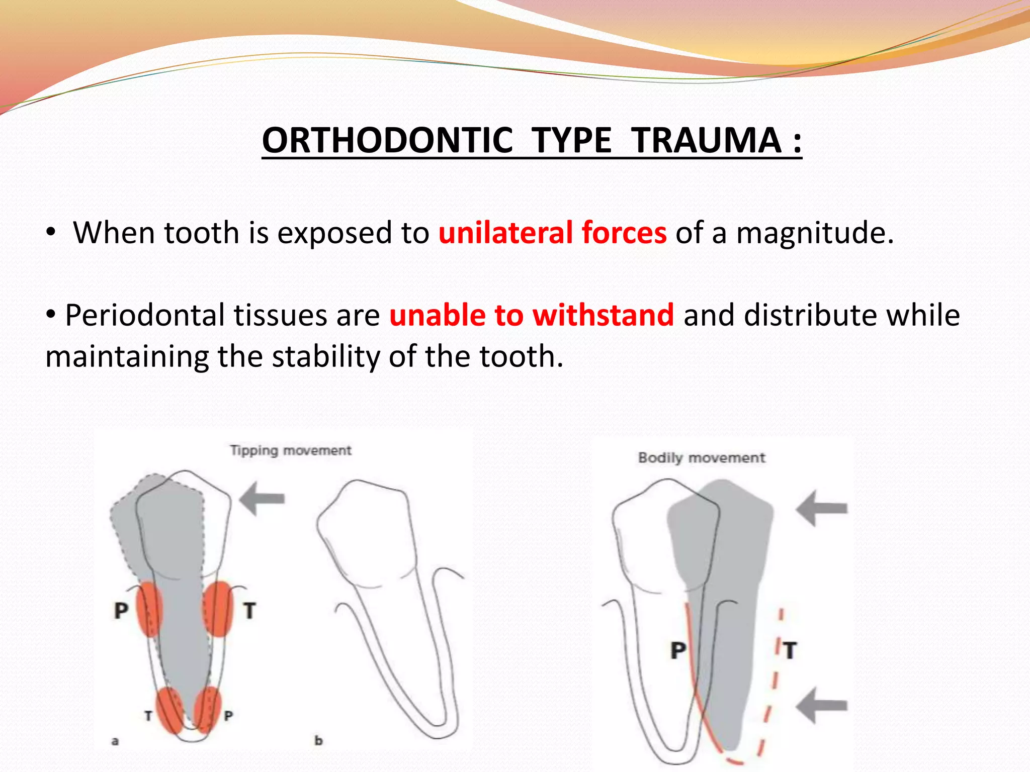
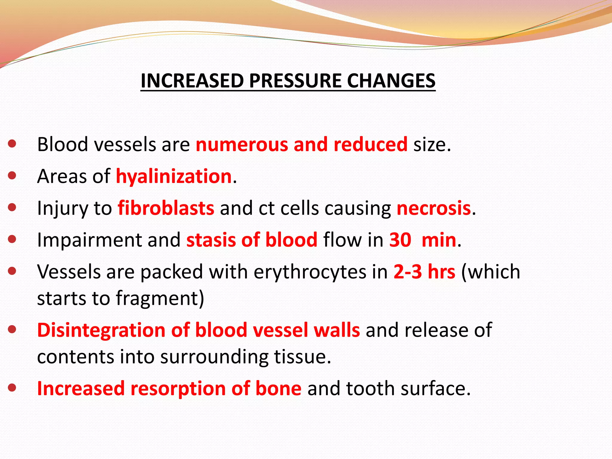
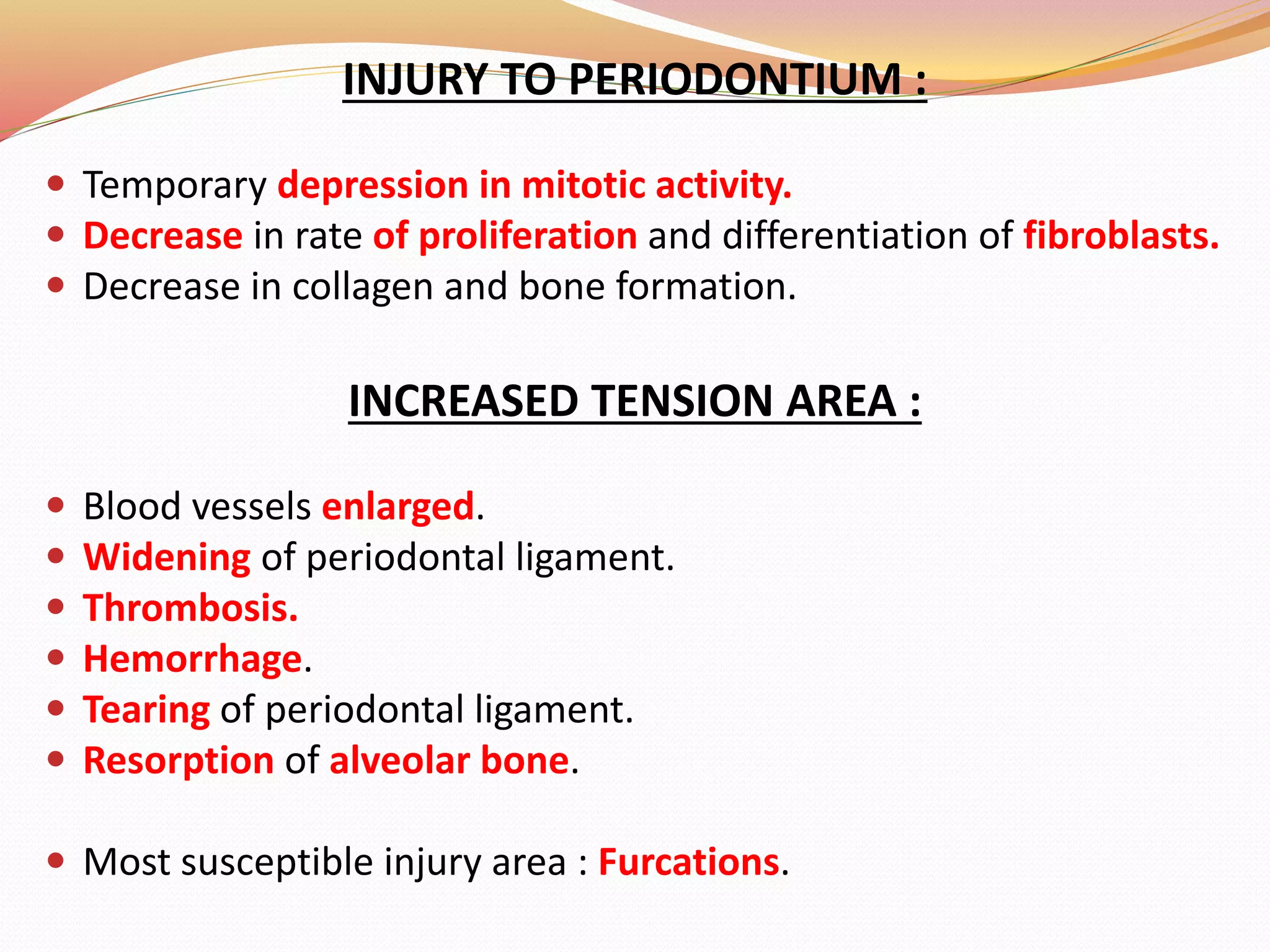
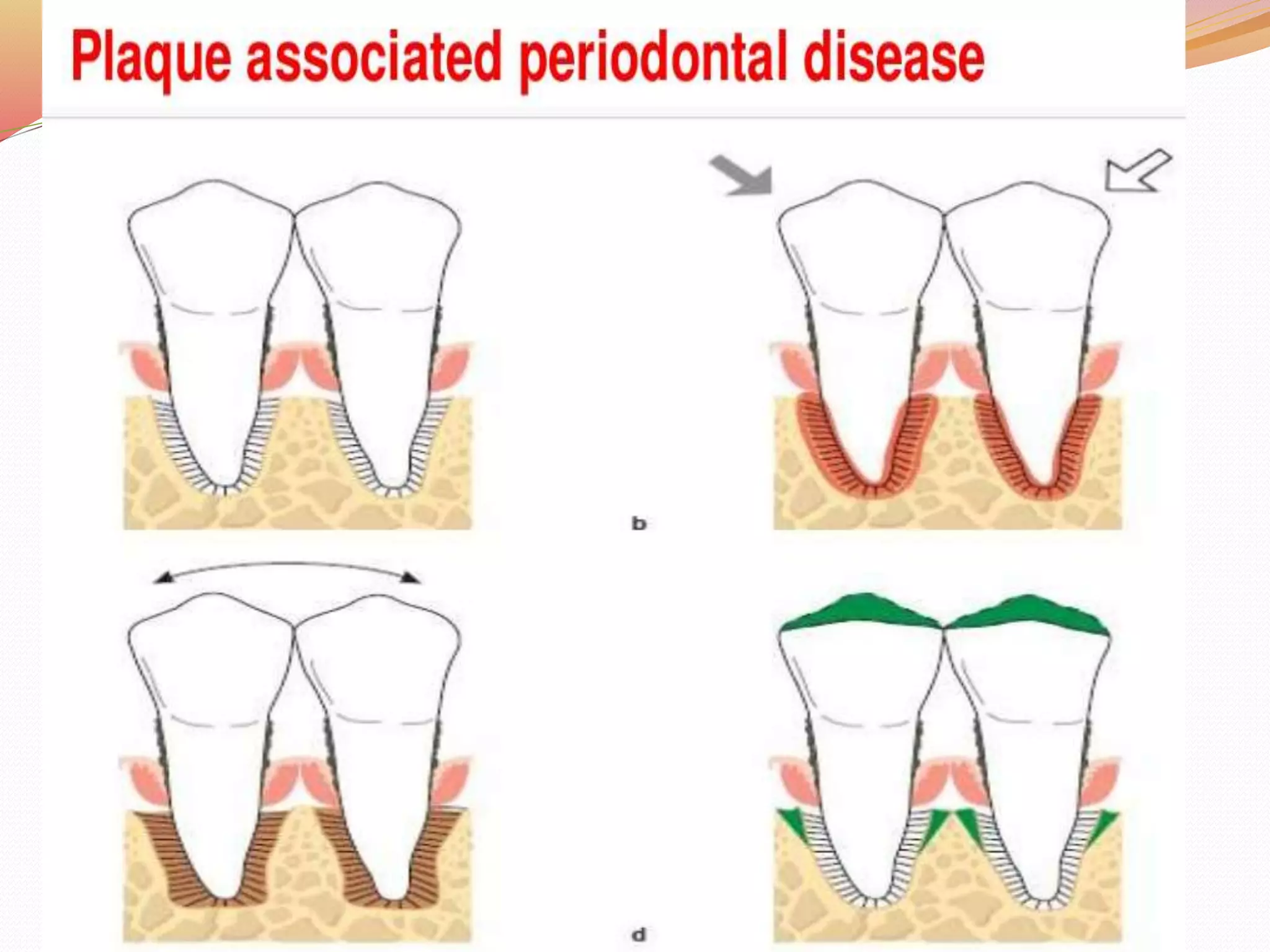
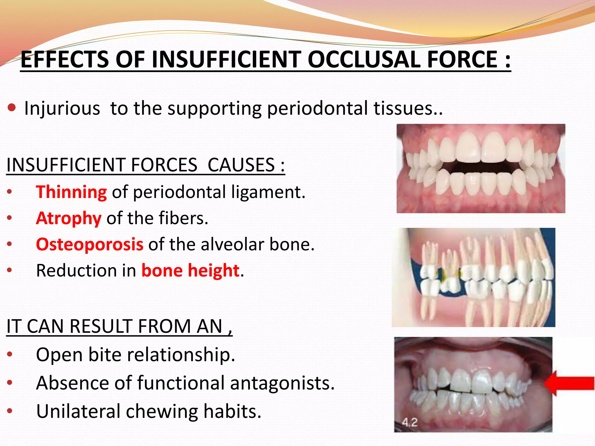
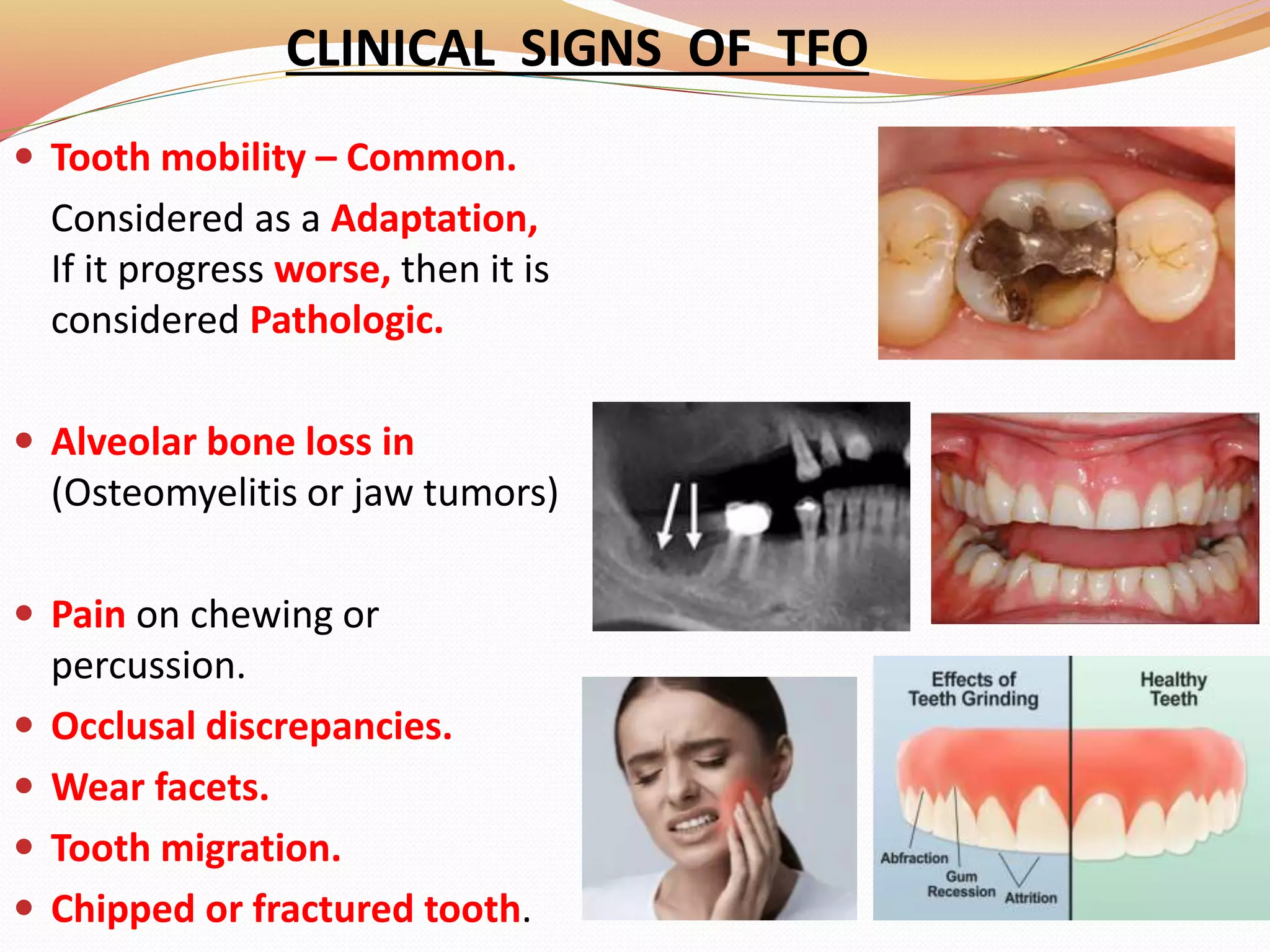
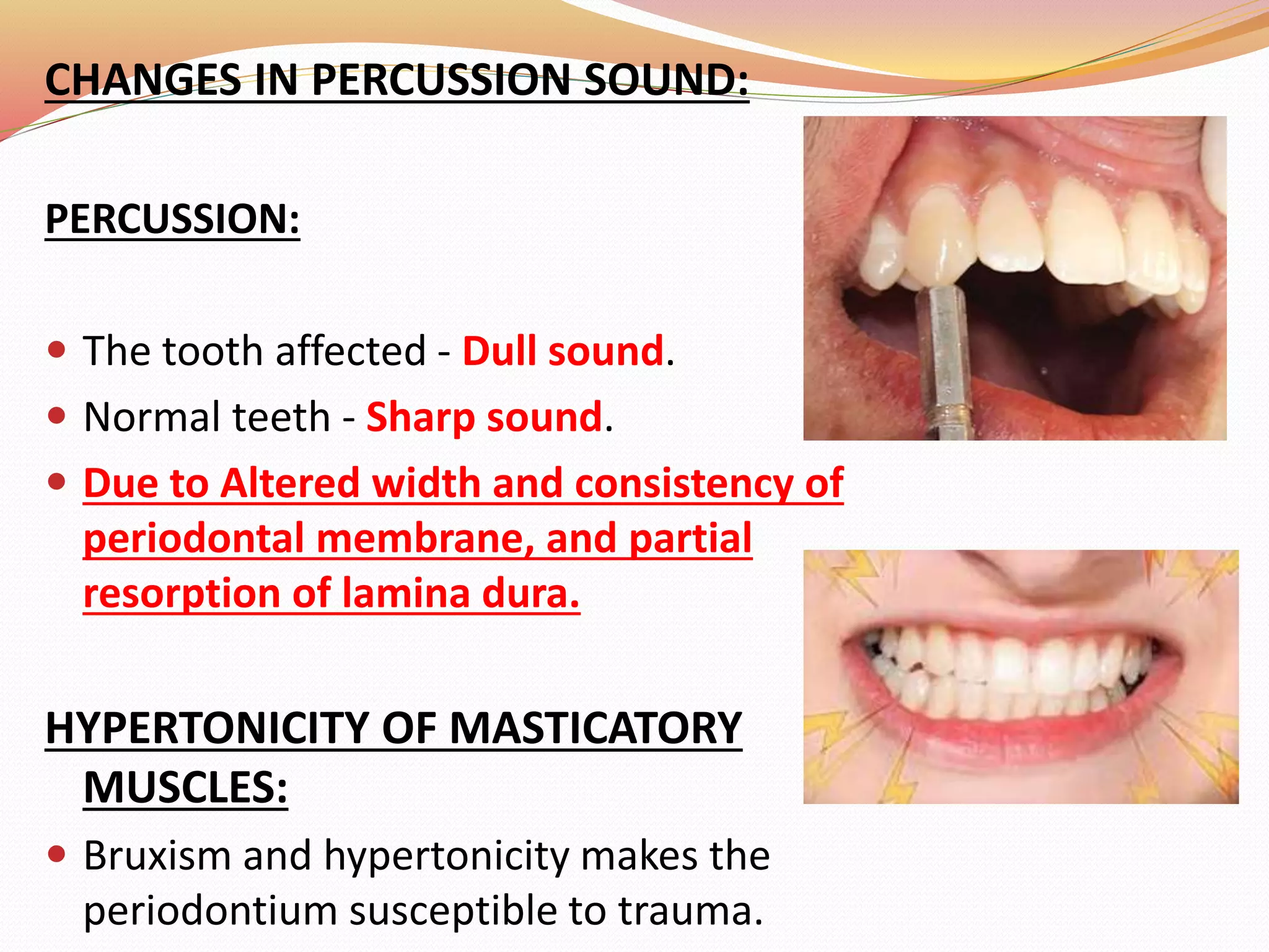
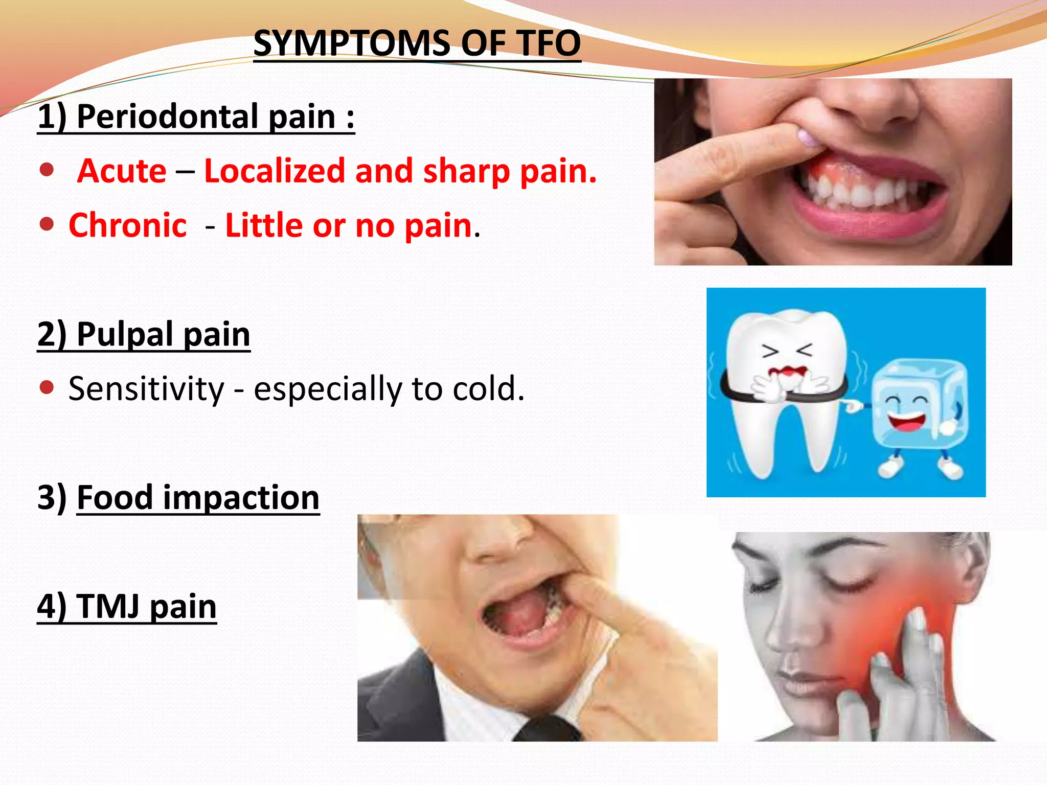
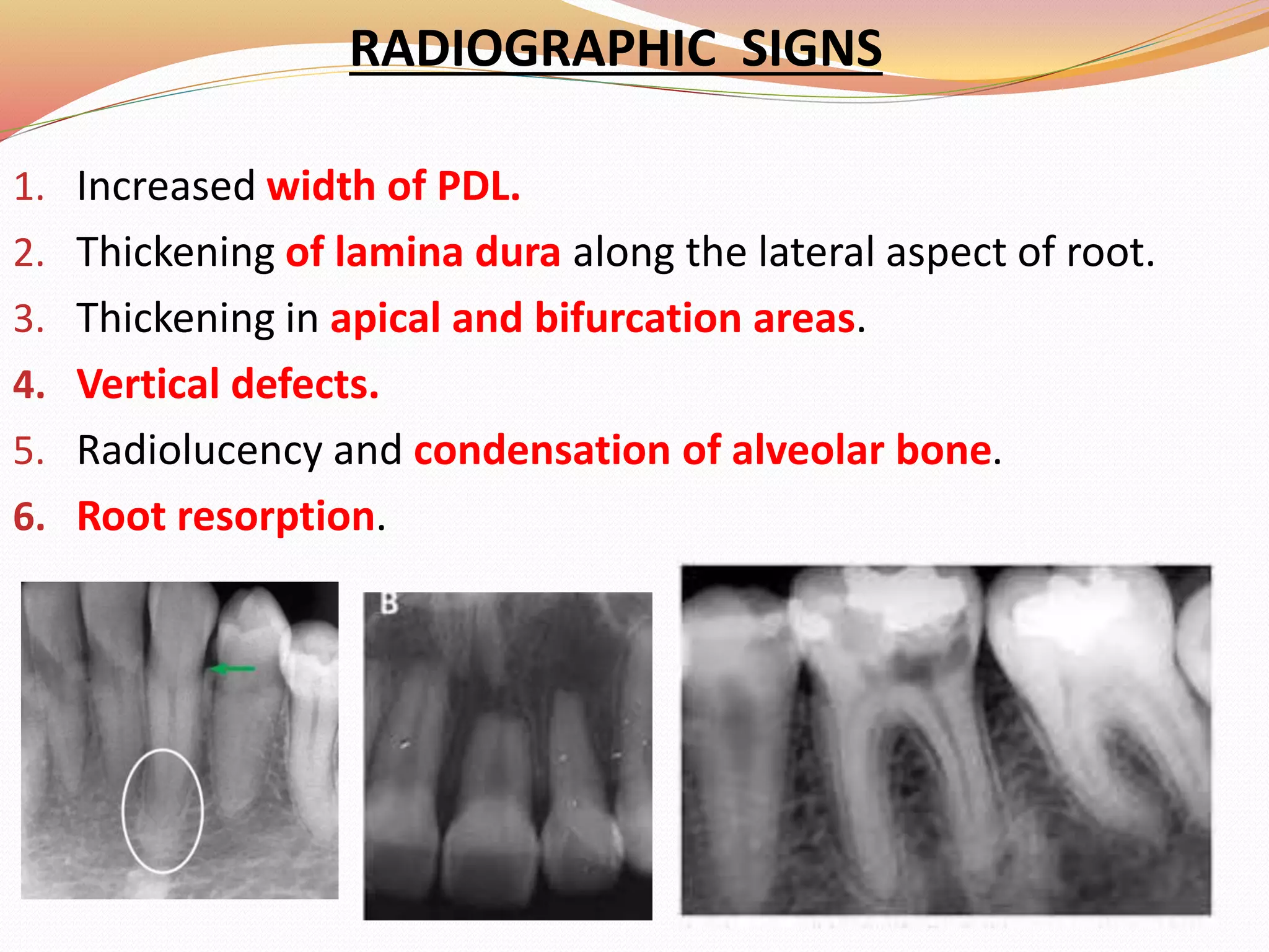
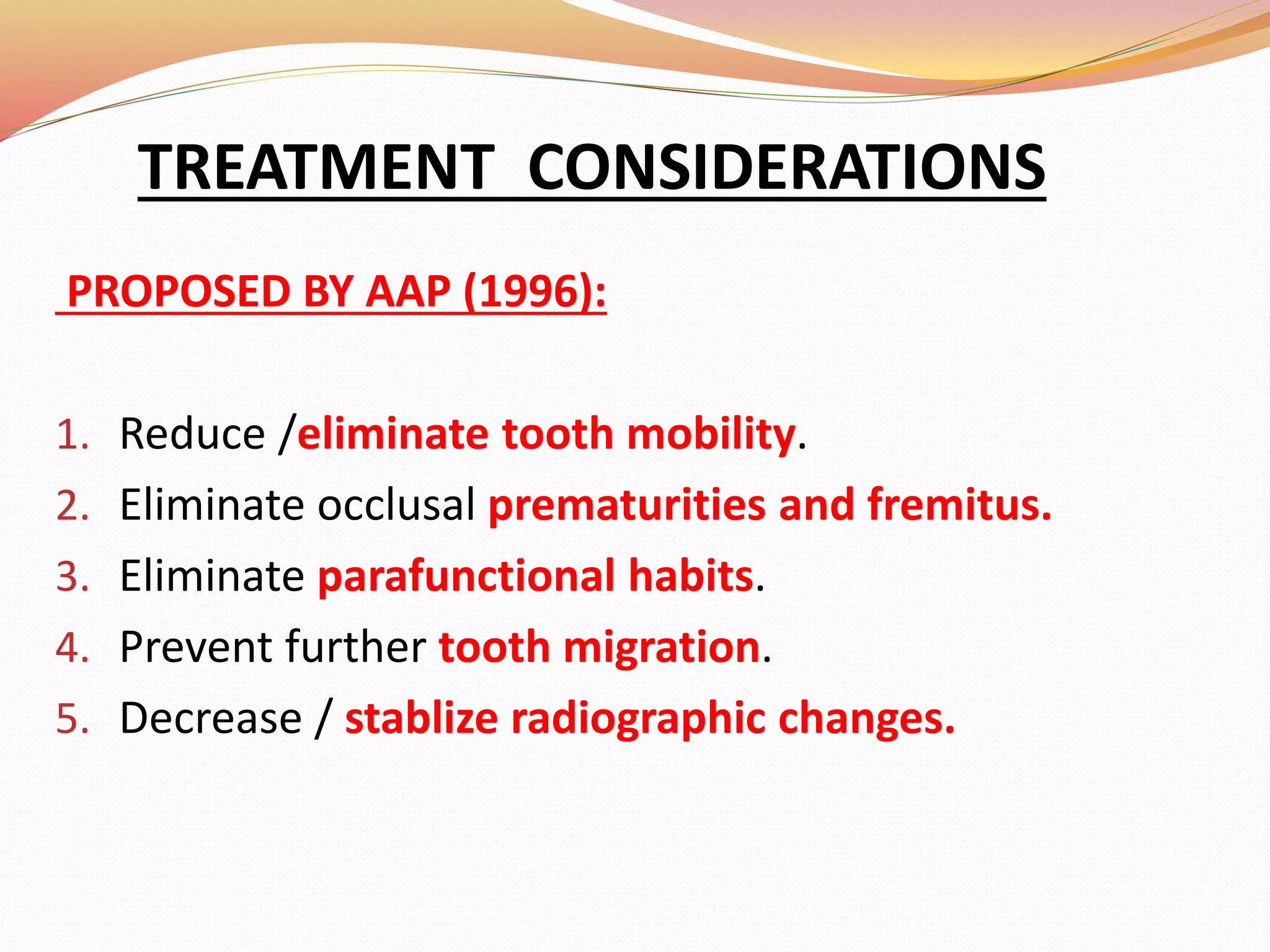
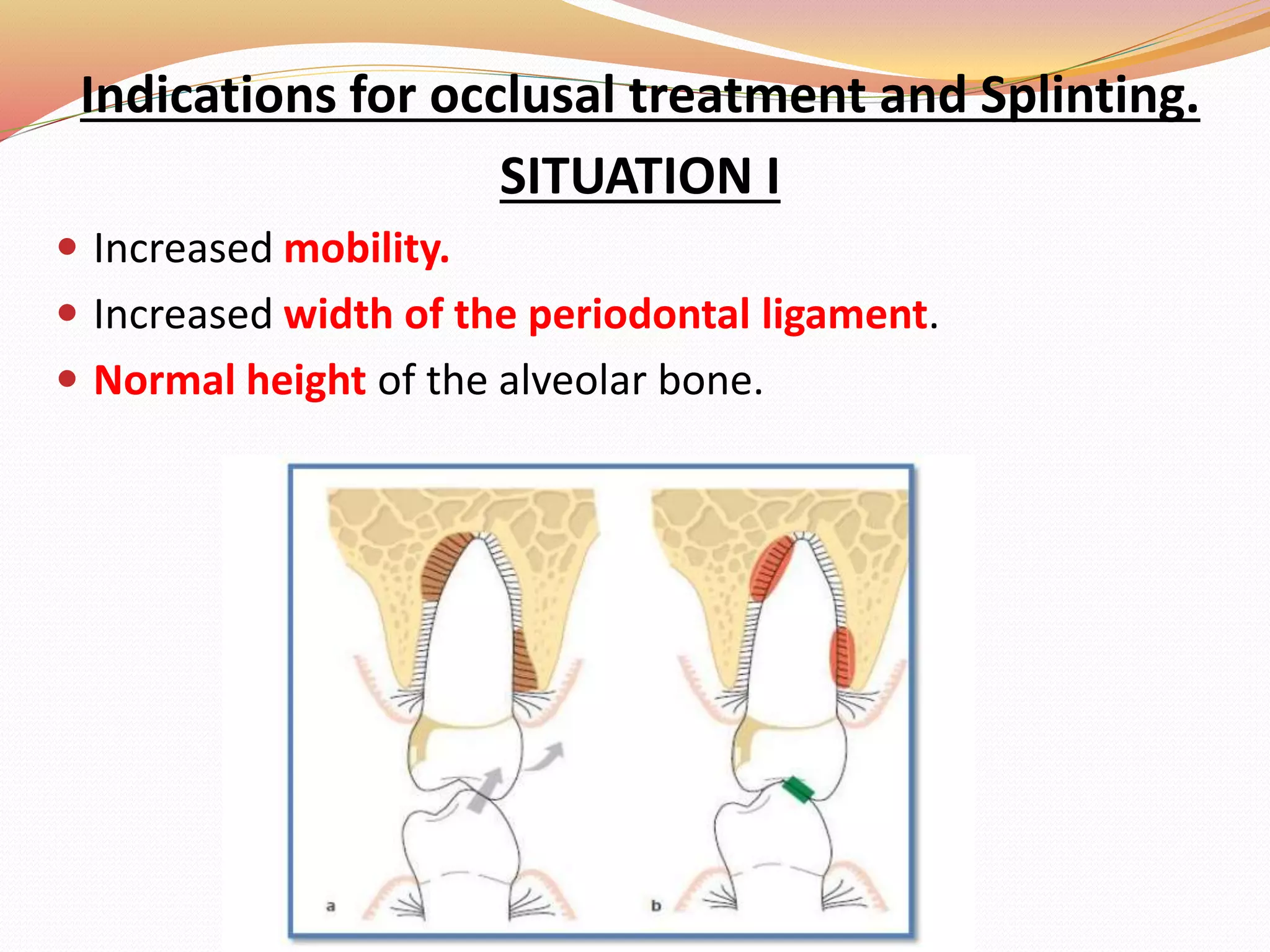
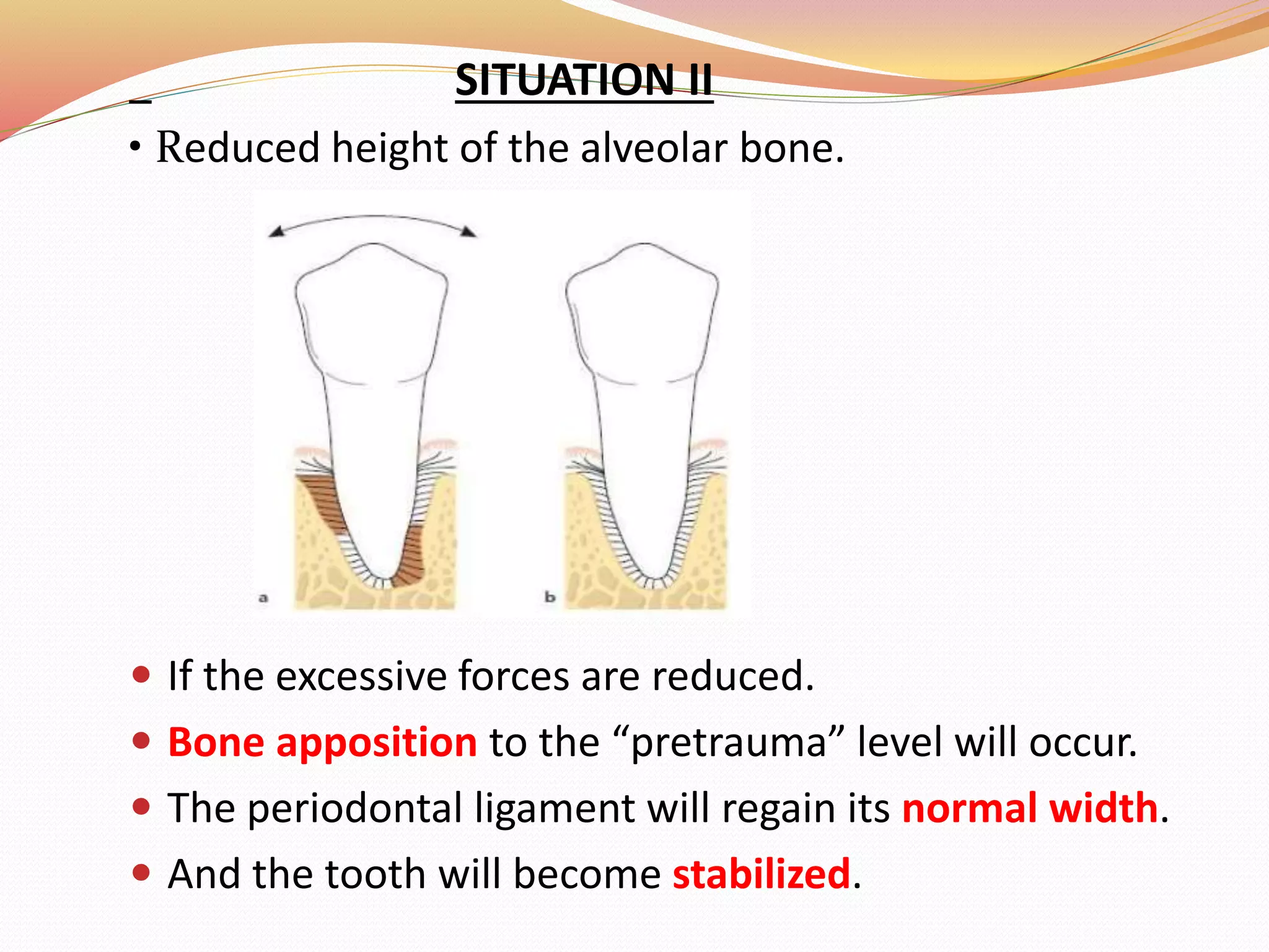
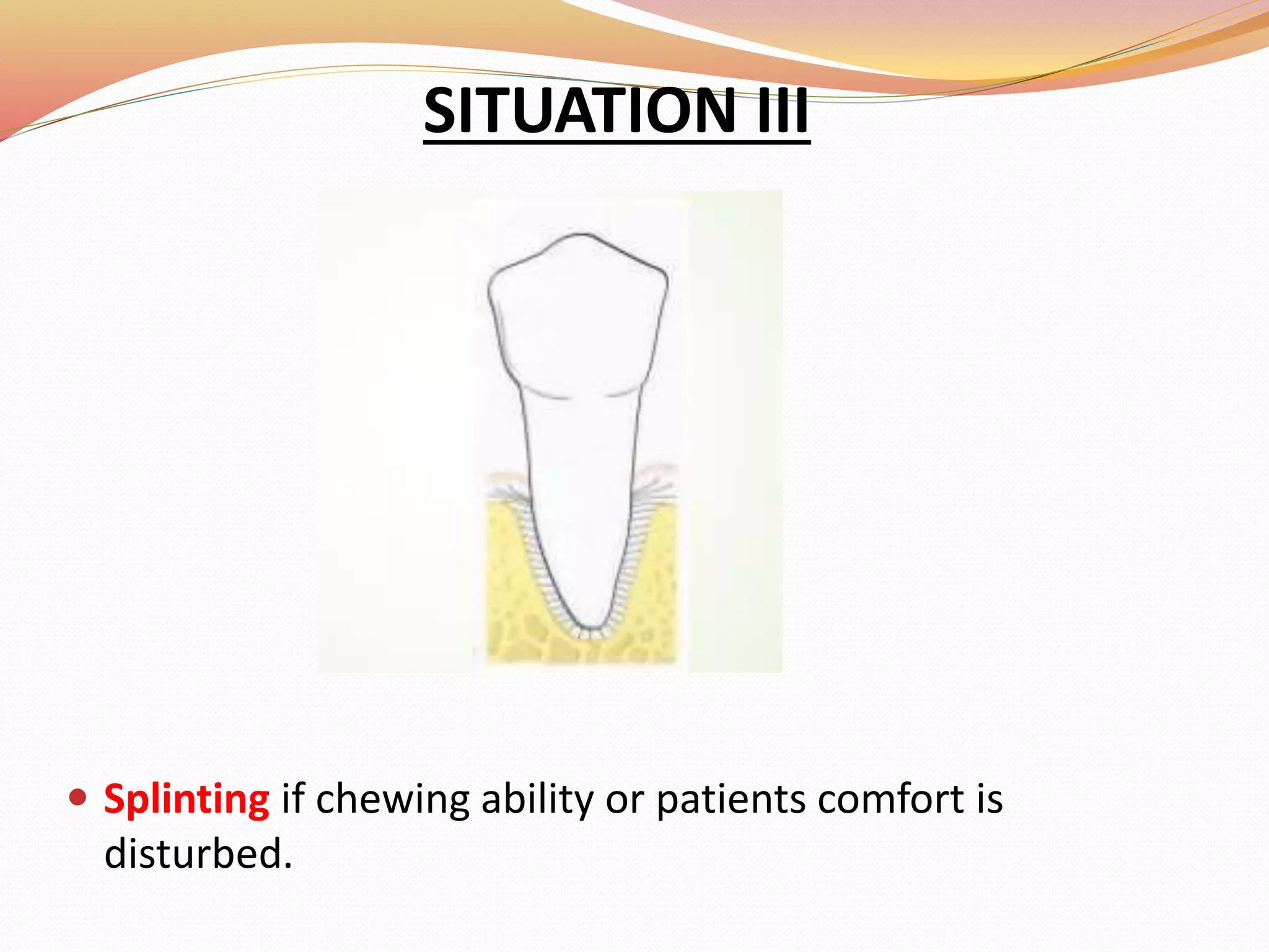
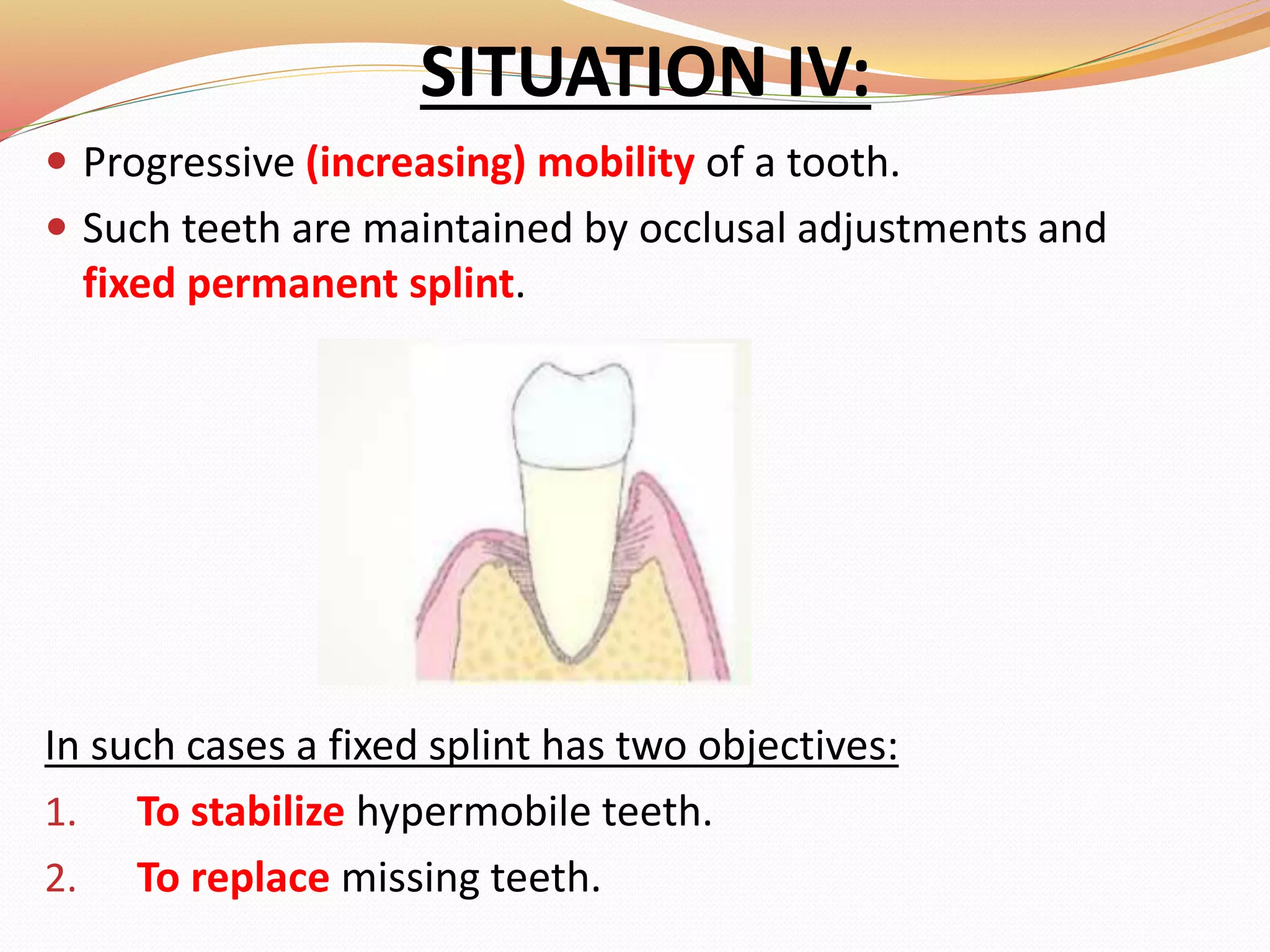
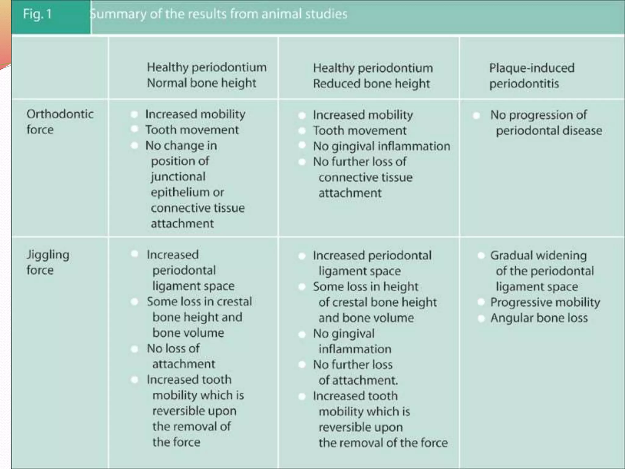
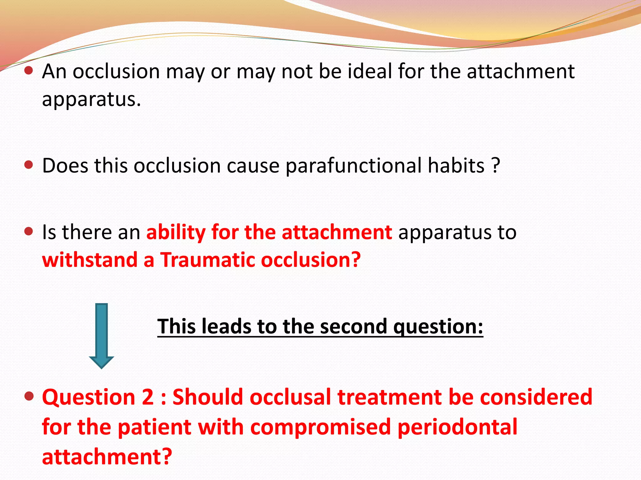
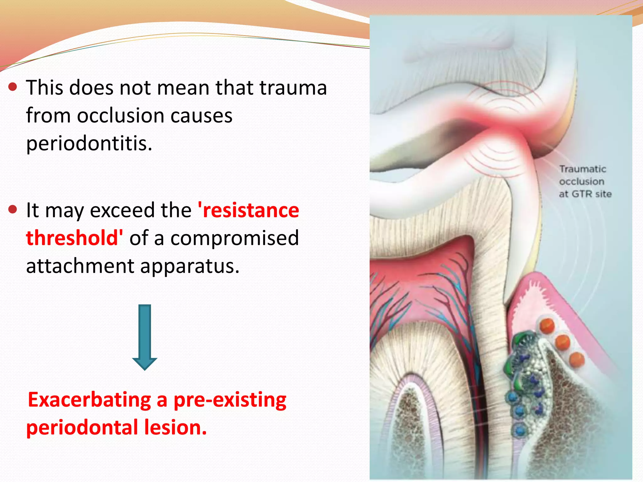
SlideShare a Scribd company logo
N . Prem Kumar
1st year PG (Periodontics)
Sri Venkateshwaraa Dental college,
Pondicherry.
CONTENTS
 Introduction and Definition.
 Diagnosis and Types.
 Glickman and Waerhaug concept.
 Stages of tissue response to injury.
 Effects of insufficient forces.
 Reversibility of traumatic lesions.
 Clinical signs and symptoms.
 Radiographic features.
 TFO and implants.
 Treatment considerations.
 Occlusal adjustments.
 Conclusion.
 References.
INTRODUCTION
 The Periodontal ligament has a cushioning effect on forces applied
to teeth.
 However occlusal force is a critical factor affecting the condition of
the periodontium.
 when occlusal forces exceed the adaptive capacity ,tissue injury
results.
 The resultant injury Trauma from occlusion. (Glickman 1974)
 Occlusion which produces such injury Traumatic occlusion.
TRAUMA FROM OCCLUSION CAN ALSO BE CALLED AS:
 Traumatizing occlusion, Occlusal trauma, Overload, Occlusal
disharmony, Functional imbalance, Occlusal dystrophy.
 Periodontal traumatism. (Orban 1958 and Prichard 1965)
 Traumatogenic occlusal situation. (Muhlemann 1956)
 Traumatogenic occlusion. (Box 1930) (Ramfjord and Ash in 1972)
 TRAUMATIC OCCLUSION (Stillman 1917) : Abnormal stress
capable of producing injury to dental or periodontal tissue.
ADAPTIVE CAPACITY OF THE PERIODONTIUM TO
OCCLUSAL FORCES
Varies in different persons and in the same person at different
times and is influenced by the:
 Magnitude.
 Direction.
 Duration.
 Frequency of the force.
INCREASED MAGNITUDE :
 Widening of PDL space.
 Increase in width of PDL fibres & an increase in density of alveolar
bone.
CHANGES IN DIRECTION:
Causes reorientation of stresses.
Lateral forces & torque more damage.
DURATION AND FREQUENCY OF OCCLUSAL FORCES:
Constant pressure on the bone is more injurious than intermittent
forces.
More frequent intermittent force, the more injury to periodontium.
DEFINITION
 “A condition where injury results to the supporting structures of the teeth by the
act of bringing the jaws into a closed position”. (Stillman 1917)
 “When occlusal forces exceed the adaptive capacity of the tissues, tissue injury
results. this resultant injury is termed TFO”.(Glickman 1974) .
 “A term used to describe pathological alterations or adaptive changes which
develop in the periodontium as a result of undue force produced by the masticatory
muscles”. (Lindhe 5th edition)
 “Damage in the periodontium caused by stress on the teeth produced directly or
indirectly by teeth of the opposing jaw”. ( WHO1978)
 “An injury to the attachment apparatus as a result of excessive occlusal force”.
(Glossary of periodontic terms ,1992)
DIAGNOSIS
TOOTH MOBILITY :
 A clinical diagnosis of occlusal trauma can only be confirmed
where progressive mobility present.
 And can be identified through a series of repeated
measurements over an extended period .
TOOTH MOBILITY CAN BE MEASURED BY :
1. Manual Examination.
2. Fremitus Test.
3.Periodontometre.
4.Periotest.
ELECTRONIC DEVICES :
USED TO DETECT MOBILITY.
 Elbrecht’s indicator (1939).
 Werner’s Oscillator (1942).
 Beyeler and Dreyfus Vibrator (1947, 1949).
 Manly’s Mobilometer (1951).
 Zwirner’s Oscillograph (1949-1952).
 Muhlemann’s Periodontometer (1954).
 Muhlemann’s Macro – Periodontometer.
 Picton’s gauge (1957).
 Parfitt’s Tranformer (1958).
 Joel’s Technique (1958).
 Goldberg’s Periodontometer (1961).
 Korber’s Transducer (1963, 1970).
 USAFSAM Periodontometer (1963).
 Pameijer’s (1973)..
 Ryden’s Laser Reflexion Method (1975).
 Dental Holographic Interferometry (1975).
 Persson and Svensson Loading/Sensing Devices (1980).
 Kaneko’s Non – Invasive Test (1986).
 Periotest (1992).
 Resonance Frequency Analysis (1994).
 Laser Vibrometry (1998).
 No Contact Vibration Device (2008).
 Zwick Method (2011).
 Konermann’s Novel Intraoral Measuring Device (2016).
 NEVD – Non – Contact electromagnetic Vibration Device
(2016).
MANUAL METHOD :
 Measured by Two dental instruments which are placed at the buccal
and lingual heights of contour of the tooth.
 Force are applied in the Bucco-lingual direction.
MILLER’S MOBILITY INDEX (1985) :
 Class I – Mobility greater than physiologic.
 Class II – Tooth can be moved upto 1mm or more in lateral direction
(buccolingually or mesiodistally). Inability to depress the tooth in
vertical direction (Apicocoronal).
 Class III – Tooth can be moved upto 1mm or more in lateral direction
(buccolingually or mesiodistally). Ability to depress the tooth in
vertical direction (Apicocoronal).
2) FREMITUS TEST/FUNCTIONAL MOBILITY :
• It is a measurement of the Vibratory pattern of the teeth.
• To measure fremitus ,a dampened index finger is placed
along the buccal and labial surface of the maxillary teeth.
• The patient is asked to tap the teeth together in the
maximum intercuspal position.
• And then grind systematically in the lateral, protrusive
contacting movements and positions.
• The teeth that are displaced shows Vibrations.
FREMITUS GRADES :
(N) : Normal.
(+) : Vibration felt.
Class 1 : Mild Vibrations or movements detected.
Class 2 : Easily palpable vibrations but no visible movements.
Class 3 : Movements visible with naked eyes.
 In the posterior teeth TFO can be detected with help of
occlusion registration strip/ articulating paper.
 High pressure points can be detected by pattern of impression
made by registration strip/articulating paper.
WEAR PATTERNS
 Bruxism - wear facets on occlusal and incisal surfaces which
can be identified as shiny and irregular surfaces.
 Abfractions - excessive lateral forces, abfractions can be seen
on pre molars.
 Thermal sensitivity.
 Muscle hyper tonicity.
 Both processes result in injury to the attachment apparatus.
• OCCLUSAL TRAUMA -
'injury to the
periodontium
resulting from
excessive occlusal
forces.
• PERIODONTITIS -
result of bacterial
factors in dental
plaque, which
exceeds the
inherent protective
mechanisms of the
host.
Let us compare the two terms here
So therefore, we Periodontist’s should ask ourselves
these questions:
1) Is there any relation between these two terms???
2) Or the occlusal trauma have any role in the etiology of
periodontal disease ???
 Before attempting to answer these questions, the different
types of trauma from occlusion need to be defined.
CLASSIFICATION
Glickman’s classification (1953)
According to the Duration of cause :
 Trauma from occlusion may be,
1. Acute.
2. Chronic.
According to Nature of the cause :
 Chronic is further classified into:
1. Primary.
2. Secondary.
Acute trauma from occlusion
 Results from abrupt occlusal impact (produced by biting a
hard object).
 Restorations or prosthetic appliances that interfere with the
direction of occlusal forces.
Clinical features :
1. Tooth pain.
2. Sensitivity to percussion.
3. Tooth mobility.
 If excess force is corrected – Injury heals.
 Otherwise it may lead to Necrosis + periodontal
abscess formation and cementum tears.
CHRONIC TRAUMA FROM OCCLUSION
 More common than acute type and has greater clinical
significance.
RATHER THAN SEQUALE OF ACUTE TYPE IT DEVELOPS FROM
FOLLOWING :
1. Occlusion produced by tooth wear,
2. Drifting movement,
3. Extrusion of teeth,
4. Parafunctional habits such as Bruxism and Clenching.
CHRONIC IS FURTHER CLASSIFIED INTO TWO TYPES:
According to nature of the cause :
PRIMARY SECONDARY
CHRONIC
Primary trauma from occlusion :
 The tissue damage around a tooth with normal height of
peridontium.
 Occurs if it is considered as the primary etiologic factor and also due
to Parafunctional habits.
Primary type happens from the following:
 Insertion of High filling.
 Prosthetic placement that creates excessive forces on abutment
and antagonist teeth.
 The drifting movement and extrusion of teeth into missing teeth.
 Orthodontic movement of teeth into functionally unacceptable
positions.
Temporary increased mobility (functional adaptation)
 No changes in the supracrestal fibres,
 No loss of periodontal attachment,
 No increased probing pocket depth.
Forces too high (above the adaptation level)
 Aseptic necrosis of the PDL.
 Root resorption occurs - resulting in shorter roots
Changes produced by primary trauma DO NOT ALTER
1. The level of connective tissue attachment.
2. Does not initiate pocket formation.
SECONDARY TRAUMA FROM OCCLUSION:
 Occurs when capacity to withstand the forces is impaired by bone
loss resulting from marginal inflammation.
 Reduces the periodonal attachment area.
 Already well tolerated forces become Traumatic.
 Recently, the distinction between primary and secondary occlusal
trauma become meaningless Changes that occur in the
periodontium are similar.
THREE DIFFERENT SITUATIONS ON WHICH EXCESSIVE OCCLUSAL
FORCES CAN BE SUPERIMPOSED ARE,
A. Normal periodontium with normal height of bone. (Primary)
B. Normal peridontium with reduced height of bone. (Secondary)
C. Marginal periodontitis with reduced height of bone. (Secondary)
TRAUMA FROM OCCLUSION
BOX’S CLASSIFICATION :
 Physiologic occlusion
A condition, in which the systems of forces acting on the
tooth during the occlusion are in a state of equilibrium, and
they do not and cannot change the normal relationship
existing between the tooth and its supporting structures.
 Traumatic occlusion
The damage produced in the periodontium is due to the
overstress produced by the occlusion.
HAMP, NYMAN, AND LINDHE’S CLASSIFICATION (1975) :
This classification is based on a horizontal component of
tissue destruction that has occurred in the interradicular
area.
Degree I: Horizontal loss of periodontal tissue support not
exceeding one-third of the width of the tooth.
Degree II: Horizontal loss of periodontal support exceeding
one-third of the width of the tooth.
Degree III : Horizontal through-and-through destruction of
the periodontal tissue in the furcation area.
EXTENSION OF GINGIVAL INFLAMMATION TO ALVEOLAR BONE
 Gingival inflammation Collagen fiber bundles
Blood vessels Alveolar bone.
 Interproximally, through the vessels perforating the crest of the
interdental septum.
 Facially & lingually , spreads along the outer periosteal surface
& penetrates into the marrow spaces through vessel channels.
 Destroys the Transseptal & Gingival fibers on the course.
ONCE BONE IS REACHED:
Fatty bone marrow replaced with
fibrous marrow and exudate.
Bone resorption proceeds from
within the marrow spaces.
Thinning of bony trabeculae &
enlargement of the marrow
spaces.
Bone destruction & a reduction in
bone height.
GLICKMAN’s CONCEPT : (1962)
 Occlusal force alters the pathway of
lesion from supragingival to
subgingival.
 Using Dogs and Rh monkeys.
 He claimed that the Pathway of the
spread of a plaque‐induced gingival
lesion can be changed.
 Glickman divided peridontium into 2
zones :
1. Zone of Irritation.
2. Zone of Co - Destruction.
ZONE OF IRRITATION
 The zone of irritation includes the Marginal and interdental papilla.
 The soft tissue of this zone is bordered by hard tissue (the tooth) only
on one side and cannot be affected by forces of occlusion.
ZONE OF CO-DESTRUCTION
 It includes the Root cementum, Periodontal ligament, and the
Alveolar bone.
 The tissues in this zone may become the seat of a lesion caused by
trauma from occlusion.
 NORMAL FORCES the pathway ALVEOLAR BONE.
 TFO (PRESENT) the pathway PERIODONTAL LIGAMENT.
WAERHAUG’S CONCEPT (1979)
 Stated a Hypothesis that Apical cells of JE and Subgingival
plaque are at different levels.
 He measured the distance between the Subgingival plaque
and :
1. The Perimeter of spread of inflammatory products.
2. The surface of the adjacent alveolar bone.
CONCEPT :
‘Plaque front’ followed the morphology of the bony defect.
 He refused ‘zone of co-destruction.
Infrabony defect occurs when subgingival plaque level has
reached more apical compared to subgingival plaque level
of adjacent tooth.
 He concluded that Angular defects and Infrabony pockets
occur equally frequently in teeth with TFO and in teeth
without TFO.
 Waerhaug's observations support findings presented by
Prichard (1965) and Manson (1976).
PRICHARD PUT FORWARD THE FOLLOWING FOUR CRITERIA
FOR THE DETERMINATION OF ADEQUATE ANGULATION OF
PERIAPICAL RADIOGRAPHS :
1. The periapical radiograph should have the ability to show the cusps
of molars with occlusal surface.
2. Enamel and pulp chambers should be seen and distinct.
3. Open interproximal spaces.
4. Contacts between the adjacent teeth should not overlap unless are
out of line.
 Paralleling technique – most accurately projects the alveolar bone.
 Bisecting angle technique – elongates the projected image.
Stages of tissue response to injury:
Tissue response occurs in three stages :
 Injury
 Repair.
 Adaptive remodelling.
STAGE I : (INJURY)
 Offending force is chronic – the periodontium is remodelled
to cushion its impact.
 The ligament is widened at expense of bone – results in
Angular bone defects without periodontal pockets and
mobility.
STAGE II REPAIR
 Repair occurs constantly in normal periodontium.
 Trauma from occlusion stimulates increased reparative
activity.
 New connective tissue cells, fibers, bone, cementum are
formed in attempt to restore the injured periodontium.
 When bone is resorbed by excess forces, the body attempts
to reinforce the thinned bony trabeculae with new bone. This
is called buttressing bone formation.
 It also occurs when bone is destroyed by inflammation or
osteolytic tumors.
CENTRAL BUTTRESSING PERIPHERALL BUTTRESSING
Occurs within the jaw. Occurs on bone surface.
Endosteal cells deposit new
bone.
In facial and lingual surfaces of
alveolar plate.
Restores bony trabaculae and
reduces the size of bone
marrow.
Based on severity it produces
shelf like thickening – Lipping.
STAGE III: ADAPATIVE REMODELING OF THE PERIDONTIUM.
 If the repair process is not enough.
 The periodontium is remodeled in an effort to create a structural
relationship in which the forces are no longer injurious.
THIS RESULTS IN :
 Widened PDL,
which is funnel shaped at the crest and Angular defects in the
bone, with no pocket formation.
 The involved teeth become loose.
 Increased vascularization.
STAGE I
Bone Resorption
Bone Formation.
STAGE II
Bone Resorption
Bone Formation.
STAGE III
Resorption and formation
return to normal.
ORTHODONTIC TYPE TRAUMA :
• When tooth is exposed to unilateral forces of a magnitude.
• Periodontal tissues are unable to withstand and distribute while
maintaining the stability of the tooth.
INCREASED PRESSURE CHANGES
 Blood vessels are numerous and reduced size.
 Areas of hyalinization.
 Injury to fibroblasts and ct cells causing necrosis.
 Impairment and stasis of blood flow in 30 min.
 Vessels are packed with erythrocytes in 2-3 hrs (which
starts to fragment)
 Disintegration of blood vessel walls and release of
contents into surrounding tissue.
 Increased resorption of bone and tooth surface.
INJURY TO PERIODONTIUM :
 Temporary depression in mitotic activity.
 Decrease in rate of proliferation and differentiation of fibroblasts.
 Decrease in collagen and bone formation.
INCREASED TENSION AREA :
 Blood vessels enlarged.
 Widening of periodontal ligament.
 Thrombosis.
 Hemorrhage.
 Tearing of periodontal ligament.
 Resorption of alveolar bone.
 Most susceptible injury area : Furcations.
BONE RESOPTION
 Osteoclasts soon appear on pressure zone “Direct bone resorption”.
 Excessive force may cause necrosis of PDL i.e. decomposition of cells,
vessels, matrix, and fibers (Hyalinization).
 Pressure severe enough to force the root against bone causes
necrosis of the periodontal ligament and bone.
 The bone is resorbed from viable periodontal ligament adjacent to
necrotic areas and from marrow spaces.
 This process is called Undermining or “Indirect bone resorption”.
EFFECTS OF INSUFFICIENT OCCLUSAL FORCE :
 Injurious to the supporting periodontal tissues..
INSUFFICIENT FORCES CAUSES :
• Thinning of periodontal ligament.
• Atrophy of the fibers.
• Osteoporosis of the alveolar bone.
• Reduction in bone height.
IT CAN RESULT FROM AN ,
• Open bite relationship.
• Absence of functional antagonists.
• Unilateral chewing habits.
REVERSIBILITY OF TRAUMATIC LESIONS :
 Trauma from occlusion is reversible.
 When the impact of artificially created force is relieved –
tissue undergoes repair.
 It does not always correct itself.
 The injurious force must be relieved for repair to occur.
 If conditions do not permit the teeth to escape from
excessive occlusal force – periodontal damage persists and
worsens.
CLINICAL SIGNS OF TFO
 Tooth mobility – Common.
Considered as a Adaptation,
If it progress worse, then it is
considered Pathologic.
 Alveolar bone loss in
(Osteomyelitis or jaw tumors)
 Pain on chewing or
percussion.
 Occlusal discrepancies.
 Wear facets.
 Tooth migration.
 Chipped or fractured tooth.
CHANGES IN PERCUSSION SOUND:
PERCUSSION:
 The tooth affected - Dull sound.
 Normal teeth - Sharp sound.
 Due to Altered width and consistency of
periodontal membrane, and partial
resorption of lamina dura.
HYPERTONICITY OF MASTICATORY
MUSCLES:
 Bruxism and hypertonicity makes the
periodontium susceptible to trauma.
SYMPTOMS OF TFO
1) Periodontal pain :
 Acute – Localized and sharp pain.
 Chronic - Little or no pain.
2) Pulpal pain
 Sensitivity - especially to cold.
3) Food impaction
4) TMJ pain
RADIOGRAPHIC SIGNS
1. Increased width of PDL.
2. Thickening of lamina dura along the lateral aspect of root.
3. Thickening in apical and bifurcation areas.
4. Vertical defects.
5. Radiolucency and condensation of alveolar bone.
6. Root resorption.
TFO AND IMPLANTS
TREATMENT CONSIDERATIONS
PROPOSED BY AAP (1996):
1. Reduce /eliminate tooth mobility.
2. Eliminate occlusal prematurities and fremitus.
3. Eliminate parafunctional habits.
4. Prevent further tooth migration.
5. Decrease / stablize radiographic changes.
 If the patient is asymptomatic - treatment of
occlusion may not be indicated.
 If the patient has occlusal discrepancies in addition to
periodontal disease - occlusal treatment can be
considered.
 Scaling and root planning is done.
 Mobility and fremitus will often be greatly reduced by
these procedures, and the need for occlusal treatment
may be diminished.
TWO BASIC APPROACHES:
1. The use of bite guard.
2. Adjusting the occlusion.
SELECTIVE GRINDING / CORONOPLASTY
 Occlusal surfaces of teeth are precisely altered to improve the
overall contact pattern.
 Tooth structure is selectively removed until the reshaped
teeth contact.
INDICATIONS FOR CORONOPLASTY
1. To reduce traumatic forces to teeth exhibiting
increasing mobility.
2. To achieve functional relationship in restorative and
orthodontic treatment.
3. To reshape teeth contributing to soft tissue injury.
4. To adjust marginal ridge relation and cusps that are
contributing to food impaction.
Indications for occlusal treatment and Splinting.
SITUATION I
 Increased mobility.
 Increased width of the periodontal ligament.
 Normal height of the alveolar bone.
SITUATION II
• Reduced height of the alveolar bone.
 If the excessive forces are reduced.
 Bone apposition to the “pretrauma” level will occur.
 The periodontal ligament will regain its normal width.
 And the tooth will become stabilized.
SITUATION III
 Splinting if chewing ability or patients comfort is
disturbed.
SITUATION IV:
 Progressive (increasing) mobility of a tooth.
 Such teeth are maintained by occlusal adjustments and
fixed permanent splint.
In such cases a fixed splint has two objectives:
1. To stabilize hypermobile teeth.
2. To replace missing teeth.
REVIEW OF STUDIES :
 Orban & Weinmann (1933) on animal models or Human
autopsy material.
 Weinmann et. al. (1941) using human autopsy material.
Result : Occlusal forces play no part in periodontal
destruction.
 Glickman et al. (1962) proposed Glickman’s concept
using dogs and Rh monkeys.
STUDIES
 Polson et.al (1976)
• Squirrel monkeys
• Mesial-distal direction of forces .
 Lindhe et. al (1977)
• Beagle dogs
• Buccal - lingual forces using high contact points.
 Compared excessive occlusal forces in absence and
presence of plaque.
S. Sangeetha et al :
 Inconclusive evidence based on well-controlled prospective
human studies.
Results :
 Removal of the anomalous occlusal forces.
 Stabilization of the affected tooth/teeth.
REVIEW OF HUMAN STUDIES AND CLINICAL TRAILS
Pihalstorm et al. (1986)
 Studied association between association of TFO and
Periodontitis.
 Teeth with TFO (hypermobility and widened PDL space)
had deeper PDs, CAL and less bone support than teeth
without these symptoms.
McQuire et al. (1996)
 Studied parafunctional habits and periodontitits
Burgett et al. (1992) :
 Studied effect of occlusal adjustment in the treatment of
periodontitis.
 occlusal adjustments showed gain in CALs Harrel and Nunn
(2001).
 Examined advanced periodontitis patients with occlusal
discrepancies.
 Slowing of progression of periodontal destruction with
occlusal therapy.
Conclusion of studies
 In healthy periodontium, no relation of occlusal trauma
in initiation of gingival recession, pocket depth and loss of
CAL.
 In teeth with progressive plaque associated disease,
enhance the rate of progression of the disease (act as a
co-destructive risk factor).
 Correction of occlusal discrepancy can restore
periodontium to normal.
QUESTION OF MYSTERY ????
1. DOES TFO HAVE ANY ROLE IN ETIOLOGY OF PERIODONTAL
DISEASE ???
2. WHETHER TFO ENHANCE THE RATE OF PERIODONTAL
DESTRUCTION???
 Human cadaver investigations. (Studies were inconclusive)
 Animal studies.
 Human clinical studies. (Lack of a reliable index for
measuring the degree of occlusal trauma)
 An occlusion may or may not be ideal for the attachment
apparatus.
 Does this occlusion cause parafunctional habits ?
 Is there an ability for the attachment apparatus to
withstand a Traumatic occlusion?
This leads to the second question:
 Question 2 : Should occlusal treatment be considered
for the patient with compromised periodontal
attachment?
 This does not mean that trauma
from occlusion causes
periodontitis.
 It may exceed the 'resistance
threshold' of a compromised
attachment apparatus.
Exacerbating a pre-existing
periodontal lesion.
CONCLUSION:
TRAUMA FROM OCCLUSION IN THE AETIOLOGY AND
TREATMENT OF PERIODONTAL DISEASE:
 No scientific evidence TFO induces Periodontitis.
 A traumatic occlusion on a healthy periodontium leads to no
attachment loss.
 Occlusal therapy – based on patient's comfort and function.
REFERENCES :
 Kundapur PP, Bhat KM, Bhat GS. Association of trauma from occlusion with localized gingival recession
in mandibular anterior teeth. Dental research journal. 2009;6(2):71.
 Fan J, Caton JG. Occlusal trauma and excessive occlusal forces: Narrative review, case definitions, and
diagnostic considerations. Journal of periodontology. 2018 Jun;89:S214-22.
 Sangeetha S, Mitra K, Yadalam U, Narayan SJ. Current concepts of trauma from occlusion-A review.
Journal of Advanced Clinical and Research Insights. 2019;6(1):14-9.
 Karolyi M . Beobachtungen uber Pyorrhoea alveolaris. Ost-Unt Vjschr Zahnheilk 1901; 17: 279.
 2Glickman I, Smulow J B . Alteration in the pathway of gingival inflammation into the underlying tissues
induced by excessive occlusal forces. J Periodontol 1962; 33: 8–13.
 Waerhaug J . The infrabony pocket and its relationship to trauma from occlusion and subgingival plaque. J
Periodontol 1979; 50: 355–365.
 Green M S, Levine D F . Occlusion and the periodontium: A review and rationale for treatment. J Calif
Dent Assoc 1996; 24: 19–27.
 Jin L J, Cao C F . Clinical diagnosis of trauma from occlusion and its relation with severity of
periodontitis. J Clin Perio 1992; 19: 92–97.
 Philstrom B L, Anderson K A, Aeppli D, Shafter E M . Association between signs of trauma from
occlusion and periodontitis. J Periodontol 1986; 57: 1–6.
Thank you !

More Related Content

What's hot

Plaque hypothesis ppt
Plaque hypothesis pptPlaque hypothesis ppt
Plaque hypothesis ppt
Perio Files
 
Furcation ppt
Furcation pptFurcation ppt
Furcation ppt
Azkah Qazi
 
4.furcation involvement and its treatment
4.furcation involvement and its treatment4.furcation involvement and its treatment
4.furcation involvement and its treatment
punitnaidu07
 
Root biomodification
Root biomodificationRoot biomodification
Root biomodification
Dr Deepu Mathews
 
Biologic width
Biologic widthBiologic width
Biologic width
Dentalorg.com DR.DEBAISH
 
local drug delivery in periodontics
local drug delivery in periodonticslocal drug delivery in periodontics
local drug delivery in periodontics
Aishwarya Hajare
 
Dento gingival unit
Dento gingival unitDento gingival unit
Dento gingival unit
sangeeta roy
 
Trauma from occlusion
Trauma from occlusionTrauma from occlusion
Trauma from occlusion
Ankita Dadwal
 
ATTACHED GINGIVA
ATTACHED GINGIVAATTACHED GINGIVA
ATTACHED GINGIVA
Dr Mushahida Anjum
 
FOOD IMPACTION AND TREATMENT
FOOD IMPACTION AND TREATMENTFOOD IMPACTION AND TREATMENT
FOOD IMPACTION AND TREATMENT
hariprasad757
 
Host modulation therapy
Host modulation therapyHost modulation therapy
Host modulation therapy
Ankita Dadwal
 
HEMISECTION
HEMISECTIONHEMISECTION
HEMISECTION
Dr AJINS CB
 
Perio - The treatment plan
Perio - The treatment planPerio - The treatment plan
Perio - The treatment plan
Sujayaa Rauniyar
 
Periodontal bone defects
Periodontal bone defectsPeriodontal bone defects
Periodontal bone defects
Heenal Adhyaru
 
Smoking and periodontal disease
Smoking and periodontal diseaseSmoking and periodontal disease
Smoking and periodontal disease
Navneet Randhawa
 
Trauma from occlusion
Trauma from occlusionTrauma from occlusion
Trauma from occlusion
Navneet Randhawa
 
POCKET ELIMINATION
POCKET ELIMINATIONPOCKET ELIMINATION
POCKET ELIMINATION
Anurag Jb
 
Aggressive Periodontitis
Aggressive PeriodontitisAggressive Periodontitis
Aggressive Periodontitis
Bhaumik Thakkar
 
Supportive Periodontal Therapy
Supportive Periodontal TherapySupportive Periodontal Therapy
Supportive Periodontal Therapy
Jignesh Patel
 

What's hot (20)

Plaque hypothesis ppt
Plaque hypothesis pptPlaque hypothesis ppt
Plaque hypothesis ppt
 
Furcation ppt
Furcation pptFurcation ppt
Furcation ppt
 
4.furcation involvement and its treatment
4.furcation involvement and its treatment4.furcation involvement and its treatment
4.furcation involvement and its treatment
 
Root biomodification
Root biomodificationRoot biomodification
Root biomodification
 
Biologic width
Biologic widthBiologic width
Biologic width
 
Gingival recession
Gingival recession Gingival recession
Gingival recession
 
local drug delivery in periodontics
local drug delivery in periodonticslocal drug delivery in periodontics
local drug delivery in periodontics
 
Dento gingival unit
Dento gingival unitDento gingival unit
Dento gingival unit
 
Trauma from occlusion
Trauma from occlusionTrauma from occlusion
Trauma from occlusion
 
ATTACHED GINGIVA
ATTACHED GINGIVAATTACHED GINGIVA
ATTACHED GINGIVA
 
FOOD IMPACTION AND TREATMENT
FOOD IMPACTION AND TREATMENTFOOD IMPACTION AND TREATMENT
FOOD IMPACTION AND TREATMENT
 
Host modulation therapy
Host modulation therapyHost modulation therapy
Host modulation therapy
 
HEMISECTION
HEMISECTIONHEMISECTION
HEMISECTION
 
Perio - The treatment plan
Perio - The treatment planPerio - The treatment plan
Perio - The treatment plan
 
Periodontal bone defects
Periodontal bone defectsPeriodontal bone defects
Periodontal bone defects
 
Smoking and periodontal disease
Smoking and periodontal diseaseSmoking and periodontal disease
Smoking and periodontal disease
 
Trauma from occlusion
Trauma from occlusionTrauma from occlusion
Trauma from occlusion
 
POCKET ELIMINATION
POCKET ELIMINATIONPOCKET ELIMINATION
POCKET ELIMINATION
 
Aggressive Periodontitis
Aggressive PeriodontitisAggressive Periodontitis
Aggressive Periodontitis
 
Supportive Periodontal Therapy
Supportive Periodontal TherapySupportive Periodontal Therapy
Supportive Periodontal Therapy
 

Similar to Trauma from occlusion.

My coronoplasty
My coronoplastyMy coronoplasty
My coronoplasty
hishashwati
 
Trauma from occlusion
Trauma from occlusionTrauma from occlusion
Trauma from occlusion
DrAtulKoundel
 
Trauma from occlusion
Trauma from occlusionTrauma from occlusion
Trauma from occlusion
Ankita Dadwal
 
Archana- occlusion in perio.pptx
Archana- occlusion in perio.pptxArchana- occlusion in perio.pptx
Archana- occlusion in perio.pptx
malti19
 
Trauma from occlusion
Trauma from occlusionTrauma from occlusion
Trauma from occlusion
romeo91
 
trauma from occlusion
trauma from occlusiontrauma from occlusion
trauma from occlusion
MEHARUNNEESA
 
Trauma from occlusion
Trauma from occlusionTrauma from occlusion
Trauma from occlusion
DrBindu Kumari
 
Trauma from occlusion
Trauma from occlusionTrauma from occlusion
Trauma from occlusion
p v k
 
Trauma from occlusion in Periodontics.pptx
Trauma from occlusion in Periodontics.pptxTrauma from occlusion in Periodontics.pptx
Trauma from occlusion in Periodontics.pptx
SUBHRADIPKAYAL
 
TRAUMA FROM OCCLUSION
TRAUMA FROM OCCLUSIONTRAUMA FROM OCCLUSION
TRAUMA FROM OCCLUSION
Vidya Vishnu
 
Trauma from Occlusion
Trauma from OcclusionTrauma from Occlusion
Trauma from Occlusion
MuhammedMNasser
 
Disorders of TMJ
Disorders of TMJDisorders of TMJ
Disorders of TMJ
Dr Nandika Babele
 
Trauma From Occlusion.pptx
Trauma From Occlusion.pptxTrauma From Occlusion.pptx
Trauma From Occlusion.pptx
DentalYoutube
 
trauma from occlusion (TFO).pptx
trauma from occlusion (TFO).pptxtrauma from occlusion (TFO).pptx
trauma from occlusion (TFO).pptx
43NehaUpreti
 
Forces acting on Dental restorations
Forces acting on Dental restorationsForces acting on Dental restorations
Forces acting on Dental restorations
Indian dental academy
 
Trauma from occlusion in periodontics
Trauma from occlusion in periodonticsTrauma from occlusion in periodontics
Trauma from occlusion in periodontics
Prabhjot Dhah
 
occlusal.pptx
occlusal.pptxocclusal.pptx
occlusal.pptx
MahrokhBordbar1
 
Occlusion basic
Occlusion basicOcclusion basic
Occlusion basic
Mehul Shinde
 
Force acting on restoration
Force acting on restorationForce acting on restoration
Force acting on restoration
Indian dental academy
 

Similar to Trauma from occlusion. (20)

My coronoplasty
My coronoplastyMy coronoplasty
My coronoplasty
 
Trauma from occlusion
Trauma from occlusionTrauma from occlusion
Trauma from occlusion
 
Trauma from occlusion
Trauma from occlusionTrauma from occlusion
Trauma from occlusion
 
Archana- occlusion in perio.pptx
Archana- occlusion in perio.pptxArchana- occlusion in perio.pptx
Archana- occlusion in perio.pptx
 
Trauma from occlusion
Trauma from occlusionTrauma from occlusion
Trauma from occlusion
 
trauma from occlusion
trauma from occlusiontrauma from occlusion
trauma from occlusion
 
Trauma from occlusion
Trauma from occlusionTrauma from occlusion
Trauma from occlusion
 
Trauma from occlusion
Trauma from occlusionTrauma from occlusion
Trauma from occlusion
 
Trauma from occlusion in Periodontics.pptx
Trauma from occlusion in Periodontics.pptxTrauma from occlusion in Periodontics.pptx
Trauma from occlusion in Periodontics.pptx
 
TRAUMA FROM OCCLUSION
TRAUMA FROM OCCLUSIONTRAUMA FROM OCCLUSION
TRAUMA FROM OCCLUSION
 
Trauma from Occlusion
Trauma from OcclusionTrauma from Occlusion
Trauma from Occlusion
 
Disorders of TMJ
Disorders of TMJDisorders of TMJ
Disorders of TMJ
 
Trauma From Occlusion.pptx
Trauma From Occlusion.pptxTrauma From Occlusion.pptx
Trauma From Occlusion.pptx
 
trauma from occlusion (TFO).pptx
trauma from occlusion (TFO).pptxtrauma from occlusion (TFO).pptx
trauma from occlusion (TFO).pptx
 
Forces acting on Dental restorations
Forces acting on Dental restorationsForces acting on Dental restorations
Forces acting on Dental restorations
 
Trauma from occlusion in periodontics
Trauma from occlusion in periodonticsTrauma from occlusion in periodontics
Trauma from occlusion in periodontics
 
occlusal.pptx
occlusal.pptxocclusal.pptx
occlusal.pptx
 
Occlusion basic
Occlusion basicOcclusion basic
Occlusion basic
 
Trauma from occlusion
Trauma from occlusionTrauma from occlusion
Trauma from occlusion
 
Force acting on restoration
Force acting on restorationForce acting on restoration
Force acting on restoration
 

Recently uploaded

How libraries can support authors with open access requirements for UKRI fund...
How libraries can support authors with open access requirements for UKRI fund...How libraries can support authors with open access requirements for UKRI fund...
How libraries can support authors with open access requirements for UKRI fund...
Jisc
 
Home assignment II on Spectroscopy 2024 Answers.pdf
Home assignment II on Spectroscopy 2024 Answers.pdfHome assignment II on Spectroscopy 2024 Answers.pdf
Home assignment II on Spectroscopy 2024 Answers.pdf
Tamralipta Mahavidyalaya
 
Lapbook sobre os Regimes Totalitários.pdf
Lapbook sobre os Regimes Totalitários.pdfLapbook sobre os Regimes Totalitários.pdf
Lapbook sobre os Regimes Totalitários.pdf
Jean Carlos Nunes Paixão
 
CLASS 11 CBSE B.St Project AIDS TO TRADE - INSURANCE
CLASS 11 CBSE B.St Project AIDS TO TRADE - INSURANCECLASS 11 CBSE B.St Project AIDS TO TRADE - INSURANCE
CLASS 11 CBSE B.St Project AIDS TO TRADE - INSURANCE
BhavyaRajput3
 
"Protectable subject matters, Protection in biotechnology, Protection of othe...
"Protectable subject matters, Protection in biotechnology, Protection of othe..."Protectable subject matters, Protection in biotechnology, Protection of othe...
"Protectable subject matters, Protection in biotechnology, Protection of othe...
SACHIN R KONDAGURI
 
The approach at University of Liverpool.pptx
The approach at University of Liverpool.pptxThe approach at University of Liverpool.pptx
The approach at University of Liverpool.pptx
Jisc
 
Introduction to AI for Nonprofits with Tapp Network
Introduction to AI for Nonprofits with Tapp NetworkIntroduction to AI for Nonprofits with Tapp Network
Introduction to AI for Nonprofits with Tapp Network
TechSoup
 
CACJapan - GROUP Presentation 1- Wk 4.pdf
CACJapan - GROUP Presentation 1- Wk 4.pdfCACJapan - GROUP Presentation 1- Wk 4.pdf
CACJapan - GROUP Presentation 1- Wk 4.pdf
camakaiclarkmusic
 
Synthetic Fiber Construction in lab .pptx
Synthetic Fiber Construction in lab .pptxSynthetic Fiber Construction in lab .pptx
Synthetic Fiber Construction in lab .pptx
Pavel ( NSTU)
 
Sha'Carri Richardson Presentation 202345
Sha'Carri Richardson Presentation 202345Sha'Carri Richardson Presentation 202345
Sha'Carri Richardson Presentation 202345
beazzy04
 
TESDA TM1 REVIEWER FOR NATIONAL ASSESSMENT WRITTEN AND ORAL QUESTIONS WITH A...
TESDA TM1 REVIEWER  FOR NATIONAL ASSESSMENT WRITTEN AND ORAL QUESTIONS WITH A...TESDA TM1 REVIEWER  FOR NATIONAL ASSESSMENT WRITTEN AND ORAL QUESTIONS WITH A...
TESDA TM1 REVIEWER FOR NATIONAL ASSESSMENT WRITTEN AND ORAL QUESTIONS WITH A...
EugeneSaldivar
 
Instructions for Submissions thorugh G- Classroom.pptx
Instructions for Submissions thorugh G- Classroom.pptxInstructions for Submissions thorugh G- Classroom.pptx
Instructions for Submissions thorugh G- Classroom.pptx
Jheel Barad
 
The Challenger.pdf DNHS Official Publication
The Challenger.pdf DNHS Official PublicationThe Challenger.pdf DNHS Official Publication
The Challenger.pdf DNHS Official Publication
Delapenabediema
 
Thesis Statement for students diagnonsed withADHD.ppt
Thesis Statement for students diagnonsed withADHD.pptThesis Statement for students diagnonsed withADHD.ppt
Thesis Statement for students diagnonsed withADHD.ppt
EverAndrsGuerraGuerr
 
Biological Screening of Herbal Drugs in detailed.
Biological Screening of Herbal Drugs in detailed.Biological Screening of Herbal Drugs in detailed.
Biological Screening of Herbal Drugs in detailed.
Ashokrao Mane college of Pharmacy Peth-Vadgaon
 
2024.06.01 Introducing a competency framework for languag learning materials ...
2024.06.01 Introducing a competency framework for languag learning materials ...2024.06.01 Introducing a competency framework for languag learning materials ...
2024.06.01 Introducing a competency framework for languag learning materials ...
Sandy Millin
 
Model Attribute Check Company Auto Property
Model Attribute  Check Company Auto PropertyModel Attribute  Check Company Auto Property
Model Attribute Check Company Auto Property
Celine George
 
Overview on Edible Vaccine: Pros & Cons with Mechanism
Overview on Edible Vaccine: Pros & Cons with MechanismOverview on Edible Vaccine: Pros & Cons with Mechanism
Overview on Edible Vaccine: Pros & Cons with Mechanism
DeeptiGupta154
 
Embracing GenAI - A Strategic Imperative
Embracing GenAI - A Strategic ImperativeEmbracing GenAI - A Strategic Imperative
Embracing GenAI - A Strategic Imperative
Peter Windle
 
Digital Tools and AI for Teaching Learning and Research
Digital Tools and AI for Teaching Learning and ResearchDigital Tools and AI for Teaching Learning and Research
Digital Tools and AI for Teaching Learning and Research
Vikramjit Singh
 

Recently uploaded (20)

How libraries can support authors with open access requirements for UKRI fund...
How libraries can support authors with open access requirements for UKRI fund...How libraries can support authors with open access requirements for UKRI fund...
How libraries can support authors with open access requirements for UKRI fund...
 
Home assignment II on Spectroscopy 2024 Answers.pdf
Home assignment II on Spectroscopy 2024 Answers.pdfHome assignment II on Spectroscopy 2024 Answers.pdf
Home assignment II on Spectroscopy 2024 Answers.pdf
 
Lapbook sobre os Regimes Totalitários.pdf
Lapbook sobre os Regimes Totalitários.pdfLapbook sobre os Regimes Totalitários.pdf
Lapbook sobre os Regimes Totalitários.pdf
 
CLASS 11 CBSE B.St Project AIDS TO TRADE - INSURANCE
CLASS 11 CBSE B.St Project AIDS TO TRADE - INSURANCECLASS 11 CBSE B.St Project AIDS TO TRADE - INSURANCE
CLASS 11 CBSE B.St Project AIDS TO TRADE - INSURANCE
 
"Protectable subject matters, Protection in biotechnology, Protection of othe...
"Protectable subject matters, Protection in biotechnology, Protection of othe..."Protectable subject matters, Protection in biotechnology, Protection of othe...
"Protectable subject matters, Protection in biotechnology, Protection of othe...
 
The approach at University of Liverpool.pptx
The approach at University of Liverpool.pptxThe approach at University of Liverpool.pptx
The approach at University of Liverpool.pptx
 
Introduction to AI for Nonprofits with Tapp Network
Introduction to AI for Nonprofits with Tapp NetworkIntroduction to AI for Nonprofits with Tapp Network
Introduction to AI for Nonprofits with Tapp Network
 
CACJapan - GROUP Presentation 1- Wk 4.pdf
CACJapan - GROUP Presentation 1- Wk 4.pdfCACJapan - GROUP Presentation 1- Wk 4.pdf
CACJapan - GROUP Presentation 1- Wk 4.pdf
 
Synthetic Fiber Construction in lab .pptx
Synthetic Fiber Construction in lab .pptxSynthetic Fiber Construction in lab .pptx
Synthetic Fiber Construction in lab .pptx
 
Sha'Carri Richardson Presentation 202345
Sha'Carri Richardson Presentation 202345Sha'Carri Richardson Presentation 202345
Sha'Carri Richardson Presentation 202345
 
TESDA TM1 REVIEWER FOR NATIONAL ASSESSMENT WRITTEN AND ORAL QUESTIONS WITH A...
TESDA TM1 REVIEWER  FOR NATIONAL ASSESSMENT WRITTEN AND ORAL QUESTIONS WITH A...TESDA TM1 REVIEWER  FOR NATIONAL ASSESSMENT WRITTEN AND ORAL QUESTIONS WITH A...
TESDA TM1 REVIEWER FOR NATIONAL ASSESSMENT WRITTEN AND ORAL QUESTIONS WITH A...
 
Instructions for Submissions thorugh G- Classroom.pptx
Instructions for Submissions thorugh G- Classroom.pptxInstructions for Submissions thorugh G- Classroom.pptx
Instructions for Submissions thorugh G- Classroom.pptx
 
The Challenger.pdf DNHS Official Publication
The Challenger.pdf DNHS Official PublicationThe Challenger.pdf DNHS Official Publication
The Challenger.pdf DNHS Official Publication
 
Thesis Statement for students diagnonsed withADHD.ppt
Thesis Statement for students diagnonsed withADHD.pptThesis Statement for students diagnonsed withADHD.ppt
Thesis Statement for students diagnonsed withADHD.ppt
 
Biological Screening of Herbal Drugs in detailed.
Biological Screening of Herbal Drugs in detailed.Biological Screening of Herbal Drugs in detailed.
Biological Screening of Herbal Drugs in detailed.
 
2024.06.01 Introducing a competency framework for languag learning materials ...
2024.06.01 Introducing a competency framework for languag learning materials ...2024.06.01 Introducing a competency framework for languag learning materials ...
2024.06.01 Introducing a competency framework for languag learning materials ...
 
Model Attribute Check Company Auto Property
Model Attribute  Check Company Auto PropertyModel Attribute  Check Company Auto Property
Model Attribute Check Company Auto Property
 
Overview on Edible Vaccine: Pros & Cons with Mechanism
Overview on Edible Vaccine: Pros & Cons with MechanismOverview on Edible Vaccine: Pros & Cons with Mechanism
Overview on Edible Vaccine: Pros & Cons with Mechanism
 
Embracing GenAI - A Strategic Imperative
Embracing GenAI - A Strategic ImperativeEmbracing GenAI - A Strategic Imperative
Embracing GenAI - A Strategic Imperative
 
Digital Tools and AI for Teaching Learning and Research
Digital Tools and AI for Teaching Learning and ResearchDigital Tools and AI for Teaching Learning and Research
Digital Tools and AI for Teaching Learning and Research
 

Trauma from occlusion.

  • 1. N . Prem Kumar 1st year PG (Periodontics) Sri Venkateshwaraa Dental college, Pondicherry.
  • 2. CONTENTS  Introduction and Definition.  Diagnosis and Types.  Glickman and Waerhaug concept.  Stages of tissue response to injury.  Effects of insufficient forces.  Reversibility of traumatic lesions.  Clinical signs and symptoms.  Radiographic features.  TFO and implants.  Treatment considerations.  Occlusal adjustments.  Conclusion.  References.
  • 3. INTRODUCTION  The Periodontal ligament has a cushioning effect on forces applied to teeth.  However occlusal force is a critical factor affecting the condition of the periodontium.  when occlusal forces exceed the adaptive capacity ,tissue injury results.  The resultant injury Trauma from occlusion. (Glickman 1974)  Occlusion which produces such injury Traumatic occlusion.
  • 4. TRAUMA FROM OCCLUSION CAN ALSO BE CALLED AS:  Traumatizing occlusion, Occlusal trauma, Overload, Occlusal disharmony, Functional imbalance, Occlusal dystrophy.  Periodontal traumatism. (Orban 1958 and Prichard 1965)  Traumatogenic occlusal situation. (Muhlemann 1956)  Traumatogenic occlusion. (Box 1930) (Ramfjord and Ash in 1972)  TRAUMATIC OCCLUSION (Stillman 1917) : Abnormal stress capable of producing injury to dental or periodontal tissue.
  • 5. ADAPTIVE CAPACITY OF THE PERIODONTIUM TO OCCLUSAL FORCES Varies in different persons and in the same person at different times and is influenced by the:  Magnitude.  Direction.  Duration.  Frequency of the force.
  • 6. INCREASED MAGNITUDE :  Widening of PDL space.  Increase in width of PDL fibres & an increase in density of alveolar bone. CHANGES IN DIRECTION: Causes reorientation of stresses. Lateral forces & torque more damage. DURATION AND FREQUENCY OF OCCLUSAL FORCES: Constant pressure on the bone is more injurious than intermittent forces. More frequent intermittent force, the more injury to periodontium.
  • 7. DEFINITION  “A condition where injury results to the supporting structures of the teeth by the act of bringing the jaws into a closed position”. (Stillman 1917)  “When occlusal forces exceed the adaptive capacity of the tissues, tissue injury results. this resultant injury is termed TFO”.(Glickman 1974) .  “A term used to describe pathological alterations or adaptive changes which develop in the periodontium as a result of undue force produced by the masticatory muscles”. (Lindhe 5th edition)  “Damage in the periodontium caused by stress on the teeth produced directly or indirectly by teeth of the opposing jaw”. ( WHO1978)  “An injury to the attachment apparatus as a result of excessive occlusal force”. (Glossary of periodontic terms ,1992)
  • 8. DIAGNOSIS TOOTH MOBILITY :  A clinical diagnosis of occlusal trauma can only be confirmed where progressive mobility present.  And can be identified through a series of repeated measurements over an extended period . TOOTH MOBILITY CAN BE MEASURED BY : 1. Manual Examination. 2. Fremitus Test. 3.Periodontometre. 4.Periotest.
  • 9. ELECTRONIC DEVICES : USED TO DETECT MOBILITY.  Elbrecht’s indicator (1939).  Werner’s Oscillator (1942).  Beyeler and Dreyfus Vibrator (1947, 1949).  Manly’s Mobilometer (1951).  Zwirner’s Oscillograph (1949-1952).  Muhlemann’s Periodontometer (1954).  Muhlemann’s Macro – Periodontometer.  Picton’s gauge (1957).  Parfitt’s Tranformer (1958).  Joel’s Technique (1958).  Goldberg’s Periodontometer (1961).  Korber’s Transducer (1963, 1970).
  • 10.  USAFSAM Periodontometer (1963).  Pameijer’s (1973)..  Ryden’s Laser Reflexion Method (1975).  Dental Holographic Interferometry (1975).  Persson and Svensson Loading/Sensing Devices (1980).  Kaneko’s Non – Invasive Test (1986).  Periotest (1992).  Resonance Frequency Analysis (1994).  Laser Vibrometry (1998).  No Contact Vibration Device (2008).  Zwick Method (2011).  Konermann’s Novel Intraoral Measuring Device (2016).  NEVD – Non – Contact electromagnetic Vibration Device (2016).
  • 11. MANUAL METHOD :  Measured by Two dental instruments which are placed at the buccal and lingual heights of contour of the tooth.  Force are applied in the Bucco-lingual direction. MILLER’S MOBILITY INDEX (1985) :  Class I – Mobility greater than physiologic.  Class II – Tooth can be moved upto 1mm or more in lateral direction (buccolingually or mesiodistally). Inability to depress the tooth in vertical direction (Apicocoronal).  Class III – Tooth can be moved upto 1mm or more in lateral direction (buccolingually or mesiodistally). Ability to depress the tooth in vertical direction (Apicocoronal).
  • 12. 2) FREMITUS TEST/FUNCTIONAL MOBILITY : • It is a measurement of the Vibratory pattern of the teeth. • To measure fremitus ,a dampened index finger is placed along the buccal and labial surface of the maxillary teeth. • The patient is asked to tap the teeth together in the maximum intercuspal position. • And then grind systematically in the lateral, protrusive contacting movements and positions. • The teeth that are displaced shows Vibrations.
  • 13. FREMITUS GRADES : (N) : Normal. (+) : Vibration felt. Class 1 : Mild Vibrations or movements detected. Class 2 : Easily palpable vibrations but no visible movements. Class 3 : Movements visible with naked eyes.
  • 14.  In the posterior teeth TFO can be detected with help of occlusion registration strip/ articulating paper.  High pressure points can be detected by pattern of impression made by registration strip/articulating paper.
  • 15. WEAR PATTERNS  Bruxism - wear facets on occlusal and incisal surfaces which can be identified as shiny and irregular surfaces.  Abfractions - excessive lateral forces, abfractions can be seen on pre molars.  Thermal sensitivity.  Muscle hyper tonicity.
  • 16.
  • 17.  Both processes result in injury to the attachment apparatus. • OCCLUSAL TRAUMA - 'injury to the periodontium resulting from excessive occlusal forces. • PERIODONTITIS - result of bacterial factors in dental plaque, which exceeds the inherent protective mechanisms of the host. Let us compare the two terms here
  • 18. So therefore, we Periodontist’s should ask ourselves these questions: 1) Is there any relation between these two terms??? 2) Or the occlusal trauma have any role in the etiology of periodontal disease ???  Before attempting to answer these questions, the different types of trauma from occlusion need to be defined.
  • 19. CLASSIFICATION Glickman’s classification (1953) According to the Duration of cause :  Trauma from occlusion may be, 1. Acute. 2. Chronic. According to Nature of the cause :  Chronic is further classified into: 1. Primary. 2. Secondary.
  • 20. Acute trauma from occlusion  Results from abrupt occlusal impact (produced by biting a hard object).  Restorations or prosthetic appliances that interfere with the direction of occlusal forces. Clinical features : 1. Tooth pain. 2. Sensitivity to percussion. 3. Tooth mobility.
  • 21.  If excess force is corrected – Injury heals.  Otherwise it may lead to Necrosis + periodontal abscess formation and cementum tears.
  • 22. CHRONIC TRAUMA FROM OCCLUSION  More common than acute type and has greater clinical significance. RATHER THAN SEQUALE OF ACUTE TYPE IT DEVELOPS FROM FOLLOWING : 1. Occlusion produced by tooth wear, 2. Drifting movement, 3. Extrusion of teeth, 4. Parafunctional habits such as Bruxism and Clenching.
  • 23. CHRONIC IS FURTHER CLASSIFIED INTO TWO TYPES: According to nature of the cause : PRIMARY SECONDARY CHRONIC
  • 24. Primary trauma from occlusion :  The tissue damage around a tooth with normal height of peridontium.  Occurs if it is considered as the primary etiologic factor and also due to Parafunctional habits. Primary type happens from the following:  Insertion of High filling.  Prosthetic placement that creates excessive forces on abutment and antagonist teeth.  The drifting movement and extrusion of teeth into missing teeth.  Orthodontic movement of teeth into functionally unacceptable positions.
  • 25.
  • 26. Temporary increased mobility (functional adaptation)  No changes in the supracrestal fibres,  No loss of periodontal attachment,  No increased probing pocket depth. Forces too high (above the adaptation level)  Aseptic necrosis of the PDL.  Root resorption occurs - resulting in shorter roots Changes produced by primary trauma DO NOT ALTER 1. The level of connective tissue attachment. 2. Does not initiate pocket formation.
  • 27. SECONDARY TRAUMA FROM OCCLUSION:  Occurs when capacity to withstand the forces is impaired by bone loss resulting from marginal inflammation.  Reduces the periodonal attachment area.  Already well tolerated forces become Traumatic.  Recently, the distinction between primary and secondary occlusal trauma become meaningless Changes that occur in the periodontium are similar.
  • 28.
  • 29. THREE DIFFERENT SITUATIONS ON WHICH EXCESSIVE OCCLUSAL FORCES CAN BE SUPERIMPOSED ARE, A. Normal periodontium with normal height of bone. (Primary) B. Normal peridontium with reduced height of bone. (Secondary) C. Marginal periodontitis with reduced height of bone. (Secondary)
  • 30. TRAUMA FROM OCCLUSION BOX’S CLASSIFICATION :  Physiologic occlusion A condition, in which the systems of forces acting on the tooth during the occlusion are in a state of equilibrium, and they do not and cannot change the normal relationship existing between the tooth and its supporting structures.  Traumatic occlusion The damage produced in the periodontium is due to the overstress produced by the occlusion.
  • 31. HAMP, NYMAN, AND LINDHE’S CLASSIFICATION (1975) : This classification is based on a horizontal component of tissue destruction that has occurred in the interradicular area. Degree I: Horizontal loss of periodontal tissue support not exceeding one-third of the width of the tooth. Degree II: Horizontal loss of periodontal support exceeding one-third of the width of the tooth. Degree III : Horizontal through-and-through destruction of the periodontal tissue in the furcation area.
  • 32. EXTENSION OF GINGIVAL INFLAMMATION TO ALVEOLAR BONE  Gingival inflammation Collagen fiber bundles Blood vessels Alveolar bone.  Interproximally, through the vessels perforating the crest of the interdental septum.  Facially & lingually , spreads along the outer periosteal surface & penetrates into the marrow spaces through vessel channels.  Destroys the Transseptal & Gingival fibers on the course.
  • 33. ONCE BONE IS REACHED: Fatty bone marrow replaced with fibrous marrow and exudate. Bone resorption proceeds from within the marrow spaces. Thinning of bony trabeculae & enlargement of the marrow spaces. Bone destruction & a reduction in bone height.
  • 34. GLICKMAN’s CONCEPT : (1962)  Occlusal force alters the pathway of lesion from supragingival to subgingival.  Using Dogs and Rh monkeys.  He claimed that the Pathway of the spread of a plaque‐induced gingival lesion can be changed.  Glickman divided peridontium into 2 zones : 1. Zone of Irritation. 2. Zone of Co - Destruction.
  • 35. ZONE OF IRRITATION  The zone of irritation includes the Marginal and interdental papilla.  The soft tissue of this zone is bordered by hard tissue (the tooth) only on one side and cannot be affected by forces of occlusion. ZONE OF CO-DESTRUCTION  It includes the Root cementum, Periodontal ligament, and the Alveolar bone.  The tissues in this zone may become the seat of a lesion caused by trauma from occlusion.
  • 36.  NORMAL FORCES the pathway ALVEOLAR BONE.  TFO (PRESENT) the pathway PERIODONTAL LIGAMENT.
  • 37. WAERHAUG’S CONCEPT (1979)  Stated a Hypothesis that Apical cells of JE and Subgingival plaque are at different levels.  He measured the distance between the Subgingival plaque and : 1. The Perimeter of spread of inflammatory products. 2. The surface of the adjacent alveolar bone.
  • 38. CONCEPT : ‘Plaque front’ followed the morphology of the bony defect.  He refused ‘zone of co-destruction. Infrabony defect occurs when subgingival plaque level has reached more apical compared to subgingival plaque level of adjacent tooth.  He concluded that Angular defects and Infrabony pockets occur equally frequently in teeth with TFO and in teeth without TFO.  Waerhaug's observations support findings presented by Prichard (1965) and Manson (1976).
  • 39. PRICHARD PUT FORWARD THE FOLLOWING FOUR CRITERIA FOR THE DETERMINATION OF ADEQUATE ANGULATION OF PERIAPICAL RADIOGRAPHS : 1. The periapical radiograph should have the ability to show the cusps of molars with occlusal surface. 2. Enamel and pulp chambers should be seen and distinct. 3. Open interproximal spaces. 4. Contacts between the adjacent teeth should not overlap unless are out of line.  Paralleling technique – most accurately projects the alveolar bone.  Bisecting angle technique – elongates the projected image.
  • 40. Stages of tissue response to injury: Tissue response occurs in three stages :  Injury  Repair.  Adaptive remodelling. STAGE I : (INJURY)  Offending force is chronic – the periodontium is remodelled to cushion its impact.  The ligament is widened at expense of bone – results in Angular bone defects without periodontal pockets and mobility.
  • 41. STAGE II REPAIR  Repair occurs constantly in normal periodontium.  Trauma from occlusion stimulates increased reparative activity.  New connective tissue cells, fibers, bone, cementum are formed in attempt to restore the injured periodontium.  When bone is resorbed by excess forces, the body attempts to reinforce the thinned bony trabeculae with new bone. This is called buttressing bone formation.  It also occurs when bone is destroyed by inflammation or osteolytic tumors.
  • 42. CENTRAL BUTTRESSING PERIPHERALL BUTTRESSING Occurs within the jaw. Occurs on bone surface. Endosteal cells deposit new bone. In facial and lingual surfaces of alveolar plate. Restores bony trabaculae and reduces the size of bone marrow. Based on severity it produces shelf like thickening – Lipping.
  • 43. STAGE III: ADAPATIVE REMODELING OF THE PERIDONTIUM.  If the repair process is not enough.  The periodontium is remodeled in an effort to create a structural relationship in which the forces are no longer injurious. THIS RESULTS IN :  Widened PDL, which is funnel shaped at the crest and Angular defects in the bone, with no pocket formation.  The involved teeth become loose.  Increased vascularization.
  • 44.
  • 45. STAGE I Bone Resorption Bone Formation. STAGE II Bone Resorption Bone Formation. STAGE III Resorption and formation return to normal.
  • 46. ORTHODONTIC TYPE TRAUMA : • When tooth is exposed to unilateral forces of a magnitude. • Periodontal tissues are unable to withstand and distribute while maintaining the stability of the tooth.
  • 47. INCREASED PRESSURE CHANGES  Blood vessels are numerous and reduced size.  Areas of hyalinization.  Injury to fibroblasts and ct cells causing necrosis.  Impairment and stasis of blood flow in 30 min.  Vessels are packed with erythrocytes in 2-3 hrs (which starts to fragment)  Disintegration of blood vessel walls and release of contents into surrounding tissue.  Increased resorption of bone and tooth surface.
  • 48. INJURY TO PERIODONTIUM :  Temporary depression in mitotic activity.  Decrease in rate of proliferation and differentiation of fibroblasts.  Decrease in collagen and bone formation. INCREASED TENSION AREA :  Blood vessels enlarged.  Widening of periodontal ligament.  Thrombosis.  Hemorrhage.  Tearing of periodontal ligament.  Resorption of alveolar bone.  Most susceptible injury area : Furcations.
  • 49. BONE RESOPTION  Osteoclasts soon appear on pressure zone “Direct bone resorption”.  Excessive force may cause necrosis of PDL i.e. decomposition of cells, vessels, matrix, and fibers (Hyalinization).
  • 50.  Pressure severe enough to force the root against bone causes necrosis of the periodontal ligament and bone.  The bone is resorbed from viable periodontal ligament adjacent to necrotic areas and from marrow spaces.  This process is called Undermining or “Indirect bone resorption”.
  • 51.
  • 52.
  • 53.
  • 54. EFFECTS OF INSUFFICIENT OCCLUSAL FORCE :  Injurious to the supporting periodontal tissues.. INSUFFICIENT FORCES CAUSES : • Thinning of periodontal ligament. • Atrophy of the fibers. • Osteoporosis of the alveolar bone. • Reduction in bone height. IT CAN RESULT FROM AN , • Open bite relationship. • Absence of functional antagonists. • Unilateral chewing habits.
  • 55. REVERSIBILITY OF TRAUMATIC LESIONS :  Trauma from occlusion is reversible.  When the impact of artificially created force is relieved – tissue undergoes repair.  It does not always correct itself.  The injurious force must be relieved for repair to occur.  If conditions do not permit the teeth to escape from excessive occlusal force – periodontal damage persists and worsens.
  • 56. CLINICAL SIGNS OF TFO  Tooth mobility – Common. Considered as a Adaptation, If it progress worse, then it is considered Pathologic.  Alveolar bone loss in (Osteomyelitis or jaw tumors)  Pain on chewing or percussion.  Occlusal discrepancies.  Wear facets.  Tooth migration.  Chipped or fractured tooth.
  • 57. CHANGES IN PERCUSSION SOUND: PERCUSSION:  The tooth affected - Dull sound.  Normal teeth - Sharp sound.  Due to Altered width and consistency of periodontal membrane, and partial resorption of lamina dura. HYPERTONICITY OF MASTICATORY MUSCLES:  Bruxism and hypertonicity makes the periodontium susceptible to trauma.
  • 58. SYMPTOMS OF TFO 1) Periodontal pain :  Acute – Localized and sharp pain.  Chronic - Little or no pain. 2) Pulpal pain  Sensitivity - especially to cold. 3) Food impaction 4) TMJ pain
  • 59. RADIOGRAPHIC SIGNS 1. Increased width of PDL. 2. Thickening of lamina dura along the lateral aspect of root. 3. Thickening in apical and bifurcation areas. 4. Vertical defects. 5. Radiolucency and condensation of alveolar bone. 6. Root resorption.
  • 61.
  • 62.
  • 63.
  • 64. TREATMENT CONSIDERATIONS PROPOSED BY AAP (1996): 1. Reduce /eliminate tooth mobility. 2. Eliminate occlusal prematurities and fremitus. 3. Eliminate parafunctional habits. 4. Prevent further tooth migration. 5. Decrease / stablize radiographic changes.
  • 65.  If the patient is asymptomatic - treatment of occlusion may not be indicated.  If the patient has occlusal discrepancies in addition to periodontal disease - occlusal treatment can be considered.  Scaling and root planning is done.  Mobility and fremitus will often be greatly reduced by these procedures, and the need for occlusal treatment may be diminished.
  • 66. TWO BASIC APPROACHES: 1. The use of bite guard. 2. Adjusting the occlusion. SELECTIVE GRINDING / CORONOPLASTY  Occlusal surfaces of teeth are precisely altered to improve the overall contact pattern.  Tooth structure is selectively removed until the reshaped teeth contact.
  • 67. INDICATIONS FOR CORONOPLASTY 1. To reduce traumatic forces to teeth exhibiting increasing mobility. 2. To achieve functional relationship in restorative and orthodontic treatment. 3. To reshape teeth contributing to soft tissue injury. 4. To adjust marginal ridge relation and cusps that are contributing to food impaction.
  • 68.
  • 69. Indications for occlusal treatment and Splinting. SITUATION I  Increased mobility.  Increased width of the periodontal ligament.  Normal height of the alveolar bone.
  • 70. SITUATION II • Reduced height of the alveolar bone.  If the excessive forces are reduced.  Bone apposition to the “pretrauma” level will occur.  The periodontal ligament will regain its normal width.  And the tooth will become stabilized.
  • 71. SITUATION III  Splinting if chewing ability or patients comfort is disturbed.
  • 72. SITUATION IV:  Progressive (increasing) mobility of a tooth.  Such teeth are maintained by occlusal adjustments and fixed permanent splint. In such cases a fixed splint has two objectives: 1. To stabilize hypermobile teeth. 2. To replace missing teeth.
  • 73. REVIEW OF STUDIES :  Orban & Weinmann (1933) on animal models or Human autopsy material.  Weinmann et. al. (1941) using human autopsy material. Result : Occlusal forces play no part in periodontal destruction.  Glickman et al. (1962) proposed Glickman’s concept using dogs and Rh monkeys. STUDIES
  • 74.  Polson et.al (1976) • Squirrel monkeys • Mesial-distal direction of forces .  Lindhe et. al (1977) • Beagle dogs • Buccal - lingual forces using high contact points.  Compared excessive occlusal forces in absence and presence of plaque.
  • 75. S. Sangeetha et al :  Inconclusive evidence based on well-controlled prospective human studies. Results :  Removal of the anomalous occlusal forces.  Stabilization of the affected tooth/teeth.
  • 76. REVIEW OF HUMAN STUDIES AND CLINICAL TRAILS Pihalstorm et al. (1986)  Studied association between association of TFO and Periodontitis.  Teeth with TFO (hypermobility and widened PDL space) had deeper PDs, CAL and less bone support than teeth without these symptoms. McQuire et al. (1996)  Studied parafunctional habits and periodontitits
  • 77. Burgett et al. (1992) :  Studied effect of occlusal adjustment in the treatment of periodontitis.  occlusal adjustments showed gain in CALs Harrel and Nunn (2001).  Examined advanced periodontitis patients with occlusal discrepancies.  Slowing of progression of periodontal destruction with occlusal therapy.
  • 78. Conclusion of studies  In healthy periodontium, no relation of occlusal trauma in initiation of gingival recession, pocket depth and loss of CAL.  In teeth with progressive plaque associated disease, enhance the rate of progression of the disease (act as a co-destructive risk factor).  Correction of occlusal discrepancy can restore periodontium to normal.
  • 79. QUESTION OF MYSTERY ???? 1. DOES TFO HAVE ANY ROLE IN ETIOLOGY OF PERIODONTAL DISEASE ??? 2. WHETHER TFO ENHANCE THE RATE OF PERIODONTAL DESTRUCTION???  Human cadaver investigations. (Studies were inconclusive)  Animal studies.  Human clinical studies. (Lack of a reliable index for measuring the degree of occlusal trauma)
  • 80.
  • 81.  An occlusion may or may not be ideal for the attachment apparatus.  Does this occlusion cause parafunctional habits ?  Is there an ability for the attachment apparatus to withstand a Traumatic occlusion? This leads to the second question:  Question 2 : Should occlusal treatment be considered for the patient with compromised periodontal attachment?
  • 82.  This does not mean that trauma from occlusion causes periodontitis.  It may exceed the 'resistance threshold' of a compromised attachment apparatus. Exacerbating a pre-existing periodontal lesion.
  • 83. CONCLUSION: TRAUMA FROM OCCLUSION IN THE AETIOLOGY AND TREATMENT OF PERIODONTAL DISEASE:  No scientific evidence TFO induces Periodontitis.  A traumatic occlusion on a healthy periodontium leads to no attachment loss.  Occlusal therapy – based on patient's comfort and function.
  • 84. REFERENCES :  Kundapur PP, Bhat KM, Bhat GS. Association of trauma from occlusion with localized gingival recession in mandibular anterior teeth. Dental research journal. 2009;6(2):71.  Fan J, Caton JG. Occlusal trauma and excessive occlusal forces: Narrative review, case definitions, and diagnostic considerations. Journal of periodontology. 2018 Jun;89:S214-22.  Sangeetha S, Mitra K, Yadalam U, Narayan SJ. Current concepts of trauma from occlusion-A review. Journal of Advanced Clinical and Research Insights. 2019;6(1):14-9.  Karolyi M . Beobachtungen uber Pyorrhoea alveolaris. Ost-Unt Vjschr Zahnheilk 1901; 17: 279.  2Glickman I, Smulow J B . Alteration in the pathway of gingival inflammation into the underlying tissues induced by excessive occlusal forces. J Periodontol 1962; 33: 8–13.  Waerhaug J . The infrabony pocket and its relationship to trauma from occlusion and subgingival plaque. J Periodontol 1979; 50: 355–365.  Green M S, Levine D F . Occlusion and the periodontium: A review and rationale for treatment. J Calif Dent Assoc 1996; 24: 19–27.  Jin L J, Cao C F . Clinical diagnosis of trauma from occlusion and its relation with severity of periodontitis. J Clin Perio 1992; 19: 92–97.  Philstrom B L, Anderson K A, Aeppli D, Shafter E M . Association between signs of trauma from occlusion and periodontitis. J Periodontol 1986; 57: 1–6.