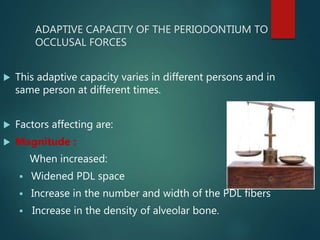
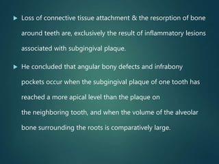
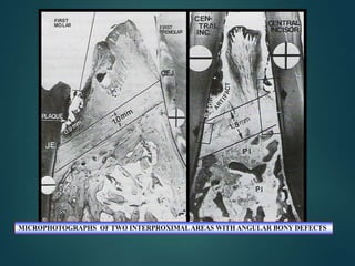
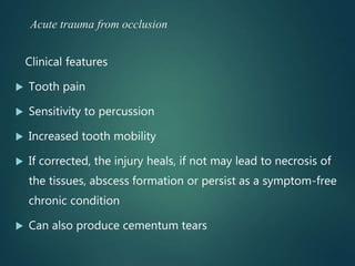
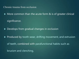
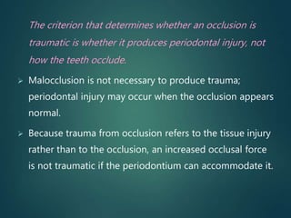
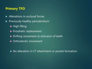
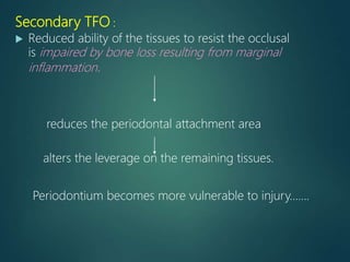
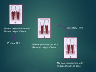
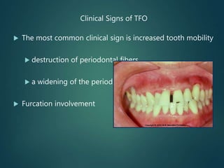
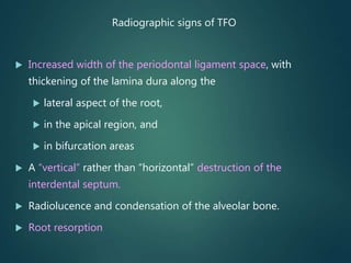
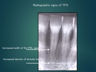
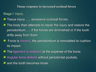
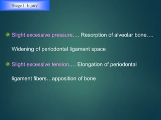
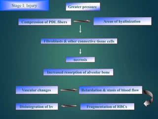
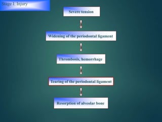
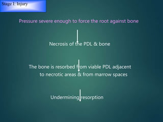
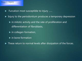
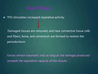
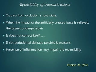
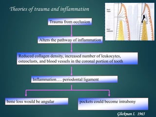
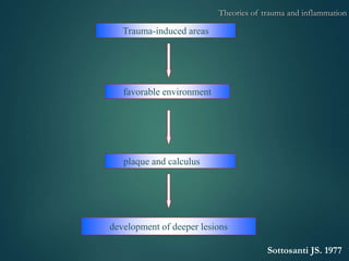
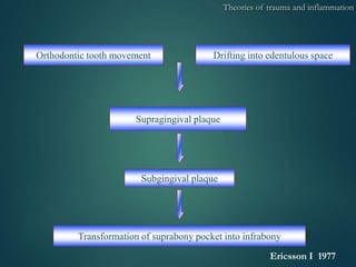
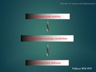
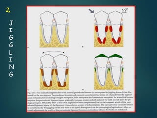
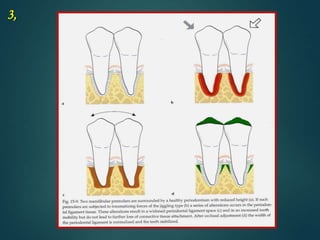
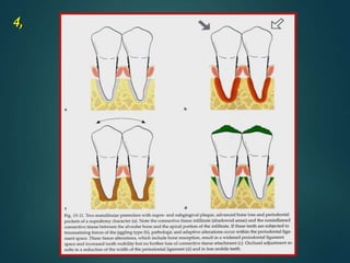
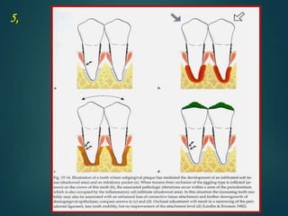
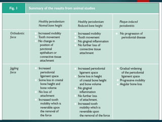
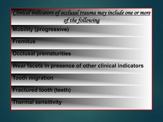
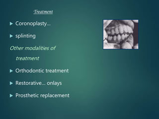
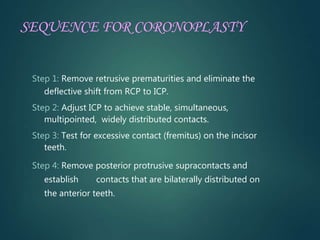
TRAUMA
FROM
OCCLUSION
Introduction
Historical Perspective
Types
Clinical & Radiographic features
Tissue Response to occlusal forces
Theories of Trauma and Inflammation
Clinical & Animal Trials
Pathological Tooth Migration
Diagnosis & Treatment
Summary & Conclusion
INTRODUCTION
ADAPTIVE CAPACITY OF THE PERIODONTIUM TO
OCCLUSAL FORCES
 This adaptive capacity varies in different persons and in
same person at different times.
 Factors affecting are:
 Magnitude :
When increased:
 Widened PDL space
 Increase in the number and width of the PDL fibers
 Increase in the density of alveolar bone.
Stress patterns around roots
changed by altreing
direction of occlusal forces (
Glickman 1970)
Lateral forces and torque
forces are more likely to
injure periodontium.
Direction :
 Reorientation of the stresses and strains
Duration & Frequency:
 Constant pressure more
injurious
 Higher the duration &frequency,
more the trauma.
DEFINITIONS
 Stillman (1917)
“ A condition where injury results to the
supporting structures of the teeth by the act of
bringing the jaws into a closed position”.
 WHO (1978)
“Damage in the periodontium caused by stress
on the teeth of the opposing jaw”.
Definition
Glossary of periodontic terms
“ An injury to the attachment apparatus as a result
excessive occlusal force”.
Glickman 1972
Trauma from occlusion is defined as when occlusal
forces exceed the adaptive capacity of the tissues.
The tissue injury results , the resultant injury is
termed as trauma from occlusion.
Occlusion that produces tissue injury is called traumatic
occlusion
Synonyms
Traumatizing occlusion,
Occlusal trauma,
Traumatogenic occlusion,
Periodontal traumatism, overload, etc.
Karolyi (1901)
Interaction may exist between "trauma from
occlusion" and "alveolar pyrrohea“.
Box (1935) and Stones (1938)
Reported experiments in sheep and monkeys,
TFO is an etiologic factor in production of that
variety of periodontal disease in which there is
vertical pocket formation associated with one or a
varying number of teeth.
Criticized :
Lacked proper controls
Experimental design did not justify
the conclusions drawn.
Glickman’s concept
 Claimed that the pathway of the spread of a plaque-
associated gingival lesion can be changed if forces of an
abnormal magnitude are acting on teeth harboring
subgingival plaque.
 Plaque-associated lesions…suprabony pockets &
horizontal bone loss.
 Sites also exposed to abnormal occlusal force…angular
bony defects & infrabony pockets
Zone of irritation
Zone of co-destruction
Glickman divided periodontal structures into two zones:
1. The zone of irritation and
2. The zone of co-destruction
Zone of irritation:
 Marginal & interdental gingiva
 Gingival lesions in non traumatized tooth propagates
 Even(horizontal) bone destruction.
Alveolar bone
Periodontal
ligament
Zone of co-destruction:
 Includes periodontal ligament, the root cementum & the
alveolar bone
 Coronally demarcated by transseptal and the dentoalveolar
collagen fiber bundles
 Fiber bundles…separate… zone of co-destruction from the
zone of irritation can be affected from two directions-
1. From The inflammatory lesion maintained by plaque in
the zone of irritation
2. From Trauma-induced changes in the zone of co-
destruction
 the fiber bundles may become dissolved and/ or
oriented in a direction parallel to the root surface.
 Gingival lesion in zone of irritation directly spread into
trauma exposed PDL.(not via bone)
 Alteration of the normal pathway of spread of plaque-
associated inflammatory lesion results in the
development of angular bony defects.
 Glickman (1967), “trauma from occlusion is an etiologic
factor of importance in situations where angular bony
defects combined with infrabony pockets are found at
one or several teeth”.
Waerhaug’s concept
 Waerhaug (1979) examined autopsy specimens similar
to Glickman’s.
 Measured in addition the distance between the
subgingival plaque and
1. Periphery of the associated inflammatory cell
infiltrate in the gingiva and
2. The surface of the adjacent bone
Refused the hypothesis that TFO played role in the
spread of a gingival lesion into the “zone of co-
destruction”.
 Loss of connective tissue attachment & the resorption of bone
around teeth are, exclusively the result of inflammatory lesions
associated with subgingival plaque.
 He concluded that angular bony defects and infrabony
pockets occur when the subgingival plaque of one tooth has
reached a more apical level than the plaque on
the neighboring tooth, and when the volume of the alveolar
bone surrounding the roots is comparatively large.
MICROPHOTOGRAPHS OF TWO INTERPROXIMAL AREAS WITH ANGULAR BONY DEFECTS
Classification of Trauma
From Occlusion
 Trauma from occlusion can be classified
 1.According to the injurious occlusal force(s) mode of
onset) –
Acute
Chronic
 2. According to the capacity of the periodontium to
resist to occlusal forces-
-Primary
- Seconday
Acute trauma from occlusion may result from an abrupt
occlusal impact
 Biting on a hard object ( e.g., an olive pit)
 Restorations or prosthetic appliances that interfere
with or alter the direction of occlusal forces on the
teeth
Acute trauma from occlusion
Clinical features
 Tooth pain
 Sensitivity to percussion
 Increased tooth mobility
 If corrected, the injury heals, if not may lead to necrosis of
the tissues, abscess formation or persist as a symptom-free
chronic condition
 Can also produce cementum tears
Chronic trauma from occlusion
 More common than the acute form & is of greater clinical
significance.
 Develops from gradual changes in occlusion
 Produced by tooth wear, drifting movement, and extrusion
of teeth, combined with parafunctional habits such as
bruxism and clenching,
The criterion that determines whether an occlusion is
traumatic is whether it produces periodontal injury, not
how the teeth occlude.
 Malocclusion is not necessary to produce trauma;
periodontal injury may occur when the occlusion appears
normal.
 Because trauma from occlusion refers to the tissue injury
rather than to the occlusion, an increased occlusal force
is not traumatic if the periodontium can accommodate it.
Primary TFO
 Alterations in occlusal forces
 Previously healthy periodontium
 High filling
 Prosthetic replacement
 Drifting movement or extrusion of teeth
 Orthodontic movement
 No alteration in CT attachment or pocket formation.
Secondary TFO :
 Reduced ability of the tissues to resist the occlusal
is impaired by bone loss resulting from marginal
inflammation.
reduces the periodontal attachment area
alters the leverage on the remaining tissues.
Periodontium becomes more vulnerable to injury…….
Normal periodontium with
Normal height of bone
Normal periodontium with
Reduced height of bone
Marginal periodontitis with
Reduced height of bone
Secondary TFO
Primary TFO
Clinical Signs of TFO
 The most common clinical sign is increased tooth mobility
 destruction of periodontal fibers
 a widening of the periodontal ligament
 Furcation involvement
Radiographic signs of TFO
 Increased width of the periodontal ligament space, with
thickening of the lamina dura along the
 lateral aspect of the root,
 in the apical region, and
 in bifurcation areas
 A “vertical” rather than “horizontal” destruction of the
interdental septum.
 Radiolucence and condensation of the alveolar bone.
 Root resorption
Increased width of the PDL space
Increased density of alveolar bone
Radiographic signs of TFO
Tissue response to increased occlusal forces
Stage I: Injury.
Tissue injury ….. excessive occlusal forces.
The body then attempts to repair the injury and restore the
periodontium….. if the forces are diminished or if the tooth
drifts away from them.
Force is chronic, the periodontium is remodeled to cushion
its impact.
The ligament is widened at the expense of the bone,
Angular bone defects without periodontal pockets,
and the tooth becomes loose.
Stage I: Injury
Slight excessive pressure…. Resorption of alveolar bone….
Widening of periodontal ligament space
Slight excessive tension…. Elongation of periodontal
ligament fibers…apposition of bone
Greater pressure
Compression of PDL fibers Areas of hyalinization
Fibroblasts & other connective tissue cells
necrosis
Vascular changes Retardation & stasis of blood flow
Fragmentation of RBCsDisintegration of bv
Increased resorption of alveolar bone
Stage I: Injury
Stage I: Injury
Widening of the periodontal ligament
Tearing of the periodontal ligament
Severe tension
Thrombosis, hemorrhage
Resorption of alveolar bone
Stage I: Injury
Pressure severe enough to force the root against bone
Necrosis of the PDL & bone
The bone is resorbed from viable PDL adjacent
to necrotic areas & from marrow spaces
Undermining resorption
Stage I: Injury
 Furcation most susceptible to injury ……
 Injury to the periodontium produces a temporary depression
 in mitotic activity and the rate of proliferation and
differentiation of fibroblasts,
 in collagen formation,
 in bone formation
 These return to normal levels after dissipation of the forces.
Stage II: Repair
 TFO stimulates increased reparative activity.
Damaged tissues are removed, and new connective tissue cells
and fibers, bone, and cementum are formed to restore the
periodontium
Forces remain traumatic only as long as the damage produced
exceeds the reparative capacity of the tissues.
Stage II: Repair
 Excessive occlusal forces…. resorption of bone…. Body reinforces
the thinned bony trabeculae with new bone…
Buttressing bone formation
Central buttressing
Endosteal cells deposit
new bone ,
Restores bony trabeculae &
reduces the size of marrow spaces
Peripheral buttressing
Shelf like thickening of
the alveolar margin…
Lipping/
Bulge in the contour
of the facial & lingual
Stage III: Adaptive Remodeling of the Periodontium.
Periodontium is remodeled in an effort to create a structural
relationship in which the forces are no longer injurious to
the tissues.
Thickened periodontal ligament, which is funnel shaped at
the crest
Angular defects in the bone, with no pocket formation.
The involved teeth become loose.
Increased vascularization
Reversibility of traumatic lesions
 Trauma from occlusion is reversible.
 When the impact of the artificially created force is relieved,
the tissues undergo repair
 It does not correct itself ……
 If not periodontal damage persists & worsens
 Presence of inflammation may impair the reversibility
Polson M 1976
Insufficient stimulation ……… thinning of the periodontal
ligament, atrophy of the fibers, osteoporosis of the
alveolar bone, and reduction in bone height.
Hypo function can result from..
Effect of excessive occlusal forces on dental pulp..
Effects Of Insufficient Occlusal Force
Effect on progression of marginal periodontitis
 Accumulation of bacterial plaque
that initiates gingivitis and
results in periodontal pocket
formation affects the marginal
gingiva, but trauma from
occlusion occurs in the
supporting tissues and does not
affect the gingiva
 blood supply is sufficient…
 Important to eliminate the marginal inflammatory
component in cases of trauma from occlusion because the
presence of inflammation affects bone regeneration after the
removal of the traumatizing contacts.
 No inflammation…the response to trauma from occlusion is
limited to adaptation to increased forces. However, in the
presence of inflammation, the changes in the shape of the
alveolar crest may be conducive to angular bone loss, and
existing pockets may become intrabony.
Effect on progression of marginal periodontitis
Theories of trauma and inflammation
Alters the pathway of inflammation
Reduced collagen density, increased number of leukocytes,
osteoclasts, and blood vessels in the coronal portion of tooth
Inflammation…. periodontal ligament
bone loss would be angular pockets could become intrabony
Trauma from occlusion
Glickman I. 1965
Trauma-induced areas
favorable environment
plaque and calculus
development of deeper lesions
Sottosanti JS. 1977
Theories of trauma and inflammation
Orthodontic tooth movement Drifting into edentulous space
Transformation of suprabony pocket into infrabony
Supragingival plaque
Subgingival plaque
Ericsson I 1977
Theories of trauma and inflammation
Increased tooth mobility
pumping effect on plaque metabolites
Increasing their diffusion
Vollmer WH 1975
Theories of trauma and inflammation
Clinical and Animal Trials
 Rosling et al. (1976)… “infrabony pocket located at hypermobile teeth
exhibited the same degree of healing as those adjacent to firm teeth”. (pdl
condtion of mobile th)
 Fleszar et al. (1980)… “pockets of clinically mobile teeth do not respond as
well to periodontal treatment as do those of firm teeth exhibiting the same
disease severity”.(mobility on healing after perio-surgery)
 Burgett et al. (1992)… Probing attachment gain was on the average about
0.5mm larger in patients who received the combined treatment, i.e. scaling and
occlusal adjustment. (occlusal adjustment on periodontitis)
Pihlstrom et al “teeth with increased mobility & widened pdl
ligament Space had , infact deeper pockets, more attachment loss &
reduced bone height”
Neidurad et al “ in beagle dogs “ probing depth at two otherwise
similar teeth one mobile & other non mobile . Probing depth
recorded 0.5 mm higher in the mobile teeth”
Orban b. smullow .weiss l , gotlieb, ramford , kohlar h, volmer ,
“ trauma from occlusion does not cause gingivitis or pocket
formation”
Erricson , lindhe, svanberg , glickman “ occusal stresses increase s
periodontal destruction induced by periodontitis”
Animal experiments:
 Reaction of normal periodontium…application of forces
inflicted on teeth in one direction only in early
experiments,
 Biopsies…tooth & periodontium;
 Tooth is exposed to unilateral forces of a magnitude,
frequency or duration…..certain well-defined reactions
develop in the periodontal structures to the altered
functional demand.
Orthodontic type trauma
forces does not cause inflammation in gingiva
1,
J
I
G
G
L
I
N
G
2,
3,
4,
5,
Clinical indicators of occlusal trauma may include one or more
of the following
Mobility (progressive)
Fremitus
Occlusal prematurities
Wear facets in presence of other clinical indicators
Tooth migration
Fractured tooth (teeth)
Thermal sensitivity
Treatment
 Coronoplasty…
 splinting
Other modalities of
treatment
 Orthodontic treatment
 Restorative… onlays
 Prosthetic replacement
SEQUENCE FOR CORONOPLASTY
Step 1: Remove retrusive prematurities and eliminate the
deflective shift from RCP to ICP.
Step 2: Adjust ICP to achieve stable, simultaneous,
multipointed, widely distributed contacts.
Step 3: Test for excessive contact (fremitus) on the incisor
teeth.
Step 4: Remove posterior protrusive supracontacts and
establish contacts that are bilaterally distributed on
the anterior teeth.
Step 5: Remove or lessen mediotrusive (balancing) interferences.
Step 6: Reduce excessive cusp steepness on the laterotrusive
(working) contacts. (BULL) Law
Step 7: Eliminate gross occlusal disharmonies.
Step 8: Recheck tooth contact relationships.
Step 9: Polish all rough tooth surfaces.
 MAINTENANCE OF OCCLUSAL STABILITY
CONCLUSION
The exact effect of TFO on the progression of human
periodontal disease remains unknown. However, all
studies performed to date strongly indicate that occlusion
is not a causative factor in periodontal disease but may be
a significant risk factor in the progression of periodontal
disease and also inhibit the potential for bone
regeneration.
On this basis, we can say that the treatment of occlusal
discrepancies should be considered as an integral part of
the overall treatment of periodontal disease and should be
included in the comprehensive treatment of this disease.
 Clinical Periodontology – Carranza 13th Edition
 Clinical Periodontology and Implant Dentistry –
Jan Lindhe 6thth Edition.
References
Thank you
StayHomeStaySafe

Trauma from occlusion

  • 1.
  • 2.
    Introduction Historical Perspective Types Clinical &Radiographic features Tissue Response to occlusal forces Theories of Trauma and Inflammation Clinical & Animal Trials Pathological Tooth Migration Diagnosis & Treatment Summary & Conclusion
  • 3.
  • 4.
    ADAPTIVE CAPACITY OFTHE PERIODONTIUM TO OCCLUSAL FORCES  This adaptive capacity varies in different persons and in same person at different times.  Factors affecting are:  Magnitude : When increased:  Widened PDL space  Increase in the number and width of the PDL fibers  Increase in the density of alveolar bone.
  • 5.
    Stress patterns aroundroots changed by altreing direction of occlusal forces ( Glickman 1970) Lateral forces and torque forces are more likely to injure periodontium. Direction :  Reorientation of the stresses and strains Duration & Frequency:  Constant pressure more injurious  Higher the duration &frequency, more the trauma.
  • 6.
  • 7.
     Stillman (1917) “A condition where injury results to the supporting structures of the teeth by the act of bringing the jaws into a closed position”.  WHO (1978) “Damage in the periodontium caused by stress on the teeth of the opposing jaw”.
  • 8.
    Definition Glossary of periodonticterms “ An injury to the attachment apparatus as a result excessive occlusal force”. Glickman 1972 Trauma from occlusion is defined as when occlusal forces exceed the adaptive capacity of the tissues. The tissue injury results , the resultant injury is termed as trauma from occlusion.
  • 9.
    Occlusion that producestissue injury is called traumatic occlusion Synonyms Traumatizing occlusion, Occlusal trauma, Traumatogenic occlusion, Periodontal traumatism, overload, etc.
  • 10.
    Karolyi (1901) Interaction mayexist between "trauma from occlusion" and "alveolar pyrrohea“.
  • 11.
    Box (1935) andStones (1938) Reported experiments in sheep and monkeys, TFO is an etiologic factor in production of that variety of periodontal disease in which there is vertical pocket formation associated with one or a varying number of teeth. Criticized : Lacked proper controls Experimental design did not justify the conclusions drawn.
  • 12.
    Glickman’s concept  Claimedthat the pathway of the spread of a plaque- associated gingival lesion can be changed if forces of an abnormal magnitude are acting on teeth harboring subgingival plaque.  Plaque-associated lesions…suprabony pockets & horizontal bone loss.  Sites also exposed to abnormal occlusal force…angular bony defects & infrabony pockets
  • 13.
    Zone of irritation Zoneof co-destruction
  • 14.
    Glickman divided periodontalstructures into two zones: 1. The zone of irritation and 2. The zone of co-destruction Zone of irritation:  Marginal & interdental gingiva  Gingival lesions in non traumatized tooth propagates  Even(horizontal) bone destruction. Alveolar bone Periodontal ligament
  • 15.
    Zone of co-destruction: Includes periodontal ligament, the root cementum & the alveolar bone  Coronally demarcated by transseptal and the dentoalveolar collagen fiber bundles  Fiber bundles…separate… zone of co-destruction from the zone of irritation can be affected from two directions- 1. From The inflammatory lesion maintained by plaque in the zone of irritation 2. From Trauma-induced changes in the zone of co- destruction
  • 16.
     the fiberbundles may become dissolved and/ or oriented in a direction parallel to the root surface.  Gingival lesion in zone of irritation directly spread into trauma exposed PDL.(not via bone)  Alteration of the normal pathway of spread of plaque- associated inflammatory lesion results in the development of angular bony defects.  Glickman (1967), “trauma from occlusion is an etiologic factor of importance in situations where angular bony defects combined with infrabony pockets are found at one or several teeth”.
  • 17.
    Waerhaug’s concept  Waerhaug(1979) examined autopsy specimens similar to Glickman’s.  Measured in addition the distance between the subgingival plaque and 1. Periphery of the associated inflammatory cell infiltrate in the gingiva and 2. The surface of the adjacent bone Refused the hypothesis that TFO played role in the spread of a gingival lesion into the “zone of co- destruction”.
  • 18.
     Loss ofconnective tissue attachment & the resorption of bone around teeth are, exclusively the result of inflammatory lesions associated with subgingival plaque.  He concluded that angular bony defects and infrabony pockets occur when the subgingival plaque of one tooth has reached a more apical level than the plaque on the neighboring tooth, and when the volume of the alveolar bone surrounding the roots is comparatively large.
  • 19.
    MICROPHOTOGRAPHS OF TWOINTERPROXIMAL AREAS WITH ANGULAR BONY DEFECTS
  • 20.
    Classification of Trauma FromOcclusion  Trauma from occlusion can be classified  1.According to the injurious occlusal force(s) mode of onset) – Acute Chronic  2. According to the capacity of the periodontium to resist to occlusal forces- -Primary - Seconday
  • 21.
    Acute trauma fromocclusion may result from an abrupt occlusal impact  Biting on a hard object ( e.g., an olive pit)  Restorations or prosthetic appliances that interfere with or alter the direction of occlusal forces on the teeth Acute trauma from occlusion
  • 22.
    Acute trauma fromocclusion Clinical features  Tooth pain  Sensitivity to percussion  Increased tooth mobility  If corrected, the injury heals, if not may lead to necrosis of the tissues, abscess formation or persist as a symptom-free chronic condition  Can also produce cementum tears
  • 23.
    Chronic trauma fromocclusion  More common than the acute form & is of greater clinical significance.  Develops from gradual changes in occlusion  Produced by tooth wear, drifting movement, and extrusion of teeth, combined with parafunctional habits such as bruxism and clenching,
  • 24.
    The criterion thatdetermines whether an occlusion is traumatic is whether it produces periodontal injury, not how the teeth occlude.  Malocclusion is not necessary to produce trauma; periodontal injury may occur when the occlusion appears normal.  Because trauma from occlusion refers to the tissue injury rather than to the occlusion, an increased occlusal force is not traumatic if the periodontium can accommodate it.
  • 25.
    Primary TFO  Alterationsin occlusal forces  Previously healthy periodontium  High filling  Prosthetic replacement  Drifting movement or extrusion of teeth  Orthodontic movement  No alteration in CT attachment or pocket formation.
  • 26.
    Secondary TFO : Reduced ability of the tissues to resist the occlusal is impaired by bone loss resulting from marginal inflammation. reduces the periodontal attachment area alters the leverage on the remaining tissues. Periodontium becomes more vulnerable to injury…….
  • 27.
    Normal periodontium with Normalheight of bone Normal periodontium with Reduced height of bone Marginal periodontitis with Reduced height of bone Secondary TFO Primary TFO
  • 28.
    Clinical Signs ofTFO  The most common clinical sign is increased tooth mobility  destruction of periodontal fibers  a widening of the periodontal ligament  Furcation involvement
  • 29.
    Radiographic signs ofTFO  Increased width of the periodontal ligament space, with thickening of the lamina dura along the  lateral aspect of the root,  in the apical region, and  in bifurcation areas  A “vertical” rather than “horizontal” destruction of the interdental septum.  Radiolucence and condensation of the alveolar bone.  Root resorption
  • 30.
    Increased width ofthe PDL space Increased density of alveolar bone Radiographic signs of TFO
  • 31.
    Tissue response toincreased occlusal forces Stage I: Injury. Tissue injury ….. excessive occlusal forces. The body then attempts to repair the injury and restore the periodontium….. if the forces are diminished or if the tooth drifts away from them. Force is chronic, the periodontium is remodeled to cushion its impact. The ligament is widened at the expense of the bone, Angular bone defects without periodontal pockets, and the tooth becomes loose.
  • 32.
    Stage I: Injury Slightexcessive pressure…. Resorption of alveolar bone…. Widening of periodontal ligament space Slight excessive tension…. Elongation of periodontal ligament fibers…apposition of bone
  • 33.
    Greater pressure Compression ofPDL fibers Areas of hyalinization Fibroblasts & other connective tissue cells necrosis Vascular changes Retardation & stasis of blood flow Fragmentation of RBCsDisintegration of bv Increased resorption of alveolar bone Stage I: Injury
  • 34.
    Stage I: Injury Wideningof the periodontal ligament Tearing of the periodontal ligament Severe tension Thrombosis, hemorrhage Resorption of alveolar bone
  • 35.
    Stage I: Injury Pressuresevere enough to force the root against bone Necrosis of the PDL & bone The bone is resorbed from viable PDL adjacent to necrotic areas & from marrow spaces Undermining resorption
  • 36.
    Stage I: Injury Furcation most susceptible to injury ……  Injury to the periodontium produces a temporary depression  in mitotic activity and the rate of proliferation and differentiation of fibroblasts,  in collagen formation,  in bone formation  These return to normal levels after dissipation of the forces.
  • 37.
    Stage II: Repair TFO stimulates increased reparative activity. Damaged tissues are removed, and new connective tissue cells and fibers, bone, and cementum are formed to restore the periodontium Forces remain traumatic only as long as the damage produced exceeds the reparative capacity of the tissues.
  • 38.
    Stage II: Repair Excessive occlusal forces…. resorption of bone…. Body reinforces the thinned bony trabeculae with new bone… Buttressing bone formation Central buttressing Endosteal cells deposit new bone , Restores bony trabeculae & reduces the size of marrow spaces Peripheral buttressing Shelf like thickening of the alveolar margin… Lipping/ Bulge in the contour of the facial & lingual
  • 39.
    Stage III: AdaptiveRemodeling of the Periodontium. Periodontium is remodeled in an effort to create a structural relationship in which the forces are no longer injurious to the tissues. Thickened periodontal ligament, which is funnel shaped at the crest Angular defects in the bone, with no pocket formation. The involved teeth become loose. Increased vascularization
  • 41.
    Reversibility of traumaticlesions  Trauma from occlusion is reversible.  When the impact of the artificially created force is relieved, the tissues undergo repair  It does not correct itself ……  If not periodontal damage persists & worsens  Presence of inflammation may impair the reversibility Polson M 1976
  • 42.
    Insufficient stimulation ………thinning of the periodontal ligament, atrophy of the fibers, osteoporosis of the alveolar bone, and reduction in bone height. Hypo function can result from.. Effect of excessive occlusal forces on dental pulp.. Effects Of Insufficient Occlusal Force
  • 43.
    Effect on progressionof marginal periodontitis  Accumulation of bacterial plaque that initiates gingivitis and results in periodontal pocket formation affects the marginal gingiva, but trauma from occlusion occurs in the supporting tissues and does not affect the gingiva  blood supply is sufficient…
  • 44.
     Important toeliminate the marginal inflammatory component in cases of trauma from occlusion because the presence of inflammation affects bone regeneration after the removal of the traumatizing contacts.  No inflammation…the response to trauma from occlusion is limited to adaptation to increased forces. However, in the presence of inflammation, the changes in the shape of the alveolar crest may be conducive to angular bone loss, and existing pockets may become intrabony. Effect on progression of marginal periodontitis
  • 45.
    Theories of traumaand inflammation Alters the pathway of inflammation Reduced collagen density, increased number of leukocytes, osteoclasts, and blood vessels in the coronal portion of tooth Inflammation…. periodontal ligament bone loss would be angular pockets could become intrabony Trauma from occlusion Glickman I. 1965
  • 46.
    Trauma-induced areas favorable environment plaqueand calculus development of deeper lesions Sottosanti JS. 1977 Theories of trauma and inflammation
  • 47.
    Orthodontic tooth movementDrifting into edentulous space Transformation of suprabony pocket into infrabony Supragingival plaque Subgingival plaque Ericsson I 1977 Theories of trauma and inflammation
  • 48.
    Increased tooth mobility pumpingeffect on plaque metabolites Increasing their diffusion Vollmer WH 1975 Theories of trauma and inflammation
  • 49.
  • 50.
     Rosling etal. (1976)… “infrabony pocket located at hypermobile teeth exhibited the same degree of healing as those adjacent to firm teeth”. (pdl condtion of mobile th)  Fleszar et al. (1980)… “pockets of clinically mobile teeth do not respond as well to periodontal treatment as do those of firm teeth exhibiting the same disease severity”.(mobility on healing after perio-surgery)  Burgett et al. (1992)… Probing attachment gain was on the average about 0.5mm larger in patients who received the combined treatment, i.e. scaling and occlusal adjustment. (occlusal adjustment on periodontitis)
  • 51.
    Pihlstrom et al“teeth with increased mobility & widened pdl ligament Space had , infact deeper pockets, more attachment loss & reduced bone height” Neidurad et al “ in beagle dogs “ probing depth at two otherwise similar teeth one mobile & other non mobile . Probing depth recorded 0.5 mm higher in the mobile teeth” Orban b. smullow .weiss l , gotlieb, ramford , kohlar h, volmer , “ trauma from occlusion does not cause gingivitis or pocket formation” Erricson , lindhe, svanberg , glickman “ occusal stresses increase s periodontal destruction induced by periodontitis”
  • 52.
    Animal experiments:  Reactionof normal periodontium…application of forces inflicted on teeth in one direction only in early experiments,  Biopsies…tooth & periodontium;  Tooth is exposed to unilateral forces of a magnitude, frequency or duration…..certain well-defined reactions develop in the periodontal structures to the altered functional demand.
  • 53.
    Orthodontic type trauma forcesdoes not cause inflammation in gingiva 1,
  • 54.
  • 55.
  • 56.
  • 57.
  • 59.
    Clinical indicators ofocclusal trauma may include one or more of the following Mobility (progressive) Fremitus Occlusal prematurities Wear facets in presence of other clinical indicators Tooth migration Fractured tooth (teeth) Thermal sensitivity
  • 60.
    Treatment  Coronoplasty…  splinting Othermodalities of treatment  Orthodontic treatment  Restorative… onlays  Prosthetic replacement
  • 61.
    SEQUENCE FOR CORONOPLASTY Step1: Remove retrusive prematurities and eliminate the deflective shift from RCP to ICP. Step 2: Adjust ICP to achieve stable, simultaneous, multipointed, widely distributed contacts. Step 3: Test for excessive contact (fremitus) on the incisor teeth. Step 4: Remove posterior protrusive supracontacts and establish contacts that are bilaterally distributed on the anterior teeth.
  • 62.
    Step 5: Removeor lessen mediotrusive (balancing) interferences. Step 6: Reduce excessive cusp steepness on the laterotrusive (working) contacts. (BULL) Law Step 7: Eliminate gross occlusal disharmonies. Step 8: Recheck tooth contact relationships. Step 9: Polish all rough tooth surfaces.  MAINTENANCE OF OCCLUSAL STABILITY
  • 64.
    CONCLUSION The exact effectof TFO on the progression of human periodontal disease remains unknown. However, all studies performed to date strongly indicate that occlusion is not a causative factor in periodontal disease but may be a significant risk factor in the progression of periodontal disease and also inhibit the potential for bone regeneration. On this basis, we can say that the treatment of occlusal discrepancies should be considered as an integral part of the overall treatment of periodontal disease and should be included in the comprehensive treatment of this disease.
  • 65.
     Clinical Periodontology– Carranza 13th Edition  Clinical Periodontology and Implant Dentistry – Jan Lindhe 6thth Edition. References
  • 66.

Editor's Notes

  • #6 Principal fibers of the PDL are arranged so that they bestaccommodate occlusal forces along the axis of the tooth .
  • #11 different opinions have been presented regarding validity of this claim.
  • #12 1930s,
  • #15 Soft tissue bordered by hard tissue only on one side & is not affected by forces of occlusion Plaque-associated lesion at a “non-traumatized” tooth propagates in apical direction by first involving the alveolar bone & only later the periodontal ligament area.