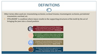
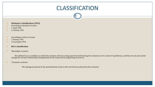
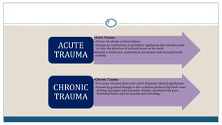
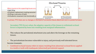
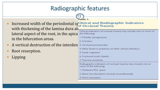
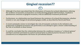
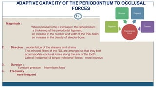
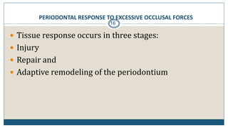
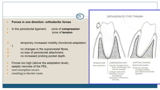
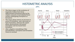
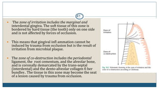
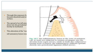
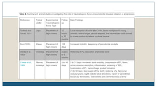
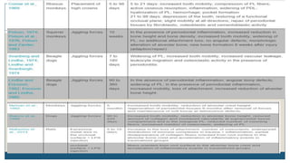
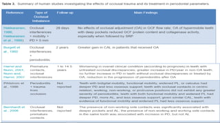
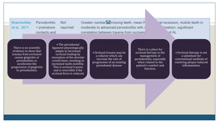
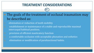
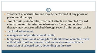
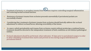
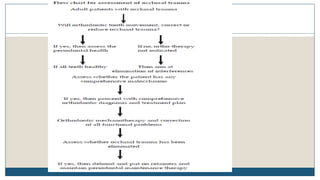
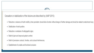
This document discusses trauma from occlusion (TFO), which refers to pathologic alterations or adaptive changes in the periodontium resulting from excessive occlusal forces. It covers the historical understanding of TFO, definitions, classifications, clinical features, and the periodontal response and adaptation to excessive forces. It also examines Glickman's concept of co-destruction between TFO and plaque-associated periodontal disease. The document provides details on injury, repair, remodeling processes in the periodontium in response to TFO.
Pathologic tooth migration (PTM) refers to tooth displacement resulting from a disturbance in factors that maintain normal tooth position. PTM is common in periodontal patients, with prevalence studies finding rates of 30-55%. The primary factor in PTM is periodontal bone loss resulting from periodontal disease. Other factors include occlusal changes from tooth loss, soft tissue pressures, oral habits, and periapical or gingival inflammation. Treatment involves periodontal therapy, sometimes with adjunctive orthodontics or prosthodontics, while prevention focuses on periodontal disease control and management of predisposing occlusal and habit factors.
This presentation describes the gingival recession, its classifications and theories of pathogenesis and different etiological factors in its progression.
This document discusses the anatomy, measurement, and clinical significance of the attached gingiva. It notes that the attached gingiva extends from the base of the gingival sulcus to the mucogingival junction. The normal width is 3-4.5mm in the maxillary anterior region but narrower in other areas. Inadequate width can facilitate subgingival plaque formation. Methods to measure width and increase width through surgery are described. The importance of keratinized, attached tissue for resisting mechanical irritation and stabilizing the gingival margin is emphasized.
This document discusses trauma from occlusion (TFO). It begins by defining TFO as pathologic alterations or adaptive changes that develop in the periodontium as a result of undue force from chewing muscles. It describes primary TFO resulting from sudden impacts and secondary TFO from gradual changes that occur with reduced bone support. Clinical features include tooth pain and mobility. Radiographic features include widened ligament space and buttressing bone. Treatment focuses on reducing tooth mobility, eliminating prematurities, and using splints. While TFO alone may increase mobility, inflammation is required for attachment loss.
In periodontology, classifications are widely used to categorize defects due to periodontitis according to their etiology, diagnosis, treatment and prognosis.
Several classifications have been proposed in the literature in order to facilitate the diagnosis of gingival recessions.
This document discusses genetics in relation to periodontitis. It provides background on genetic study designs like segregation analysis, twin studies, and linkage/association studies that are used to identify genes associated with periodontal diseases. Specific genes linked to aggressive periodontitis are mentioned, including mutations in the alkaline phosphatase, cathepsin C, and CD18/CD11 genes. Studies finding autosomal dominant and recessive inheritance of aggressive periodontitis in different populations are summarized. The role of HLA antigens and IL-1 gene polymorphisms in periodontitis susceptibility is also briefly covered.
Gingival biotype refers to the thickness and width of gingiva and alveolar bone morphology. This study aimed to understand the association between biotype and the dentopapillary complex to define objective determinants of biotype classification. 50 subjects were examined, measuring crown length, width, papillary height and width to classify biotype as thin or thick. Statistical analysis found crown length was the best determinant of biotype, with thin biotype having longer crowns and thinner papillae compared to thick biotype. The results provide objective guidelines for determining biotype based on correlations with dentopapillary complex dimensions.
Scaling and root planing (SRP) is a non-surgical treatment for periodontitis that aims to remove dental plaque and calculus from tooth surfaces. It involves scaling to remove deposits and root planing to smooth root surfaces. The goals are to eliminate periodontitis by removing irritants and restoring a healthy environment for tissue healing. The long-term effectiveness depends on factors like patient compliance, disease severity, and anatomical challenges. Overhanging restorations can interfere with cleaning and disturb the ecological balance, allowing disease-causing bacteria to proliferate.
Bleeding on probing is an early sign of gingival inflammation and is commonly used to assess periodontal disease status. It occurs when increased crevicular fluid and breakdown of gingival tissues due to inflammation allows blood vessels to rupture upon gentle probing. Local factors like poor oral hygiene and systemic conditions like vitamin deficiencies or coagulation disorders can contribute to abnormal gingival bleeding. The bleeding point index is used to evaluate gingival inflammation by recording the number of bleeding sites after probing specific areas in the mouth.
The document discusses the microbiology of dental plaque and periodontal infections. It notes that after birth, infants acquire oral bacteria that establish a mature microbiota in the mouth within two weeks. Dental plaque is a biofilm that forms on teeth and consists of over 500 microbial species living in an extracellular matrix. Key points include that plaque maturation involves an ecological shift from primarily aerobic bacteria to anaerobic species over time. The microbial composition of plaque can influence the transition from periodontal health to disease if pathogens overgrow due to changes in the local environment.
This document discusses trauma from occlusion (TFO), defined as pathological alterations or adaptive changes that develop in the periodontium due to excessive occlusal forces. It provides historical context on TFO research dating back to 1901, classifications of TFO, stages of tissue response to TFO including injury and repair, and factors that can increase occlusal forces or decrease the periodontium's resistance to forces. TFO can be acute or chronic and primary (due to occlusal factors) or secondary (due to reduced periodontal support). Excessive forces can cause tissue injury through thrombosis, hemorrhage or necrosis while the body attempts repair through new tissue formation and bone remodeling.
Dr. Nael Al Masri provides a comprehensive overview of periodontal examination and diagnosis. The summary includes:
1. A periodontal examination involves assessing the patient's medical and dental history, examining plaque, gingiva, probing depths, clinical attachment levels, bleeding, bone loss, tooth mobility, and furcation involvement.
2. Key findings that help determine a diagnosis include probing depths above 3mm, clinical attachment loss, bleeding on probing, recession, furcation involvement, and tooth mobility.
3. The examination is used to establish a diagnosis, prognosis, and develop a customized treatment plan for the patient.
Hold oxygen mask
Monitor vital signs
IV fluids if needed
45 8/31/2012 Dr. Nitika Jain
46 Dr. nitika jain 31 August 2012
Local anesthetic and analgesic
administration during pregnancy
Local anesthetics are safe to use during pregnancy.
Lignocaine is the local anesthetic of choice during
pregnancy.
Use the minimum effective dose.
Avoid repeated administration of local anesthetics.
Use aspirin or acetaminophen for analgesia during
pregnancy.
Avoid NSAIDs during pregnancy
Classification of periodontal diseases /certified fixed orthodontic courses ...Indian dental academy
This document discusses the classification of periodontal diseases. It outlines the need for classification to aid in diagnosis, treatment planning, and communication. It then details the historical evolution of classification systems from the 1870s to present day. Early systems were based on clinical characteristics, while later systems incorporated concepts of pathology and the infectious etiology of diseases. The current paradigm recognizes periodontitis as an inflammatory disease caused by bacterial plaque and host responses. Classification systems continue to be refined as understanding improves.
Chronic periodontitis is the most common form of periodontitis and is characterized by microbial biofilm formation, periodontal inflammation, and attachment and bone loss. It is diagnosed based on clinical attachment level assessment and radiographs showing bone loss. Risk factors include local factors like plaque and calculus that retain bacteria, and systemic factors like diabetes which impair the immune response and increase the severity of periodontitis. The condition progresses slowly over time with increased attachment and bone loss with age.
This document provides an overview of endodontic-periodontal interactions. It discusses the pathways connecting endodontic and periodontal tissues, the etiology of endo-perio lesions, classifications of endo-perio lesions, diagnostic procedures, differences between periodontal and periapical abscesses, the endo-perio controversy, and management of pulpal and periodontal diseases. The key relationships covered are the anatomical and pathological connections between the pulp and periodontium, the bacteria commonly found in both tissues, and the debate around whether periodontal or endodontic disease can cause the other.
The document discusses the historical development and current classification of periodontal diseases. It outlines several past classification systems from the 19th century based on clinical characteristics to more recent systems from the late 20th century incorporating etiology and pathogenesis. The current 1999 classification system from the International Workshop for a Classification of Periodontal Diseases and Conditions is explained in detail, categorizing diseases based on factors like plaque-induced vs. non-plaque induced gingival diseases, chronic vs. aggressive periodontitis, and periodontitis as a manifestation of systemic diseases.
This document discusses various techniques for non-surgical periodontal therapy, focusing on root planing. It defines root planing as the removal of plaque, calculus, and contaminated cementum and dentin from root surfaces. It discusses the rationale for root planing, including the removal of diseased cementum that may contain toxins. It evaluates different root planing instruments like curettes and ultrasonic scalers. While some studies found root smoothness and cementum removal were unnecessary, most support root planing to remove toxins and prepare the surface for new attachment. The document analyzes debates around techniques and their role in resolving inflammation and facilitating healing.
This document discusses risk assessment in periodontal disease. It defines key terms like risk, risk factors, risk determinants, and risk markers. It categorizes different types of risk elements and describes several risk assessment tools that use various clinical and historical parameters to provide a risk score. The document concludes that while identification of risk factors has furthered understanding of periodontitis, predicting individual risk remains limited.
The document discusses periodontal-endodontic lesions, which occur when inflammation spreads between the pulp and periodontium. It identifies three categories of pathways for communication: developmental, pathologic, and iatrogenic. Microorganisms like Porphyromonas gingivalis and Treponema denticola have been found in endo-perio lesions. Diagnosis involves tests like radiographs, probing, and pulp vitality tests. Treatment aims to address both the pulpal and periodontal involvement through approaches like root canal therapy, scaling and root planing, and sometimes extraction.
At the end of this session, the student should be able to describe:
What is Periodontium and its role
Ecology of Dental Crevice and its role
Conditions that affect Periodontal tissue
Role of Microorganisms in Periodontal Disease
Complex relationship between Plaque and periodontal disease
1) There is debate around the role of occlusion in periodontal disease, with some studies finding a relationship between excessive occlusal forces and periodontal destruction, while others finding no such relationship.
2) Animal studies show that occlusal forces can lead to increased tooth mobility and minor bone loss in healthy periodontium, but do not enhance progression of plaque-induced periodontitis.
3) Occlusal adjustment may be indicated when occlusal forces cause trauma, aggravate parafunction, or to aid splint therapy, but its role in periodontal treatment is unclear.
This document provides an overview of periodontal abscesses. It defines a periodontal abscess as a localized collection of pus within the periodontal tissues. Periodontal abscesses are classified based on duration, number, location and etiology. The main causes are periodontitis, trauma from foreign objects, and certain dental procedures. Microbiologically, periodontal abscesses involve anaerobic bacteria normally found in the oral cavity like P. gingivalis, P. intermedia and F. nucleatum. Clinical features include pain, swelling and exudate from the gingiva. Management involves incision and drainage of the acute lesion along with antibiotics and treatment of the underlying condition like scaling or surgery.
This document discusses soft tissue grafting procedures used in periodontal plastic surgery. It provides an overview of common grafting techniques like free gingival grafts and connective tissue grafts used to treat mucogingival defects. Details are given on the indications, surgical protocols, advantages/disadvantages of each technique. Post-operative healing times and expectations are reviewed. The goal is to understand how and when these procedures can be used to correct mucogingival defects and improve periodontal health and aesthetics.
This document discusses gingival recession, including its definitions, classifications, etiology, factors affecting treatment outcomes, and treatments. It provides an overview of several classification systems for gingival recession, including those proposed by Sullivan and Atkins, Miller, Mahajan, Cairo, and Ashish Kumar. Miller's classification is the most widely used but has limitations, so modifications have been suggested. The document also proposes a new comprehensive classification system that aims to address the limitations of previous systems.
Supportive periodontal therapy (SPT) involves long-term maintenance programs following active periodontal treatment to maintain periodontal health. SPT involves periodic examination, motivation and instrumentation of sites showing inflammation, treatment of reinfected sites, and polishing. It begins after active treatment and is aimed at preventing recurrence through early detection of disease. The frequency of SPT visits depends on the patient's periodontal risk assessment but generally occurs every 3-4 months. It can be performed by general dentists or specialists depending on the extent of original periodontal destruction. Adjunctive use of antimicrobials may also be included in SPT.
The document discusses the role of occlusion in periodontal disease. It defines occlusion and classifies occlusion types. It explores the biological basis of occlusal function and the relationship between occlusal disharmony and periodontal disease. Occlusal trauma from hyperfunction, hypofunction, and parafunctions like bruxism can impact periodontal tissues. The document outlines methods for clinical diagnosis and developing treatment plans involving occlusal therapy, adjustment, and splinting to address occlusal factors and support periodontal treatment.
The document provides information on trauma from occlusion and coronoplasty. It defines trauma from occlusion as damage to the periodontium caused by excessive occlusal forces. Coronoplasty involves selective reduction of occlusal surfaces to influence mechanical contact conditions and sensory input, with the aim of reducing excessive tooth mobility and providing functional stimulation for periodontal health. The document discusses the diagnosis, classification, and clinical features of trauma from occlusion, as well as the objectives, methods, and techniques used in performing coronoplasty.
This document discusses trauma from occlusion (TFO), which refers to injury to the periodontium resulting from excessive occlusal forces. It defines key terms, classifies TFO, and describes the stages of tissue response and role of occlusion in periodontal disease pathogenesis. Several human and animal studies are summarized that investigated the relationship between occlusal forces and periodontal disease, generally finding that excessive forces alone do not cause attachment loss but may alter the pathway of destruction in the presence of plaque. The document provides an overview of TFO and its relationship to periodontal disease.
This document defines trauma from occlusion (TFO) and outlines its causes, classification, clinical features, radiographic findings, and treatment. TFO occurs when occlusal forces exceed the adaptive capacity of the periodontium, causing tissue injury. It can be acute from a sudden impact or chronic from gradual changes in occlusion. Factors that increase traumatic forces are magnitude, direction, and duration of forces. TFO is classified as primary, secondary, or combined based on causative factors. Clinical features include tooth mobility and pain. Radiographic findings show increased periodontal ligament space and bone loss. Treatment goals are to maintain periodontal health and function through occlusal adjustment, habit management, stabilization, orthodontics, reconstruction,
This document discusses trauma from occlusion (TFO) and its effects on the periodontium. It defines TFO as injury to the periodontal tissues caused by excessive occlusal forces. It describes the different types of occlusal forces and their effects, including acute vs chronic TFO, and primary vs secondary TFO. It also discusses the stages of tissue response to increased occlusal forces: injury, repair, and adaptive remodeling. The document examines various theories on the interaction between TFO and plaque-associated periodontal disease.
Occlusal Considerations For Implant Supported Prostheses Implant Protectes O...Mohammed Alshehri
Trauma from occlusion refers to pathological changes in the periodontium caused by excessive force from chewing muscles. While excessive force alone does not cause tissue breakdown, it may act as a co-factor in plaque-induced periodontal disease by enhancing the rate of progression. Proper treatment of plaque is important to arrest tissue destruction, even if occlusal trauma persists. Treating occlusal trauma alone through adjustment or splinting may reduce mobility but not stop further breakdown from untreated plaque.
This document discusses trauma from occlusion, or occlusal trauma. It begins by outlining different types of occlusal forces, including physiological forces and various traumatic forces. It then describes the adaptive capacity of the periodontium to withstand occlusal forces up to a point before injury occurs, which is termed trauma from occlusion. The document outlines the stages of tissue response to increased occlusal forces: injury, repair through reparative processes, and potential adaptive remodeling of the periodontium. It also discusses classifications of trauma from occlusion and the effects of both insufficient and excessive occlusal forces.
This document discusses occlusion and its role in periodontics. It defines occlusion and describes ideal occlusion. It discusses differing occlusal schemes and questions regarding occlusion's role in periodontitis, dental implants, and abfraction. It covers trauma from occlusion, including classifications and tissue response. It reviews historical studies on occlusion's role and their shortcomings. Animal studies demonstrated that jiggling trauma can aggravate periodontal disease. Clinical studies provide some evidence occlusion may be a risk factor in disease progression. The role of occlusion in implant dentistry aims to protect implants from biomechanical overload.
This document provides an overview of occlusion and occlusal therapy. It discusses the forces involved in jaw movement, the biologic basis of normal occlusion and occlusion-related dysfunction. It covers clinical examination techniques used to evaluate occlusion and various occlusal therapies including occlusal appliances, occlusal adjustment, splint therapy and orthodontic treatment. The goal of occlusal therapy is to establish stable functional relationships that are favorable for oral health by reducing excessive occlusal forces and correcting occlusal disharmonies.
Trauma from occlusion in Periodontics.pptxSUBHRADIPKAYAL
Contents
1. Definitions
2. Introduction
3. Classification of Trauma from occlusion
4. Stages of tissue response
5. Clinical features
6. Radiological features
7. Trauma from occlusion and plaque associated periodontal disease
8. Treatment of TFO
9. References
Definitions
• When occlusal forces exceed the adaptive capacity of tissues, tissue injury results. The resultant injury is termed as trauma from occlusion. - Carranza 10th edition
• Trauma from occlusion is a term used to describe pathologic alterations or adaptive changes which develop in the periodontium as a result of undue force produced by the masticatory muscles. - Lindhe 6th edition
• Stillman (1917) as “a condition where injury results to the supporting structures of the teeth by the act of bringing the jaws into a closed position”.
• WHO (1978) defined trauma from occlusion as “damage in the periodontium caused by stress on the teeth produced directly or indirectly by teeth of the opposing jaw”.
• Injury resulting in tissue changes within the attachment apparatus as a result of occlusal force(s). - AAP Glossary of periodontal terms 2001; 4th Edition
Introduction
• The periodontal ligament has a cushioning effect on forces applied to teeth as means to accommodate forces exerted on the crown.
• When there is increase in occlusal forces, changes occur in the periodontium in order to accommodate such forces.
• Changes occur in magnitude, direction, duration and frequency of increased occlusal forces.
Increased magnitude of occlusal forces
• Widening of periodontal ligament space.
• An increase in number and width of periodontal ligament fibers.
• An increase in the density of alveolar bone.
Changes in direction of occlusal forces
• Reorientation of the stresses and strains within the periodontium.
• The principal fibers of the periodontal ligament are arranged so that they best accommodate occlusal forces along the long axis of the tooth.
• Lateral (horizontal) and torque (rotational) forces are more likely to injure the periodontium.
Duration and frequency of occlusal forces
• Constant pressure on the bone is more injurious than intermittent forces.
• The more frequent the application of an intermittent force, the more injurious the force is to the periodontium.
Classification
According to mode of onset
1. Acute
2. Chronic
According to the capacity of the periodontium to resist to occlusal forces
1. Primary
2. Secondary
Acute trauma from occlusion
• Acute trauma from occlusion results from an abrupt occlusal impact such as that produced by biting on a hard object. Restorations or prosthetic appliances that interfere with or alter the direction of occlusal forces on the teeth may also induce acute trauma.
• Clinical features
1. Tooth pain
2. Sensitivity to percussion
3. Tooth mobility
Chronic trauma from occlusion
• It is more common than acute trauma from occlusion and is of greater clinical significance.
INTRODUCTION
DEFINITION
TYPES OF TRAUMA FROM OCCLUSION
GLICKMAN CONCEPT
WAERHAUG CONCEPT
STAGES OF TISSUE RESPONSE TO INJURY
CLINICAL AND RADIOGRAPHIC FEATURES OF TFO
CLINICAL DIAGNOSIS OF TFO
TFO AND IMPLANTS
TREATMENT OF TFO
CONCLUSION
REFRENCES
INTRODUCTION
DEFINITION
TYPES OF TRAUMA FROM OCCLUSION
GLICKMAN CONCEPT
WAERHAUG CONCEPT
STAGES OF TISSUE RESPONSE TO INJURY
CLINICAL AND RADIOGRAPHIC FEATURES OF TFO
CLINICAL DIAGNOSIS OF TFO
TFO AND IMPLANTS
TREATMENT OF TFO
CONCLUSION
REFRENCES
Trauma from occlusion occurs when occlusal forces exceed the tolerance of the periodontal tissues, causing injury. The document defines occlusion and discusses the forces involved. It describes physiologic, non-physiologic, and therapeutic occlusion and outlines the stages of tissue response to increased forces - injury, repair, and adaptive remodeling. Clinical signs of trauma from occlusion include increased tooth mobility, which can be initial or secondary mobility. Both physiologic adaptation and pathological response to excessive forces present as increased mobility clinically.
Trauma from occlusion occurs when excessive occlusal forces exceed the adaptive capacity of the periodontium, causing injury. It can be acute from a sudden impact or chronic from gradual changes in occlusion over time. Clinical signs include increased tooth mobility, bone loss, and widening of the periodontal ligament space seen radiographically. Theories suggest trauma alters the pathway of inflammation and may increase periodontal destruction. Treatment involves eliminating traumatic occlusal contacts through procedures like occlusal adjustment and splinting to allow the tissues to heal.
Tissue reaction to orthodontic tooth movement-a new paradigmAngela Cahua Cruz
This document discusses tissue reaction to orthodontic tooth movement and proposes a new hypothesis. It reviews literature on intruding teeth with periodontal breakdown and concludes that low, continuous forces can lead to improved attachment levels. It also describes tissue reaction in monkeys undergoing orthodontic translation, finding differing responses in areas subject to varying stress/strain distributions. Based on these results and finite element modeling, it suggests perceiving direct resorption as lowered strain initiating remodeling and indirect remodeling as attempting to remove ischemic bone, with intrusion bending the alveolar wall via Sharpey's fibers pull.
This document provides an overview of occlusion evaluation and therapy. It defines key terminology related to occlusion and mandibular movements. It describes the components of the masticatory system and discusses normal occlusion and occlusal dysfunction. The document outlines clinical evaluation procedures for occlusion including TMD screening, tooth mobility testing, and cast analysis. It discusses occlusal appliance therapy and requirements for occlusal stability. The summary emphasizes evaluation of occlusion, use of appliances to encourage tooth tightening, and progressive occlusal adjustment.
The adaptive capacity of the periodontium allows it to accommodate forces from occlusion. When occlusal forces exceed this capacity, trauma from occlusion occurs, potentially leading to periodontal injury. The magnitude, direction, duration and frequency of forces influence the periodontium's response. Signs of trauma from occlusion include pain, mobility, increased periodontal pocketing, bone loss and root resorption. Trauma can alter the progression of periodontal disease from a suprabony to an infrabony pattern and increase the rate of attachment loss. Pathologic tooth migration may result when the balance of factors maintaining normal position is disturbed by periodontal disease.
TRAUMA FROM OCCLUSION- APOORVA DHOPTE.pptxdrapoorvand
Trauma from occlusion refers to tissue injury caused when occlusal forces exceed the adaptive capacity of the tissues. It can be classified as acute or chronic depending on the mode of onset, and primary or secondary depending on whether occlusion is the primary cause of periodontal destruction. Clinical features include increased tooth mobility while radiographic features show widened periodontal spaces and bone loss. The tissue response progresses through injury, repair, and adaptive remodeling stages. Plaque initiates gingivitis and pocket formation independently of trauma from occlusion. Management involves relieving injurious forces for permanent repair.
1) Trauma from occlusion refers to injury to the periodontium caused by excessive occlusal forces. It can be either acute due to sudden impact or chronic due to gradual excessive forces.
2) Primary trauma results from changes in occlusal forces, while secondary trauma occurs when the periodontium has a reduced capacity to withstand normal occlusal forces, such as in cases of periodontitis.
3) Signs of trauma from occlusion include mobility, pain, bone loss, and widening of the periodontal ligament space seen radiographically. Management involves identifying and eliminating the source of trauma, such as through occlusal adjustments.
When occlusal forces exceed the adaptive capacity of the tissues, tissue injury results. The resultant injury is termed as trauma from occlusion.
TFO refers to tissue injury, not the occlusal force. An occlusion that produces such injury is termed as traumatic occlusion.
Are you looking for a long-lasting solution to your missing tooth?
Dental implants are the most common type of method for replacing the missing tooth. Unlike dentures or bridges, implants are surgically placed in the jawbone. In layman’s terms, a dental implant is similar to the natural root of the tooth. It offers a stable foundation for the artificial tooth giving it the look, feel, and function similar to the natural tooth.
Know the difference between Endodontics and Orthodontics.Gokuldas Hospital
Your smile is beautiful.
Let’s be honest. Maintaining that beautiful smile is not an easy task. It is more than brushing and flossing. Sometimes, you might encounter dental issues that need special dental care. These issues can range anywhere from misalignment of the jaw to pain in the root of teeth.
Computer in pharmaceutical research and development-Mpharm(Pharmaceutics)MuskanShingari
Statistics- Statistics is the science of collecting, organizing, presenting, analyzing and interpreting numerical data to assist in making more effective decisions.
A statistics is a measure which is used to estimate the population parameter
Parameters-It is used to describe the properties of an entire population.
Examples-Measures of central tendency Dispersion, Variance, Standard Deviation (SD), Absolute Error, Mean Absolute Error (MAE), Eigen Value
These lecture slides, by Dr Sidra Arshad, offer a simplified look into the mechanisms involved in the regulation of respiration:
Learning objectives:
1. Describe the organisation of respiratory center
2. Describe the nervous control of inspiration and respiratory rhythm
3. Describe the functions of the dorsal and respiratory groups of neurons
4. Describe the influences of the Pneumotaxic and Apneustic centers
5. Explain the role of Hering-Breur inflation reflex in regulation of inspiration
6. Explain the role of central chemoreceptors in regulation of respiration
7. Explain the role of peripheral chemoreceptors in regulation of respiration
8. Explain the regulation of respiration during exercise
9. Integrate the respiratory regulatory mechanisms
10. Describe the Cheyne-Stokes breathing
Study Resources:
1. Chapter 42, Guyton and Hall Textbook of Medical Physiology, 14th edition
2. Chapter 36, Ganong’s Review of Medical Physiology, 26th edition
3. Chapter 13, Human Physiology by Lauralee Sherwood, 9th edition
Travel vaccination in Manchester offers comprehensive immunization services for individuals planning international trips. Expert healthcare providers administer vaccines tailored to your destination, ensuring you stay protected against various diseases. Conveniently located clinics and flexible appointment options make it easy to get the necessary shots before your journey. Stay healthy and travel with confidence by getting vaccinated in Manchester. Visit us: www.nxhealthcare.co.uk
STUDIES IN SUPPORT OF SPECIAL POPULATIONS: GERIATRICS E7shruti jagirdar
Unit 4: MRA 103T Regulatory affairs
This guideline is directed principally toward new Molecular Entities that are
likely to have significant use in the elderly, either because the disease intended
to be treated is characteristically a disease of aging ( e.g., Alzheimer's disease) or
because the population to be treated is known to include substantial numbers of
geriatric patients (e.g., hypertension).
Osvaldo Bernardo Muchanga-GASTROINTESTINAL INFECTIONS AND GASTRITIS-2024.pdfOsvaldo Bernardo Muchanga
GASTROINTESTINAL INFECTIONS AND GASTRITIS
Osvaldo Bernardo Muchanga
Gastrointestinal Infections
GASTROINTESTINAL INFECTIONS result from the ingestion of pathogens that cause infections at the level of this tract, generally being transmitted by food, water and hands contaminated by microorganisms such as E. coli, Salmonella, Shigella, Vibrio cholerae, Campylobacter, Staphylococcus, Rotavirus among others that are generally contained in feces, thus configuring a FECAL-ORAL type of transmission.
Among the factors that lead to the occurrence of gastrointestinal infections are the hygienic and sanitary deficiencies that characterize our markets and other places where raw or cooked food is sold, poor environmental sanitation in communities, deficiencies in water treatment (or in the process of its plumbing), risky hygienic-sanitary habits (not washing hands after major and/or minor needs), among others.
These are generally consequences (signs and symptoms) resulting from gastrointestinal infections: diarrhea, vomiting, fever and malaise, among others.
The treatment consists of replacing lost liquids and electrolytes (drinking drinking water and other recommended liquids, including consumption of juicy fruits such as papayas, apples, pears, among others that contain water in their composition).
To prevent this, it is necessary to promote health education, improve the hygienic-sanitary conditions of markets and communities in general as a way of promoting, preserving and prolonging PUBLIC HEALTH.
Gastritis and Gastric Health
Gastric Health is one of the most relevant concerns in human health, with gastrointestinal infections being among the main illnesses that affect humans.
Among gastric problems, we have GASTRITIS AND GASTRIC ULCERS as the main public health problems. Gastritis and gastric ulcers normally result from inflammation and corrosion of the walls of the stomach (gastric mucosa) and are generally associated (caused) by the bacterium Helicobacter pylor, which, according to the literature, this bacterium settles on these walls (of the stomach) and starts to release urease that ends up altering the normal pH of the stomach (acid), which leads to inflammation and corrosion of the mucous membranes and consequent gastritis or ulcers, respectively.
In addition to bacterial infections, gastritis and gastric ulcers are associated with several factors, with emphasis on prolonged fasting, chemical substances including drugs, alcohol, foods with strong seasonings including chilli, which ends up causing inflammation of the stomach walls and/or corrosion. of the same, resulting in the appearance of wounds and consequent gastritis or ulcers, respectively.
Among patients with gastritis and/or ulcers, one of the dilemmas is associated with the foods to consume in order to minimize the sensation of pain and discomfort.
- Video recording of this lecture in English language: https://youtu.be/Pt1nA32sdHQ
- Video recording of this lecture in Arabic language: https://youtu.be/uFdc9F0rlP0
- Link to download the book free: https://nephrotube.blogspot.com/p/nephrotube-nephrology-books.html
- Link to NephroTube website: www.NephroTube.com
- Link to NephroTube social media accounts: https://nephrotube.blogspot.com/p/join-nephrotube-on-social-media.html
Promoting Wellbeing - Applied Social Psychology - Psychology SuperNotesPsychoTech Services
A proprietary approach developed by bringing together the best of learning theories from Psychology, design principles from the world of visualization, and pedagogical methods from over a decade of training experience, that enables you to: Learn better, faster!
2. CONTENTS
INTRODUCTION
HISTORICAL ASPECTS
DEFINITIONS
CLASSIFICATION
CLINICAL FEATURES
PERIODONTAL RESPONSE TO EXCESSIVE OCCLUSAL FORCES
INJURY
REPAIR
ADAPTIVE REMODELLING OF THE PERIODONTIUM
TRAUMA FROM OCCLUSION AND PLAQUE-ASSOCIATED PERIODONTAL DISEASE
GLICKMANS’ CONCEPT (THEORY OF CO-DESTRUCTION)
WAERHAUG CONCEPT
CLINICAL TRIALS
HUMAN TRIALS
ANIMAL TRIALS
TFO AROUND IMPLANTS
PATHOLOGICAL TOOTH MIGRATION
EFFECT OF TFO ON DENTAL PULP
EFFECETS OF INSUFFICIENT OCCLUSAL FORCES
REVERSIBILITY OF TRAUMATIC LESIONS
LITERATURE REVIEWS
DIAGNOSIS
TREATMENT ASPECTS
CONCLUSION
REFERENCES
3. INTRODUCTION
For many years, the role of occlusion and its dynamic interactive impact on the periodontium has been an
issue of controversy and extensive debate. Although a variety of occlusal conditions have purportedly
been related to this interaction, the central focus has been on occlusal trauma resulting from excessive
forces applied to the periodontium
Trauma from occlusion is a term used to describe pathologic alterations or adaptive changes which develop
in the periodontium as a result of undue force produced by the masticatory muscles.
Trauma from occlusion is only one of many terms that have been used to describe such alterations in the
periodontium.
In addition to producing damage in the periodontal tissues, excessive occlusal force may also cause injury
in, for example, the temporomandibular joint, the masticatory muscles, and the pulp tissue.
4. HISTORICAL ASPECT
4
Karolyi (1901) –
Postulated interaction
between TFO &
“alveolar pyrrohea”
Stillman (1917 &
1926) – Advocated
use of occlusal
adjustment for
treatment of TFO
Box & Stones
(1930’s ) - Animal
experiments TFO
etiologic factor in
periodontal disease
Orban & Weinman
(1933) - Occlusal
forces did not have a
major effect on
periodontal
destruction.
Glickman and co-
workers (1960s ) -
Performed a series
of animal model and
human autopsy
studies
Goldman et al in an
experimental animal
study, evaluated the
effects of occlusal
trauma on gingival
blood flow.
Stahl (1968) –
Evaluated the
interaction between
occlusal trauma and
plaque-induced
periodontal
inflammation.
Waerhaug (1979)-
Evaluated the
relationship of the
morphology of the
bone & pocket with
the plaque level and
presence or absence
of excessive occlusal
forces.
5. DEFINITIONS
5
Other terms often used are: traumatizing occlusion, occlusal trauma, traumatogenic occlusion, periodontal
traumatism, overload, etc.
STILLMANN “a condition where injury results to the supporting structures of the teeth by the act of
bringing the jaws into a closed position
When occlusal forces exceed the adaptive
capacity of the tissues, tissue injury results.this
resultant injury is termed TFO. (Glickman)
STILLMANN “a condition where injury results to
the supporting structures of the teeth by the act
of bringing the jaws into a closed position
Damage in the periodontium caused by stress
on the teeth produced directly or indirectly by
teeth of the opposing jaw. (WHO,1978)
An injury to the attachment apparatus as a
result of excessive occlusal force.
(Glossary of periodontic terms AAP,1992)
6. CLASSIFICATION
6
Glickman’s classification (1953)
According to duration of cause:
i. Acute TFO
ii. Chronic TFO.
According to nature of cause:
i. Primary TFO
ii. Secondary TFO.
Box’s classification
Physiologic occlusion
Box defined it as a condition, in which the systems of forces acting upon the tooth during the occlusion are in a state of equilibrium, and they do not and cannot
change the normal relationship existing between the tooth and its supporting structures.
Traumatic occlusion
The damage produced in the periodontium is due to the overstress produced by the occlusion
7. 7
•Acute Trauma
•Occurs on abrupt occlusal impact.
•Caused by restorations or prosthetic appliances that interfere with
or alter the direction of occlusal forces on the teeth.
•Results in tooth pain, sensitivity to percussion and increased tooth
mobility
ACUTE
TRAUMA
•Chronic Trauma
•It is more common than acute and is of greater clinical significance.
•Caused by gradual changes in the occlusion produced by tooth wear,
drifting movement and extrusion of teeth combined with para-
functional habits such as bruxism and clenching.
CHRONIC
TRAUMA
8. 8
When trauma to the supporting tissues occurs as a result of alterations in occlusal forces, it is called primary TFO.
•High filling
•Prosthetic replacement creating excessive forces
•Drifting or extrusion of teeth
•Orthodontic movement into functionally unacceptable oppositions
In primary TFO there are no changes in the level of connective tissue attachment and no pocket formation
Secondary TFO Occurs when, the adaptive capacity of the tissues to withstand occlusal
forces is impaired by bone loss resulting from marginal inflammation.
This reduces the periodontal attachment area and alters the leverage on the remaining
tissues.
The periodontium becomes vulnerable to injury, and previously well tolerated forces
become traumatic.
Combined occlusal trauma refers to injury resulting from abnormal occlusal forces applied
to a tooth or teeth with inadequate (abnormal) periodontal support
9. 9
1. Normal periodontium with normal height of bone
2. Normal periodontium with reduced height of bone
3. Marginal periodontitis with reduced height of bone
It has been found in experimental animals that systemic disorders can reduce tissue resistance and that previously
tolerable forces may become excessive. This could theoretically represent
another mechanism by which tissue resistance to increased forces is lowered, thereby resulting in secondary
trauma from occlusion
10. The signs and symptoms of chronic trauma from
occlusion include (AAP, 2000).10
Tooth mobility
Tooth migration
Tooth pain or discomfort on chewing or percussion
Widening of the PDL, disruption of the lamina dura, radiolucency in the furcation or at the apex of
vital teeth, root resorption as observed in intra-oral X-rays images
Tenderness of mastication muscles and/or TMJ dysfunction
Presence of wear facets beyond levels expected for patient’s age or diet
Chipped enamel or crown/root factures
Increased width of periodontal ligament space, often with thickening of the lamina dura along the
lateral aspect of the root, in the apical and furcation areas,
Angular bone loss, mainly at the coronal level of interdental septum, with a funnel-shaped
morphology
Radiolucency lamina dura surrounding the angular bone loss, resembling an infrabony defect
Condensation of the alveolar bone can be produced after widening of the periodontal ligament
Root resorption
11. INFLUENCE OF OCCLUSION ON THE
PERIODONTIUM11
CLINICAL FEATURES
Progressive mobility
Fremitus
Wear facets
Occlusal discrepencies
Fractured tooth
Thermal sensivity
12. 12
Fremitus is a measurement of the vibratory patterns of the teeth when
the teeth are placed in contacting positions and movements.
A dampened index finger is placed along the buccal and labial surfaces
of the maxillary teeth, and patient is asked to tap the teeth together in
the maximum ICP and grind systemically in the lateral, protrusive, and
lateral protrusive contacting movements and positions
• Class 1: Mild vibration or movement detected
• Class II: Easily palpable vibration but no visible movement
• Class III: Movement visible with the naked eye.
13. Radiographic features
13
Increased width of the periodontal space, often
with thickening of the lamina dura along the
lateral aspect of the root, in the apical region, and
in the bifurcation areas.
A vertical destruction of the interdental septum
Root resorption.
Lipping
14. Gingival recession??
14
Although it has been speculated that the elimination of trauma by occlusal adjustment, relieving
the incidence of excessive occlusal force on teeth showing gingival cleft formation, resulted in
spontaneous remission of gingival recession, with no additional treatment performed
Furthermore, no relationship was found between the existence of occlusal discrepancies, whether
treated or not, and changes in keratinized gingiva width, as described by Harrel and Nunn
A recent systematic review to investigate the role of traumatic occlusion in the initiation and
progression of gingival recessions was performed, in an attempt to gain a clearer understanding of
the role of occlusion in these lesions
It could be concluded that the relationship between the conditions remains as “a clinical opinion”
and that it seemed convenient to control occlusal factor in clinical practice because it can be
considered as an independent risk factor for periodontal disease
15. ADAPTIVE CAPACITY OF THE PERIODONTIUM TO OCCLUSAL
FORCES
15
Magnitude :
When occlusal force is increased, the periodontium
a thickening of the periodontal ligament,
an increase in the number and width of the PDL fibers
an increase in the density of alveolar bone.
2. Direction : reorientation of the stresses and strains
The principal fibers of the PDL are arranged so that they best
accommodate occlusal forces along the axis of the tooth .
Lateral (horizontal) & torque (rotational) forces more injurious
3. Duration :
Constant pressure Intermittent force
4 . Frequency
more frequent
16. PERIODONTAL RESPONSE TO EXCESSIVE OCCLUSAL FORCES
16
Tissue response occurs in three stages:
Injury
Repair and
Adaptive remodeling of the periodontium
17. Tissue injury
17
Tissue injury is produced by excessive occlusal forces. The body then
attempts to repair the injury and restore the periodontium.
This can occur if the forces are diminished or if the tooth drifts away from
them. If the offending force is chronic, however, the periodontium is
remodeled to cushion its impact.
The ligament is widened at the expense of the bone, which results in
angular bone defects without periodontal pockets, and the tooth becomes
loose.
18. 18
Under the forces of occlusion, a tooth rotates around a fulcrum or axis of
rotation, which in single-rooted teeth is located in the junction between the
middle third and the apical third of the clinical root and in multirooted teeth in
the middle of the interradicular bone .
This creates areas of pressure and tension on opposite sides of the fulcrum.
Different lesions are produced by different degrees of pressure and tension. If
jiggling forces are exerted, these different lesions may coexist in the same area.
19. 19
Slightly excessive pressure stimulates resorption of the alveolar bone, with a
resultant widening of the periodontal ligament space.
Slightly excessive tension causes elongation of the periodontal ligament fibers and
the apposition of alveolar bone.
In areas of increased pressure, the blood vessels are numerous and reduced in
size; in areas of increased tension, they are enlarged.
23. 23
The areas of the periodontium that are most susceptible to injury from
excessive occlusal forces are the furcations.
Injury to the periodontium produces a temporary depression in mitotic
activity, in the rate of proliferation and differentiation of fibroblasts,in
collagen formation, and in bone formation
These return to normal levels after the dissipation of the forces.
24. ORTHODONTIC TYPE TRAUMA
24
When a tooth is exposed to unilateral forces of a magnitude, frequency or duration that its
periodontal tissues are unable to withstand and distribute while maintaining stability of the tooth,
certain well-defined reactions develop in the periodontal structures to the altered functional
demand
When horizontally directed forces are applied, the tooth tilts in direction of the force resulting in
the development of pressure and tension zones within the marginal and apical parts the tooth
becomes hypermobile temporarily, moves to a new position and healing takes place.
If the crown of the tooth is effected by such horizontally directed forces, eventually results in
adaptation of periodontal structures to functional demand.
In this tissue reaction, bone resorbing osteoclasts soon appear on the bone surface of the alveolus
in the pressure zone. A process of bone resorption is initiated. This phenomenon is called direct
bone resorption.
25. 25
Forces in one direction: orthodontic forces
In the periodontal ligament, zone of compression
zone of tension
temporary increased mobility (functional adaptation
).
no changes in the supracrestal fibres,
no loss of periodontal attachment,
no increased probing pocket depth.
Forces too high (above the adaptation level),
aseptic necrosis of the PDL.
root resorption occurs
resulting in shorter roots.
28. INDIRECT BONE RESRPTION
28
If the force applied is of higher magnitude, the result may be necrosis of the
periodontal ligament tissue in the pressure zone, i.e. decomposition of cells, vessels,
matrix, and fi bers (hyalinization). “
Direct bone resorption” therefore cannot occur. Instead, osteoclasts appear in marrow
spaces within the adjacent bone tissue where the stress concentration is lower than in
the periodontal ligament and a process of undermining or “indirect bone resorption” is
initiated.
Through this reaction the surrounding bone is resorbed until there is a breakthrough to
the hyalinized tissue within the pressure zone.
Irrespective of whether the bone resorption is of a direct or an indirect nature the
tooth moves (tilts) further in the direction of the force.
29. TRAUMA JIGGLING TYPE
29
Alternate traumatic forces are applied in buccal and lingual directions or
mesial and distal directions so that the teeth are not allowed to move away
from the force.
There is a combination of pressure and tension zones associated with
increased width of the PDL space on both sides, leading to inflammatory
changes, active bone resorption and progressive mobility.
(Wentz et al. 1958;Glickman & Smulow 1968; Svanberg & Lindhe 1973; Meitner 1975; Ericsson &
Lindhe 1982).
At one stage, when the increasing width reaches a level where it compensates for
the forces, the teeth are hypermobile but mobility is no longer progressive
33. 33 In presence of plaque-associated periodontal disease, the tissues in
the pressure/tension zones could not become adapted and the
injury in the zones of co-destruction had a more permanent
character.
Continuous alveolar bone destruction
Mobility of the teeth remained progressive
Merging of the zone of irritation and the zone of co-destruction
Dentogingival epithelium proliferated apically and periodontal
disease was aggravated.
In conjunction with “jiggling type of trauma” no clear cut pressure
and tension sites can be identified but rather there a combination of
pressure and tension on both sides of the jiggled tooth
34. STAGE 11 REPAIR
34
Repair occurs constantly in the normal periodontium, and trauma from
occlusion stimulates increased reparative activity.
The damaged tissues are removed, and new connective tissue cells and
fibers, bone, and cementum are formed in an attempt to restore the
injured periodontium .
Forces remain traumatic only as long as the damage produced exceeds the
reparative capacity of the tissues.
35. Buttressing bone formation
35 Excessive occlusal forces…. resorption of bone…. Body reinforces the thinned bony trabeculae with new
bone…Buttressing bone formation
CENTRAL
Endosteal cells
deposit
new bone
Restores bony
trabeculae &
reduces the size of
marrow spaces
PERIPHERAL
Shelf like
thickening of the
alveolar margin
LIPPING
Bulge in the
contour of the
facial & lingual
bone
36. Stage III: Adaptive Remodeling of the
Periodontium36
If the repair process cannot keep pace with the destruction caused by the
occlusion, the periodontium is remodeled in an effort to create a structural
relationship in which the forces are no longer injurious to the tissues.
This results in a widened periodontal ligament, which is funnel shaped at
the crest, and angular defects in the bone, with no pocket formation. The
involved teeth become loose.
Increased vascularization has also been reported
37. HISTOMETRIC ANALYSIS
37
The three stages in the evolution of
traumatic lesions have been
differentiated histometrically by the
relative amounts of periodontal bone
surface undergoing resorption or
formation
The injury phase shows an increase in
areas of resorption and a decrease in
bone formation, whereas the repair
phase demonstrates decreased
resorption and increased bone
formation.
After adaptive remodeling of the
periodontium, resorption and formation
return to normal.
38. Relationship Between Plaque-InducedPeriodontal Diseases and Trauma
From Occlusion
38
The clinical impressions of early investigators and clinicians
assigned an important role to TFO in the etiology of periodontal
lesions. Numerous studies have since been performed to determine
the mechanisms by which TFO may affect periodontal disease.
These studies can be classified broadly into three categories: -
Human autopsy material
Clinical trials
Animal experiments
39. HUMAN AUTOPSY MATERIAL
39
Certain criteria were evaluated using human autopsy material.
They were
Histopathology of the lesions
Presence of apical extension of microbial deposits
Mobility
Occlusion
Based on these findings, two concepts were proposed: -
Glickmans concept- Theory of Co-destruction.
Waerhaug concept
40. Glickman’s concept
40
Glickman (1965, 1967) claimed
that the pathway of the spread of a
plaque-associated gingival lesion
can be changed if forces of an
abnormal magnitude are acting on
teeth harboring subgingival plaque.
Based on this concept, the
periodontal structures can be
divided into two zones: -
Zone of irritation
Zone of co-destruction
Zone of irritation and co-destruction
41. 41
The zone of irritation includes the marginal and
interdental gingiva. The soft tissue of this zone is
bordered by hard tissue (the tooth) only on one side
and is not affected by forces of occlusion.
This means that gingival infl ammation cannot be
induced by trauma from occlusion but is the result of
irritation from microbial plaque.
The zone of co-destruction includes the periodontal
ligament, the root cementum, and the alveolar bone,
and is coronally demarcated by the trans-septal
(interdental) and the dento-alveolar collagen fi ber
bundles . The tissue in this zone may become the seat
of a lesion caused by trauma from occlusion.
42. 42
Through this exposure from two different directions the fiber bundles may
become dissolved and/or orientated in a direction parallel to the root
surface.
The spread of an infl ammatory lesion from the zone of irritation directly
down into the periodontal ligament (i.e. not via the interdental bone) may
hereby be facilitated
This alteration of the “normal” pathway of spread of the plaque-associated
infl ammatory lesion results in the development of angular bony defects.
43. Waerhaugs concept
43
Waerhaug (1979) examined autopsy specimens similar to Glickman’s, but in addition
measured
the distance between the subgingival plaque and
(1) the periphery of the associated infl ammatory cell infi ltrate in the gingiva, and
(2) the surface of the adjacent alveolar bone.
He concluded from his analysis that angular bony defects and infrabony pockets occur
equally often at periodontal sites of teeth which are not affected by trauma from
occlusion as in traumatized teeth.
In other words, he refuted the hypothesis that trauma from occlusion played a role in
the spread of a gingival lesion into the “zone of codestruction”.
44. 44
Loss of connective tissue attachment and the resorption of bone
around teeth are exclusively the result of inflammatory lesions
associated with subgingival plaque.
Angular bony defects and infrabony pockets occur when the
subgingival plaque of tooth has reached a more apical level than the
microbiota of the neighboring tooth and the volume of the alveolar
bone surrounding the roots is comparatively large.
45. HUMAN TRIALS
45Rosling et al, 1976: The infrabony pocket located at hypermobile teeth exhibited the same degree healing as
those adjacent to firm teeth.
Fleszar et al, 1980: Pockets of clinically mobile teeth do not respond as well to periodontal treatment as those of
firm teeth exhibiting the same disease activity
Richard A. Reinhardt et.al, 1984, used a mathematical system (finite element anaiysis) to calculate principal
periodontal ligament stresses in primary and secondary occlusal trauma. The periodontal ligament appears to
play a significant role in modulating the stresses transmitted from the root to the bone.
Philstrom et al, 1986: teeth with increased mobility and widened PDL space had, in fact, deeper pockets, more
attachment loss and less bone support than teeth without these symptoms.
Burgett et al: Probing attachment gain was on the average about 0.5mm larger in patients who received scaling
and occlusal adjustment than in patients in whom the occlusal adjustment was not included.
Neiderud et al, 1992: Tissue alterations which occur at mobile teeth with clinically healthy gingival may reduce
the resistance offered by the periodontal tissues to probing.
A recent retrospective study (Harrel et al. 2001) showed that: teeth with occlusal discrepancies had deeper PDs
and worse prognosis occlusal adjustment improved the prognosis
46. ANIMAL TRIALS
46
Rochester Group (Polson and co workers)
Used squirrel monkeys
Used mesial-distal compression forces (orthodontic type)
Experimental times up to 10 weeks
Occlusal trauma does not influence periodontal disease
progression (No evidence of attachment loss in the presence of
plaque and occlusal forces)
47. 47
Gothenburg Group (Lindhe and co workers)
Used beagle dogs
Applied buccal-lingual forces using a cap splint (jiggling type)
Experimental times up to one year
Occlusal trauma could accelerate the progression of periodontal disease (Evidence
of attachment loss when plaque and occlusal forces were both present)
This group found that occlusal stresses increase the periodontal destruction
induced by periodontitis.
48. Other theories
48 Trauma from occlusion may alter the pathway of the extension of gingival inflammation to
the underlying tissues. This may be favored by the reduced collagen density and the
increased number of leukocytes, osteoclasts, and blood vessels in the coronal portion of
increasingly mobile teeth. inflammation may then proceed to the periodontal ligament rather
than to the bone. Resulting bone loss would be angular, and pockets could become intrabony.
• Trauma-induced areas of root resorption uncovered by apical migration of the inflamed
gingival attachment may offer a favorable environment for the formation and attachment of
plaque and calculus and therefore may be responsible for the development of deeper lesions.
• Supragingival plaque can become subgingival if the tooth is tilted orthodontically or if it
migrates into an edentulous area, which results in the transformation of a suprabony pocket
into an intrabony pocket.
• Increased mobility of traumatically loosened teeth may have a pumping effect on plaque
metabolites, thereby increasing their diffusion.
52. 52
There is no scientific
evidence to show that
trauma from occlusion
causes gingivitis or
periodontitis or
accelerates the
progression of gingivitis
to periodontitis.
• The periodontal
ligament physiologically
adapts to increased
occlusal loading by
resorption of the alveolar
crestal bone, resulting in
increased tooth mobility.
This is occlusal trauma
and is reversible if the
occlusal force is reduced.
• Occlusal trauma may be
a cofactor which can
increase the rate of
progression of an existing
periodontal disease
There is a place for
occlusal therapy in the
management of
periodontitis, especially
when related to the
patient's comfort and
function.
• Occlusal therapy is not
a substitute for
conventional methods of
resolving plaque-induced
inflammation.
53. MALOCCLUSION ARE TRAUMATIC???
53
The criterion that determines whether an occlusion is traumatic is whether it produces periodontal injury, not
how the teeth occlude.
Malocclusion is not necessary to produce trauma; periodontal injury may occur when the occlusion
appears normal.
Because trauma from occlusion refers to the tissue injury rather than to the occlusion, an increased
occlusal force is not traumatic if the periodontium can accommodate it.
54. EFFECTS OF EXCESSIVE OCCLUSAL FORCESON DENTAL PULP
54
The effects of excessive occlusal forces on the dental pulp have not been
established. Some clinicians report the disappearance of pulpal symptoms after
the correction of excessive occlusal forces.
Pulpal reactions have been noted in animals subjected to increased occlusal forces,
but these did not occur when the forces were minimal and occurred over short
periods
55. PATHOLOGICAL TOOTH MIGRATION
55
Refers to tooth displacement that results when the balance among the factors
that maintain physiologic tooth position is disturbed by periodontal disease.
may occur in association with gingival inflammation and pocket formation
as the disease progresses.
most frequently in the anterior region…
usually accompanied by mobility and rotation.
migration in the occlusal or incisal direction is termed extrusion.
56. 56
Pathogenesis
Two major factors play a role in maintaining the normal position of the teeth:
1. Health and normal height of the periodontium and
2. Forces exerted on the teeth.
latter includes the forces of occlusion and pressure from the lips, cheeks, and
tongue
57. 57
FAILURE TO REPLACE FIRST MOLARS.
1. Second and third molars tilt, resulting in a decrease in vertical dimension
2. PM move distally, and the mandibular incisors tilt or drift lingually. While drifting distally, the
mandibular premolars lose their intercuspating relationship with the maxillary teeth and may
tilt distally.
3. Anterior overbite is increased. Mandibular incisors strike the maxillary incisors near the
gingiva or traumatize the gingiva.
4. The maxillary incisors are pushed labially and laterally
5. The anterior teeth extrude because the incisal apposition has largely disappeared.
6. Diastemata are created by the separation of the anterior teeth
58. TFO AROUND IMPLANTS
58
An osseointegrated implant is direct contact with the sarrounding bone
It has been suggested that functional load on dental implant may enhance
osseointegration and not result in the marginal bone loss when the occlusal
load is adequately distributed
But occlusal overload adversly effects the implant stability
It has been suggested by human and animal studies that occlusal overload may
cause the loss of osseointegration and early implant failure
59. Effects of Insufficient Occlusal Force
59
Insufficient occlusal force may also be injurious to the supporting
periodontal tissues.
Insufficient stimulation causes thinning of the periodontal ligament,
atrophy of the fibers, osteoporosis of the alveolar bone, and a reduction in
bone height.
Hypofunction can result from an open-bite relationship, an absence of
functional antagonists, or unilateral chewing habits that neglect one side
of the mouth
60. Reversibility of Traumatic Lesions
60
Trauma from occlusion is reversible.
When trauma is artificially induced in experimental animals, the teeth move away or intrude into
the jaw.
When the impact of the artificially created force is relieved, the tissues undergo repair.
Although trauma from occlusion is reversible under such conditions, it does not always correct
itself, and therefore it is not always temporary or of limited clinical significance.
The injurious force must be relieved for repair to occur.
If conditions in humans do not permit the teeth to escape from or adapt to excessive occlusal force,
periodontal damage persists and worsens.
The presence of inflammation in the periodontium as a result of plaque accumulation may impair
the reversibility of traumatic lesions.
61. 61
Trauma from occlusion also tends to change the shape of the alveolar crest. The change
in shape consists of a widening of the marginal periodontal ligament space, a narrowing
of the interproximal alveolar bone, and a shelflike thickening of the alveolar margin.
Therefore although trauma from occlusion does not alter the inflammatory process, it
changes the architecture of the area around the inflamed site.
Thus in the absence of inflammation, the response to trauma from occlusion is limited
to adaptation to the increased forces.
In the presence of inflammation, however, the changes in the shape of the alveolar crest
may be conducive to angular bone loss, and existing pockets may become intrabony.
62. Diagnosis
62
Cardinal manifestation of
primary TFO is increased tooth mobility.
2. Tilting and migration of individual teeth or of complete segments.
Careful palpation of the muscles of mastication to ascertain whether there
is hypertrophy or sign of hypertonicity with possible spasm of one group
of muscle
4. Palpation of TMJ and observation of any deviation of the mandible in
various paths of closure
5. Fremitus test
63. MANAGEMENT OF TRAUMA FROM OCCLUSION63
Occlusal Analysis: Occlusion analysis is the study of the
relationship of the occlusal surfaces of opposing teeth and
their associated functional harmonies
Occlusal registration steps
Occlusal indicator wax
Articulating paper, blue.(40-80 microns)
Occlusal Contact Detection Instruments
Occlusal sonography
65. SEQUENCE FOR CORONOPLASTY
65
Step 1: Remove
retrusive
prematurities
and eliminate
the deflective
shift from RCP
to ICP
Step 2: Adjust
ICP to achieve
stable,
simultaneous,
multipointed,
widely
distributed
contacts.
Step 3: Test for
excessive
contact
(fremitus) on
the incisor
teeth.
Step 4: Remove
posterior
protrusive
supracontacts
and establish
contacts that
are bilaterally
distributed on
the anterior
teeth.
Step 6: Reduce
excessive cusp
steepness on
the
laterotrusive
(worki
ng) contacts.
Step 7:
Eliminate gross
occlusal
disharmonies.
Step 8:
Recheck
tooth contact
relationships.
Step 9: Polish
all rough
tooth
surfaces
66. TREATMENT CONSIDERATIONS
66
The goals of the treatment of occlusal traumatism may
be described as:
1. elimination or reduction of tooth mobility
2. establishment or maintenance of a stable and reproducible maximal
intercuspal habitual position;
3. provision of efficient masticatory function
4. a comfortable occlusion with acceptable phonation and esthetics
5. elimination or modification of parafunctional habits.
67. 67
Treatment of occlusal trauma may be performed at any phase of
periodontal therapy.
For chronic periodontitis, treatment efforts are directed toward
elimination or minimization of excessive forces, and occlusal
therapy may be accomplished through several differentapproaches
occlusal adjustment;
management of parafunctional habits;
temporary, provisional, or long-term stabilization of mobile teeth;
orthodontic tooth movement; and occlusal reconstruction or
extraction of selected teeth, depending on the case.
68. 68
Treatment of primary or secondary trauma from occlusion requires controlling marginal inflammation
and restoring normal occlusal function.
Remission of primary trauma from occlusion proceeds uneventfully if periodontal pockets are
successfully treated.
Considering that, treatment of primary trauma from occlusion should basically address the occlusal
adjustment to redirect occlusal forces, allowing reversibility of the lesion.
In cases n which periodontitis and trauma from occlusion coexist without producing a combined lesion,
treatment should be directed to the independent treatment of both conditions as two distinct pathological
entities.
Occlusal adjustment should be performed, when indicated, 45 days prior to periodontal surgery in order
to allow rebuilding of existent bone matrix and prevent its loss during surgical debridement of
periodontal defects
72. CONCLUSION
72
In the field of periodontics, it seems crucial to prevent, treat, and maintain the periodontal
supporting apparatus, which is responsible for the long-term stability of teeth.
Accepting this concept, periodontal treatment should be directed at the preservation of
periodontal supporting tissues, the homeostatic behavior of which may be dependent on the
proper distribution and neutralization of applied mechanical forces.
Most of the research to investigate the role of occlusion in periodontics has been performed
in animal models or cadavers and hence does not reflect the real influence of trauma from
occlusion in humans.
As a consequence, aggravation of plaque-related inflammatory periodontal disease by
trauma from occlusion is still under question, and further investigations are necessaryto
elucidate such an association.
73. REFERENCES
73
Carranza’s Clinical Periodontology; 8th editi; 10 and 13th edition
Clinical Periodontology and Implantology;4th and fifth and 6 thedition; Jan Lindhe
Trauma from Occlusion: The Overstrain of the Supporting Structures of the Teeth Dhirendra
Kumar Singh, Md. Jalaluddin, Rajeev Ranjan Indian Journal of Dental Sciences 2017
Role of occlusion in periodontal disease Euloir Passanezi , Adriana Campos Passanezi
Sant'Ana Periodontology 2000. 2019;79:129–150.
Trauma from occlusion — An orthodontist’s perspective R. Saravanan, Prajeeth J. Babu, P.
Rajakumar Journal of Indian Society of Periodontology - Vol 14, Issue 2, Apr-Jun 2010
Occlusal considerations in periodontics S. J. Davies,1 R. J. M. Gray,2 G. J. Linden,3and J. A.
James, BRITISH DENTAL JOURNAL, VOLUME 191, NO. 11, DECEMBER 8 2001
Editor's Notes
Karolyi was the first one to start the most controversial issue by introducing in 1901 the concept of bruxism as a significant factor in the pathogenesis of periodontitis. It is known as the “Karolyi effect
the first comprehensive study of the role of occlusal
stress on teeth in relation to periodontal disease was made by
Talbot, who pointed out that man is predisposed to disease of
the supporting tissues of the teeth because jaw function has
been greatly decreased by modern methods of food preparation.
Box et al. did study on sheeps’ tooth suggesting that TFO
produces vertical bone defect.
Stillman[8] was the first toemphasize traumatic occlusion as a cause of periodontaldisease. Repeated abnormal pressures of one tooth on anotherproduce traumatic injury. He pointed out that there arenoninfectious changes that are directly produced by traumaticocclusion.
Glickman and Smulow[9‑11] proposed the theory in the early1960s that a traumatogenic occlusion could act as a cofactor inthe progression of periodontitis. This theory is known as the “codestructive theory.”
Goldman[12] proved that occlusal traumawas not the cause of soft tissue lesions such as Stillman’s clefts and McCall’s festoons
Waerhaug[13,14] proved the involvement of TFO in the pathogenesis of Infrabony pockets.
Polson[15,16] used squirrel monkeys as their animal model.
Houston et al.[17] concluded that there is no correlation between periodontal
disease and bruxism; they seldom occurred in the same
individual, and bruxism and occlusal status are not closely
associated.
Burgett et al.[18] found no significant difference in the in tooth mobility between the adjusted and the nonadjustedgroups.
Wolffe et al.[19] stated that “a periodontium remained healthy despite the persistent forces that caused the drifting of the teeth and significant changes in occlusion
Ericsson et al.[20] showed that splinting failed to retard attachment loss or to inhibit plaque down growth. He showed that despite healthy gingival tissues, jiggled teeth lost marginal bone and had more probing depth when compared to the nonjiggled.
Cardinal manifestation of primary TFO is increased tooth
mobility. The mobility can be assessed by mechanical and
electronic instrument
Increased mobility is seen in two conditions:
Destruction of periodontal fibers in the injury stage
Widening of the PDL space in the final phase
PERIOTEST , PERIODONTOMETER
Occlusal or incisal surface worn out by attrition is called facet. When active tooth gnashing occurs, the enamel rods are fractured and become highly reflective to light.
Thus curvy, smooth, and curviplanar facets are indicative of ongoing frictional activity.
Facets can be classified into two types:
Horizontal facets
Vertical facets
Changes in
the periodontium depend on the magnitude, direction, duration,
and frequency of increased occlusal forces.
When the magnitude of occlusal forces is increased, the
periodontium responds with a widening of the periodontal
ligament space, an increase in the number and width of periodontal
ligament fibers, and an increase in the density of alveolar bone.
Changing the direction of occlusal forces causes a reorientation of
the stresses and strains within the periodontium (Fig. 25.1).23 The
principal fibers of the periodontal ligament are arranged so that
they best accommodate occlusal forces along the long axis of the
tooth. Lateral (horizontal) and torque (rotational) forces are more
likely to injure the periodontium.
The response of alveolar bone is also affected by the duration and
frequency of occlusal forces. Constant pressure on the bone is more
injurious than intermittent forces. The more frequent the
application of an intermittent force, the more injurious the force is
to the periodontium
FIG.
Greater pressure produces a gradation of changes in the
periodontal ligament, starting with compression of the fibers,
which produces areas of hyalinization.54-56 Subsequent injury to the
fibroblasts and other connective tissue cells leads to necrosis of
areas of the ligament.52,56 Vascular changes are also produced:
within 30 minutes, impairment and stasis of blood flow occur; at 2
to 3 hours, blood vessels appear to be packed with erythrocytes,
which start to fragment; and between 1 and 7 days, disintegration
of the blood vessel walls and release of the contents into the
surrounding tissue occur.53,63 In addition, increased resorption of
alveolar bone and resorption of the tooth surface occur
Severe tension causes widening of the periodontal ligament,
thrombosis, hemorrhage, tearing of the periodontal ligament, and
resorption of alveolar bone.
Pressure severe enough to force the root against bone causes necrosis
of the periodontal ligament and bone. The bone is resorbed from
viable periodontal ligament adjacent to necrotic areas and from
marrow spaces; this process is called undermining resorption.
These studies involved the placement of high crowns or restorations on the teeth of dogs or monkeys, resulting in a continuous or intermittent force in one direction.
This breakthrough results in a reduction of the stress in this area, and cells from the neighboring bone or adjacent areas of the periodontal ligament can proliferate into the pressure zone and replace the previously hyalinized tissue, thereby re-establishing prerequisites for “direct bone resorption”.
Trauma from occlusion in humans, however, occurs as a result of forces that act alternatively in opposite directions. These were analyzed in experimental animals with jiggling forces, usually produced by means of a high crown combined with an orthodontic appliance. In another method, the teeth were separated by wooden or elastic material wedged interproximally to displace a tooth toward the opposite proximal side. After 48 hours the wedge was removed, and the procedure repeated on the opposite side
When bone is resorbed by excessive occlusal forces, the body
attempts to reinforce the thinned bony trabeculae with new bone
(Fig. 25.6). This attempt to compensate for lost bone is called
buttressing bone formation, and it is an important feature of the
reparative process associated with trauma from occlusion.16 It also
occurs when bone is destroyed by inflammation or osteolytic
tumors.
Buttressing bone formation occurs within the jaw (central buttressing) and on the bone surface (peripheral buttressing).
During central buttressing, the endosteal cells deposit new bone,
which restores the bony trabeculae and reduces the size of the
marrow spaces. Peripheral buttressing occurs on the facial and
lingual surfaces of the alveolar plate. Depending on its severity,
peripheral buttressing may produce a shelflike thickening of the
alveolar margin, which is referred to as lipping (Fig. 25.7), or a
pronounced bulge in the contour of the facial and lingual bone
Evolution of traumatic lesions as depicted
experimentally in rats by variations in relative amounts
of areas of bone formation and bone resorption in
periodontal bone surfaces. The horizontal axis shows
the number of days after the initiation of traumatic
interference. The vertical axis shows the percentage of
bone surface undergoing resorption or formation. The
stages in the evolution of the lesions are represented
in the top drawings, which show the average amount of
bone activity for each group
The clinical impressions of early investigators and clinicians
assigned an important role to trauma from occlusion in the etiology
of periodontal lesions. Since then, numerous studies have been
performed that have attempted to determine the mechanisms by
which trauma from occlusion may affect periodontal disease
The interaction between trauma from occlusion
and plaque-associated periodontal disease in humans was frequently discussed in the period 1955–1970
estruction
from the zone of irritation can be affected
from two different directions:
1. From the infl ammatory lesion maintained by
plaque in the zone of irritation
2. From trauma-induced changes in the zone of
co-destruction.
bundles. It is the seat of a lesion caused by trauma from occlusion.
The fiber bundles which separate the zone of co-destruction from the zone of irritation can be affected from two different directions: -
from the inflammatory lesion maintained by plaque in the zone of irritation
from trauma induced changes in the zone of co-destruction
Alteration of the normal pathway of spread results in development of angular bony defects
(Fig. 14-1):
The periodontal
structures can be divided into two zones1. The zone of irritation
2. The zone of co-destruction.
Waerhaug’s observations support findings presented by Prichard (1965) and Manson (1976) which imply that the pattern of loss of supporting structures is the result of an interplay between the form and volume of the alveolar bone and the apical extension of the microbial plaque on the adjacent root surfaces
The Eastman Dental Center group in Rochester, New York, used
squirrel monkeys, produced trauma by repetitive interdental
wedging, and added mild to moderate gingival inflammation;
experimental times were up to 10 weeks. They reported that the
presence of trauma did not increase the loss of attachment induced
by periodontitis.42,45
The University of Gothenburg group in
Sweden used beagle dogs, produced trauma by placing cap splints
and orthodontic appliances, and induced severe gingival
inflammation; experimental times were up to 1 year.
By analyzing data from the literature, several points were
stressed by the authors, including
Any occlusion that produces periodontal
injury is traumatic. Malocclusion is not necessary to produce
trauma; periodontal injury may occur when the occlusion appears
normal. The dentition may be anatomically and aesthetically
acceptable but functionally injurious. Similarly, not all
malocclusions are necessarily injurious to the periodontium
Tooth with weakened support….unable to maintain its normal position in the arch and moves away from the opposing force unless it is restrained by proximal contact.
Force that moves the weakly supported tooth……created by factors such as occlusal contacts or pressure from the tongue.
Changes in the Forces Exerted on the Teeth
Changes in the magnitude, direction, or frequency of the forces exerted on the teeth can induce pathologic migration of a tooth or group of teeth.
Changes in the forces may occur as a result of unreplaced missing teeth, failure to replace first molars, or other causes
The primary purpose of occlusion analysis is to reveal interferences in articulation which cannot be observed directly in the mouth.
Once the simple diagnostic casts are transferred to an adjustable articulator via face bow and bite fork.
retruded cuspal position (RCP) to intercuspal position (ICP