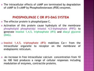
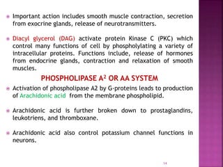
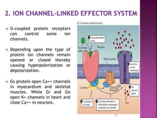
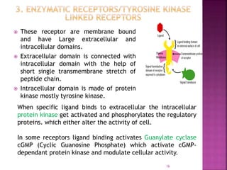
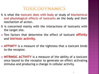
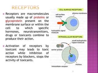
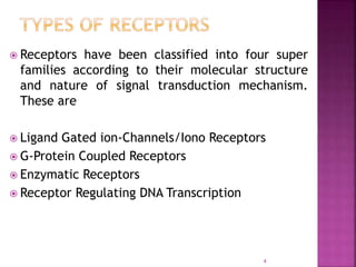
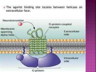
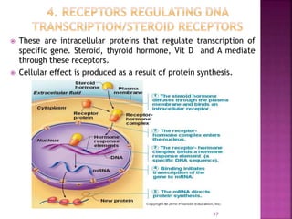
AMIR SOHAIL
1
It is what the toxicant does with body or study of biochemical
and physiological effects of toxicants on the body and their
mechanism of action.
It is concerned mainly with the interactions of toxicants with
the target site.
 Two factors that determine the effect of toxicant affinity
and intrinsic activity.
 AFFINITY is a measure of the tightness that a toxicant binds
to the receptor.
 INTRINSIC ACTIVITY is a measure of the ability of a toxicant
once bound to the receptor to generate an effect activating
stimulus and producing a change in cellular activity.
2
 Receptors are macromolecules
usually made up of proteins or
glycoprotein present on the
membrane surface or within the
cell to which specific
hormones, neurotransmitters,
drugs or toxicants combine to
produce their action.
 Activation of receptors by
toxicant may leads to toxic
action while inhibition of
receptors by blockers, stops the
activity of toxicants.
3
 Receptors have been classified into four super
families according to their molecular structure
and nature of signal transduction mechanism.
These are
 Ligand Gated ion-Channels/Iono Receptors
 G-Protein Coupled Receptors
 Enzymatic Receptors
 Receptor Regulating DNA Transcription
4
 These are cell surface receptors that enclose selective
ion-channels which allow the free movement of certain
ions like sodium, potassium, calcium and chlorides.
 Receptors of this type control the fastest synaptic events
in nervous system where lag time between ligand binding
and cellular response is only in milliseconds.
 Examples includes nicotinic-cholinergic receptors, GABA
receptors.
STRUCTURE
All ligand gated ion channels are made up of subunit
proteins. (Alpha, beta, gamma, delta kappa).
These subunit penetrate all the way through the
membrane laying side by side in a circle to form a tubular
channel (Like petals of lily).
5
6
SIGNALING MECHANISM
 The toxicant molecule attached
with the ligand binding site
located on subunit protein
directly.
 The channel remain activated
until the ligand attached to the
extracellular binding site.
 Due to the concentration
gradient flow of ions the electric
potential across the membrane
changed causing depolarization
(Na+ Channels) or hyper
polarization (Cl- Channels) of cell
7
 Large super family of membrane bound receptors which
regulate distinct effector proteins (Enzymes, Channels, or
Carriers) through mediation of transducers, a group of
guanine nucleotides (GTP and GDP) binding proteins known
as G-Proteins. These are heterotrimeric molecules
consisting of three subunits (α, β, and γ).
 Example of G-Protein Coupled receptors include
muscarinic-cholinergic receptors, Histamine receptors,
Adrenergic receptors and many peptide hormone
receptors.
STRUCTURE
 They consist of 400-500 chains of polypeptides, The amino
terminus followed by amino acid sequence having 7 Alpha
helical membrane spanning hydrophobic region and 3
extracellular and 3 intracellular loops.
 The agonist binding site locates between helicase on
extracellular face.
9
SIGNALING MECHANISM
 In the resting stage the G-Proteins exists as an unattached α,
β, and γ trimer with Guanosine diphosphate (GDP) occupying
the site on α subunit.
 When the agonist binds to G-Protein receptor, the agonist-
receptor complex facilitates displacement of Guanosine
diphosphate (GDP) by Guanosine triphosphate (GTP).
 Binding of GTP activates the α subunit and the α-GTP
complex then dissociate from β and γ subunit and interact
with membrane bound effector protein(Ion channel, enzyme,
or transport protein).
 The activation process terminated when hydrolysis of
GTP to GDP occurs through GTPase activity of α-subunit.
 It has been estimated that initial agonist-receptor
interaction lasts only for a few seconds but once a G-
protein is activated it remain active for about 10
seconds. Which amplify the signals. Thus in G-Coupled
system occupancy of only a fraction of receptor
population is enough to produce maximal response.
 Gs, Gi, Go, Gq and G13 are different type of G-Proteins
 G-Coupled Effector system function by
1. ENZYME LINKED EFFECTOR SYSTEM
2. ION CHANNEL LINKED EFFECTOR SYSTEM
11
 Three Effector systems through which these receptors function.
 Adenylate Cyclase or cAMP system
 Phospholipase C or IP3-DAG system
 Phospholipase A2 or AA System
ADENYLATE CYCLASE OR cAMP SYSTEM
 The effector protein is Adenylate cyclase which is activated by
α-GTP complex.
 Activation of Adenylate Cyclase enzyme by stimulatory G-
Protein (Gs) leads to increased synthesis of cAMP (3-5 cyclic
adenisine monophopshate).
 Accumulation of cAMP within the cell activate cAMP-dependant
Protein Kinase.
 These Protein kinases phosphorylate and alter the function of
cell.
 The intracellular effects of cAMP are terminated by degradation
of cAMP to 5-cAMP by Phosphodiesterases (PDE) enzymes.
PHOSPHOLIPASE C OR IP3-DAG SYSTEM
 The effector protein is phospholipase C.
 Activation of this protein cause hydrolysis of the membrane
phospholipid phosphatidyl inisotol 4,5-biphosphate (PIP2) to
generate Inositol 1,4,5, triphosphate (IP3) and diacyl glycerol
(DAG).
 Inositol 1,4,5, triphosphate (IP3) mobilizes Ca++ from the
intracellular organelle to receptor on the membrane of
endoplasmic reticulum.
 An increase in free intracellular calcium concentration from 10
to 100 fold produces a range of cellular responses including
modulation of enzymes, contractile proteins.
 Important action includes smooth muscle contraction, secretion
from exocrine glands, release of neurotransmitters.
 Diacyl glycerol (DAG) activate protein Kinase C (PKC) which
control many functions of cell by phospholylating a variety of
intracellular proteins. Functions include, release of hormones
from endocrine glands, contraction and relaxation of smooth
muscles.
PHOSPHOLIPASE A2 OR AA SYSTEM
 Activation of phospholipase A2 by G-proteins leads to production
of Arachidonic acid from the membrane phospholipid.
 Arachidonic acid is further broken down to prostaglandins,
leukotriens, and thromboxane.
 Arachidonic acid also control potassium channel functions in
neurons.
14
 G-coupled protein receptors
can control some ion
channels.
 Depending upon the type of
protein ion channels remain
opened or closed thereby
causing hyperpolerization or
depolarization.
 Gs protein open Ca++ channels
in myocardium and skeletal
muscles. While Gi and Go
open K+ channels in heart and
close Ca++ in neurons.
15
 These receptor are membrane bound
and have Large extracellular and
intracellular domains.
 Extracellular domain is connected with
intracellular domain with the help of
short single transmembrane stretch of
peptide chain.
 Intracellular domain is made of protein
kinase mostly tyrosine kinase.
16
When specific ligand binds to extracellular the intracellular
protein kinase get activated and phosphorylates the regulatory
proteins. which either alter the activity of cell.
In some receptors ligand binding activates Guanylate cyclase
cGMP (Cyclic Guanosine Phosphate) which activate cGMP-
dependant protein kinase and modulate cellular activity.
 These are intracellular proteins that regulate transcription of
specific gene. Steroid, thyroid hormone, Vit D and A mediate
through these receptors.
 Cellular effect is produced as a result of protein synthesis.
17

Toxicodynamics or pharmacodynamics

  • 1.
  • 2.
    It is whatthe toxicant does with body or study of biochemical and physiological effects of toxicants on the body and their mechanism of action. It is concerned mainly with the interactions of toxicants with the target site.  Two factors that determine the effect of toxicant affinity and intrinsic activity.  AFFINITY is a measure of the tightness that a toxicant binds to the receptor.  INTRINSIC ACTIVITY is a measure of the ability of a toxicant once bound to the receptor to generate an effect activating stimulus and producing a change in cellular activity. 2
  • 3.
     Receptors aremacromolecules usually made up of proteins or glycoprotein present on the membrane surface or within the cell to which specific hormones, neurotransmitters, drugs or toxicants combine to produce their action.  Activation of receptors by toxicant may leads to toxic action while inhibition of receptors by blockers, stops the activity of toxicants. 3
  • 4.
     Receptors havebeen classified into four super families according to their molecular structure and nature of signal transduction mechanism. These are  Ligand Gated ion-Channels/Iono Receptors  G-Protein Coupled Receptors  Enzymatic Receptors  Receptor Regulating DNA Transcription 4
  • 5.
     These arecell surface receptors that enclose selective ion-channels which allow the free movement of certain ions like sodium, potassium, calcium and chlorides.  Receptors of this type control the fastest synaptic events in nervous system where lag time between ligand binding and cellular response is only in milliseconds.  Examples includes nicotinic-cholinergic receptors, GABA receptors. STRUCTURE All ligand gated ion channels are made up of subunit proteins. (Alpha, beta, gamma, delta kappa). These subunit penetrate all the way through the membrane laying side by side in a circle to form a tubular channel (Like petals of lily). 5
  • 6.
  • 7.
    SIGNALING MECHANISM  Thetoxicant molecule attached with the ligand binding site located on subunit protein directly.  The channel remain activated until the ligand attached to the extracellular binding site.  Due to the concentration gradient flow of ions the electric potential across the membrane changed causing depolarization (Na+ Channels) or hyper polarization (Cl- Channels) of cell 7
  • 8.
     Large superfamily of membrane bound receptors which regulate distinct effector proteins (Enzymes, Channels, or Carriers) through mediation of transducers, a group of guanine nucleotides (GTP and GDP) binding proteins known as G-Proteins. These are heterotrimeric molecules consisting of three subunits (α, β, and γ).  Example of G-Protein Coupled receptors include muscarinic-cholinergic receptors, Histamine receptors, Adrenergic receptors and many peptide hormone receptors. STRUCTURE  They consist of 400-500 chains of polypeptides, The amino terminus followed by amino acid sequence having 7 Alpha helical membrane spanning hydrophobic region and 3 extracellular and 3 intracellular loops.
  • 9.
     The agonistbinding site locates between helicase on extracellular face. 9
  • 10.
    SIGNALING MECHANISM  Inthe resting stage the G-Proteins exists as an unattached α, β, and γ trimer with Guanosine diphosphate (GDP) occupying the site on α subunit.  When the agonist binds to G-Protein receptor, the agonist- receptor complex facilitates displacement of Guanosine diphosphate (GDP) by Guanosine triphosphate (GTP).  Binding of GTP activates the α subunit and the α-GTP complex then dissociate from β and γ subunit and interact with membrane bound effector protein(Ion channel, enzyme, or transport protein).
  • 11.
     The activationprocess terminated when hydrolysis of GTP to GDP occurs through GTPase activity of α-subunit.  It has been estimated that initial agonist-receptor interaction lasts only for a few seconds but once a G- protein is activated it remain active for about 10 seconds. Which amplify the signals. Thus in G-Coupled system occupancy of only a fraction of receptor population is enough to produce maximal response.  Gs, Gi, Go, Gq and G13 are different type of G-Proteins  G-Coupled Effector system function by 1. ENZYME LINKED EFFECTOR SYSTEM 2. ION CHANNEL LINKED EFFECTOR SYSTEM 11
  • 12.
     Three Effectorsystems through which these receptors function.  Adenylate Cyclase or cAMP system  Phospholipase C or IP3-DAG system  Phospholipase A2 or AA System ADENYLATE CYCLASE OR cAMP SYSTEM  The effector protein is Adenylate cyclase which is activated by α-GTP complex.  Activation of Adenylate Cyclase enzyme by stimulatory G- Protein (Gs) leads to increased synthesis of cAMP (3-5 cyclic adenisine monophopshate).  Accumulation of cAMP within the cell activate cAMP-dependant Protein Kinase.  These Protein kinases phosphorylate and alter the function of cell.
  • 13.
     The intracellulareffects of cAMP are terminated by degradation of cAMP to 5-cAMP by Phosphodiesterases (PDE) enzymes. PHOSPHOLIPASE C OR IP3-DAG SYSTEM  The effector protein is phospholipase C.  Activation of this protein cause hydrolysis of the membrane phospholipid phosphatidyl inisotol 4,5-biphosphate (PIP2) to generate Inositol 1,4,5, triphosphate (IP3) and diacyl glycerol (DAG).  Inositol 1,4,5, triphosphate (IP3) mobilizes Ca++ from the intracellular organelle to receptor on the membrane of endoplasmic reticulum.  An increase in free intracellular calcium concentration from 10 to 100 fold produces a range of cellular responses including modulation of enzymes, contractile proteins.
  • 14.
     Important actionincludes smooth muscle contraction, secretion from exocrine glands, release of neurotransmitters.  Diacyl glycerol (DAG) activate protein Kinase C (PKC) which control many functions of cell by phospholylating a variety of intracellular proteins. Functions include, release of hormones from endocrine glands, contraction and relaxation of smooth muscles. PHOSPHOLIPASE A2 OR AA SYSTEM  Activation of phospholipase A2 by G-proteins leads to production of Arachidonic acid from the membrane phospholipid.  Arachidonic acid is further broken down to prostaglandins, leukotriens, and thromboxane.  Arachidonic acid also control potassium channel functions in neurons. 14
  • 15.
     G-coupled proteinreceptors can control some ion channels.  Depending upon the type of protein ion channels remain opened or closed thereby causing hyperpolerization or depolarization.  Gs protein open Ca++ channels in myocardium and skeletal muscles. While Gi and Go open K+ channels in heart and close Ca++ in neurons. 15
  • 16.
     These receptorare membrane bound and have Large extracellular and intracellular domains.  Extracellular domain is connected with intracellular domain with the help of short single transmembrane stretch of peptide chain.  Intracellular domain is made of protein kinase mostly tyrosine kinase. 16 When specific ligand binds to extracellular the intracellular protein kinase get activated and phosphorylates the regulatory proteins. which either alter the activity of cell. In some receptors ligand binding activates Guanylate cyclase cGMP (Cyclic Guanosine Phosphate) which activate cGMP- dependant protein kinase and modulate cellular activity.
  • 17.
     These areintracellular proteins that regulate transcription of specific gene. Steroid, thyroid hormone, Vit D and A mediate through these receptors.  Cellular effect is produced as a result of protein synthesis. 17