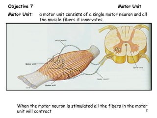
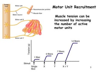
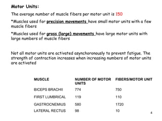
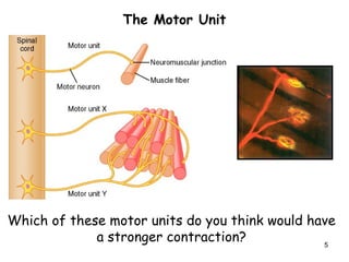
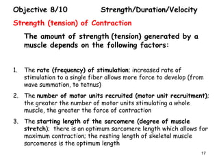
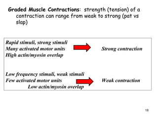
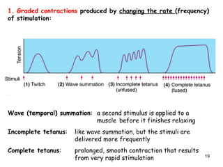
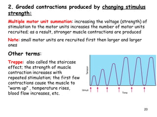
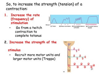
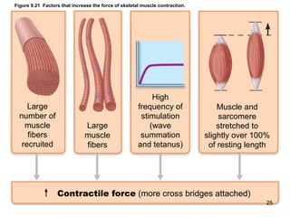
Objective 7: Define the term and discuss how
motor units are varying sizes are recruited to
produce movements of varying strength and
precision.
1
Objective 7 Motor Unit
Motor Unit: a motor unit consists of a single motor neuron and all
the muscle fibers it innervates.
When the motor neuron is stimulated all the fibers in the motor
unit will contract 2
Motor Unit Recruitment
Muscle tension can be
increased by increasing
the number of active
motor units
3
Motor Units:
The average number of muscle fibers per motor unit is 150
*Muscles used for precision movements have small motor units with a few
muscle fibers
*Muscles used for gross (large) movements have large motor units with
large numbers of muscle fibers
Not all motor units are activated asynchoronously to prevent fatigue. The
strength of contraction increases when increasing numbers of motor units
are activated
MUSCLE NUMBER OF MOTOR
UNITS
FIBERS/MOTOR UNIT
BICEPS BRACHII 774 750
FIRST LUMBRICAL 119 110
GASTROCNEMIUS 580 1720
LATERAL RECTUS 98 10
4
The Motor Unit
Which of these motor units do you think would have
a stronger contraction? 5
Objective 8: Describe the following types of muscle
responses to stimulation: twitch, graded muscle
contraction (wave summation, incomplete tetanus,
complete tetanus, multiple motor unit summation), treppe,
isotonic contraction and isometric contraction.
Muscle tone: refers to the fact that all muscles are in a slightly
contracted state, even if they appear to be relaxed;
this partial contraction is maintained by the nervous
system and keeps muscles from atrophy; it also
helps to maintain stability and posture
6
Contraction: the process of generating force in a muscle as a
result of cross bridge activity; contraction may or may not lead
to shortening
Tension (effort): the force exerted by a muscle on a load
Load (resistance): any force that opposes the tension (effort)
generated by muscle contraction
Muscle twitch/contraction: the response of a skeletal muscle to a single
brief stimulus
7
Latent (lag) period: short period of time after stimulation when
excitation/coupling is occurring; or from AP in nerve to
Ca++
binding to troponin
Contraction period: cross bridge activity leads to tension development;
the amount of tension depends on the number of
motor units recruited (activated)
Relaxation period: cross bridge activity stops as Ca2+
is pumped back
into the sarcoplasmic reticulum; muscle tension falls
Myogram: the graph of a muscles mechanical contractile activity
(tension or shortening) vs. time (t vs. T)
8
Types of Contraction: Isotonic vs. Isometric
Isotonic contraction: muscle changes length and moves a load
Tension generated by the muscle exceeds resistance (load)
and the muscle shortens, or T > L
Concentric Action:
muscle shortens
Eccentric action:
muscle lengthens
9
Isometric Contraction: muscle generates tension but
does not shorten or lengthen (isometric means same
length)
The resistance (load) is greater than the tension
developed by the muscle or L > T
10
Objective 9: Describe the role of the following in
supplying energy for muscle contraction: ATP,
creatine phosphate, anaerobic respiration and
aerobic respiration. Define the following related
terms: aerobic endurance, anaerobic threshold,
muscle fatigue, contractures and oxygen debt.
11
Objective 9 Metabolism
ATP is the preferred fuel for muscle contraction; it is needed for muscle
contraction and for muscle relaxation:
1. ATP fuels the power stroke: moves the cross bridges from low energy
to high energy positions
2. ATP is needed to detach cross bridges after the power stroke; new
ATP binds to cross bridges, allowing them to break free from the actin
filaments
3. ATP is needed for sarcomere relaxation since it is used to pump
calcium back into the sarcoplasmic reticulum
12
Where does a muscle cell get all of this ATP?
1) Muscle fibers store very little ATP; when activity starts, stored
ATP is hydrolyzed (1st
8 seconds of contraction)
Adenosine P-P-P
ATPase
Adenosine P-P + Pi + energy
Three additional sources of ATP for contracting
muscle fibers:
2) Creatine phosphate (8-10 seconds of contraction)
creatine
creatine P + ADP
kinase
creatine + ATP
13
3. Anaerobic glycolysis: quick but low yield (up to 3 minutes of
contraction)
glucose lactic acid + 2 ATP
- takes place in the sarcoplasm
- little ATP is generated from each glucose
- relatively quick pathway
- used primarily for short term, vigorous activity
(sprinting, diving, tennis, soccer, etc)
4. Aerobic respiration: slow but high yield (endurance activities)
glucose + O2 CO2 + H2O + 36 ATP
- takes place in mitochondria
- slower pathway
- used for prolonged activities, such as jogging
14
Definitions:
Aerobic endurance: the length of time that a muscle can continue to
contract using aerobic pathways
Anaerobic threshold: the point at which muscle metabolism switches
from aerobic to anaerobic
Muscle fatigue: a state in which a muscle is physiologically unable
to contract; in this state, the rate of ATP
production is less than the rate of ATP consumption
Oxygen debt: the amount of oxygen needed to restore muscle to
its resting metabolic state
In the muscle cell:
lactic acid + O2 pyruvic acid ATP + CO2 + H2O
-the ATP generated will restore resting levels of ATP and CP
In the liver:
lactic acid glucose glycogen
15
16
Objective 8/10 Strength/Duration/Velocity
Strength (tension) of Contraction
The amount of strength (tension) generated by a
muscle depends on the following factors:
1. The rate (frequency) of stimulation; increased rate of
stimulation to a single fiber allows more force to develop (from
wave summation, to tetnus)
2. The number of motor units recruited (motor unit recruitment);
the greater the number of motor units stimulating a whole
muscle, the greater the force of contraction
3. The starting length of the sarcomere (degree of muscle
stretch); there is an optimum sarcomere length which allows for
maximum contraction; the resting length of skeletal muscle
sarcomeres is the optimum length
17
Graded Muscle Contractions: strength (tension) of a
contraction can range from weak to strong (pat vs
slap)
Rapid stimuli, strong stimuli
Many activated motor units Strong contraction
High actin/myosin overlap
Low frequency stimuli, weak stimuli
Few activated motor units Weak contraction
Low actin/myosin overlap
18
1. Graded contractions produced by changing the rate (frequency)
of stimulation:
Wave (temporal) summation: a second stimulus is applied to a
muscle before it finishes relaxing
Incomplete tetanus: like wave summation, but the stimuli are
delivered more frequently
Complete tetanus: prolonged, smooth contraction that results
from very rapid stimulation 19
2. Graded contractions produced by changing stimulus
strength:
Multiple motor unit summation: increasing the voltage (strength) of
stimulation to the motor units increases the number of motor units
recruited; as a result, stronger muscle contractions are produced
Note: small motor units are recruited first then larger and larger
ones
Other terms:
Treppe: also called the staircase
effect; the strength of muscle
contraction increases with
repeated stimulation; the first few
contractions cause the muscle to
“warm up” , temperature rises,
blood flow increases, etc.
20
So, to increase the strength (tension) of a
contraction:
1. Increase the rate
(frequency) of
stimulation
– Go from a twitch
contraction to
complete tetanus
2. Increase the strength of the
stimulus
– Recruit more motor units and
larger motor units (Treppe)
21
3. Length-Tension Relationship
A maximum tetanic contraction is produced at an optimal overlap of thick
and thin filaments
22
By decreasing
filament overlap
no tension can
develop
By increasing fiber
(actin/mysosin) overlap,
Less tension can develop
Optimal overlap,
maximal tension can
develop
http://labyrinth.mvm.ed.ac.uk/mnode.asp?id=644
23
Velocity/Duration:
Velocity: the rate of contraction, speed over unit time (how fast)
Duration: how long the contraction lasts before fatigue sets in
Velocity and duration depend on characteristics of the load:
As load increases velocity decreasesAs load increases duration decreases
The Bottom Line
As load increases, velocity and duration decrease, or, the
heavier the weight the slower the lift and shorter the time
you can hold the weight 24
Figure 9.21 Factors that increase the force of skeletal muscle contraction.
Large
number of
muscle
fibers
recruited
Large
muscle
fibers
High
frequency of
stimulation
(wave
summation
and tetanus)
Muscle and
sarcomere
stretched to
slightly over 100%
of resting length
Contractile force (more cross bridges attached)
25
Figure 9.23 Factors influencing velocity and duration of skeletal muscle contraction.
Predominance
of fast glycolytic
(fatigable) Type III
fibers
Contractile
velocity
Small load Predominance
of slow oxidative
(fatigue-resistant)
Type I fibers
Contractile
duration
26
Velocity and duration also depends on the
characteristics of the muscle fiber
27
Fiber type (Type I) Slow Oxidative (Type II) Fast Oxidative (Type III) Fast Glycolytic
Structure slow myosin ATPase fast myosin ATPase fast mysosin ATPase
Aerobic adapted Aerobic adapted Anaerobic adapted
- myoglobin (red fibers) - some myoglobin (pinks) - no myoglobin (white fibers)
- well vascularized - well vascularized -poorly vascularized
-many mitochodnria - many mitochondria -few mitochondria
-small diameter fibers - intermediate diameter -large diameter
Functional -slow speed of contraction fast speed of contraction fast speed of contraction
Properties -fatigue resistant fatigue resistant fatigable
-used for endurance and used for endurance used for intense, short
prolonged contraction and rapid contraction term movements
28
-Repeated low-level
contractions e.g. walking
or low intensity cycling for
30 mins.
-Postural muscles, e.g. of
the neck and spine, & leg
muscles (which have Type
I & Type IIa fibres).
-Activities involving
speed, strength and
power, e.g. moderately
weight training and fast
running e.g. 400 metres.
-Leg muscles have large
quantities of both Type I
and Type IIa fibers.
Short, fast, bursts of
power (but rapid
fatigue) e.g. heavy
weight training,
power lifting, and
100 metre sprints.
-Arm muscles. N.B.
Type IIb fibres can
be converted into
IIa fibres by
resistance training.
Contraction: the process of generating force in a muscle as a
result of cross bridge activity; contraction may or may not lead
to shortening
Tension (effort): the force exerted by a muscle on a load
Load (resistance): any force that opposes the tension (effort)
generated by muscle contraction
Fulcrum (joint):elbow, shoulder…etc
Objective 12: Lever Systems
29
• Basic principle of levers
Mechanical Advantage: Mechanical Disadvantage:
Effort farther/load closer to fulcrum Effort closer/load farther from
fulcrum
• Three classes of levers
– Depends on relative position of effort, fulcrum, load
Effort
Load
Fulcrum
Effort
Load
Fulcrum
30
Figure 10.4a Lever systems. (1 of 2)
First-class lever
Arrangement:
load-fulcrum-effort
Load
L
Effort
Fulcrum
Load
L
Effort
Fulcrum
Load
Fulcrum
Effort
In the body: A first-class lever system
raises your head off your chest. The
posterior neck muscles provide the effort,
the atlanto-occipital joint is the fulcrum,
and the weight to be lifted is the facial
skeleton.
Example: scissors, seesaw
First-class lever
31
Figure 10.4b Lever systems. (1 of 2)
Second-class lever
Arrangement:
fulcrum-load-effort
Load
L
EffortFulcrum
Load
L
Effort
Fulcrum
Load
Fulcrum
Effort
In the body: Second-class leverage is
exerted when you stand on tip-toe. The
effort is exerted by the calf muscles
pulling upward on the heel; the joints of
the ball of the foot are the fulcrum; and
the weight of the body is the load.
Example: wheelbarrow, standing on toes
Second-class lever
32
Figure 10.4c Lever systems. (1 of 2)
Third-class lever
Arrangement:
load-effort-fulcrum
Load
L
Effort
Fulcrum
Load
L
Effort
Fulcrum
Load
Fulcrum
Effort
In the body: Flexing the forearm by the
biceps brachii muscle exemplifies
third-class leverage. The effort is exerted
on the proximal radius of the forearm, the
fulcrum is the elbow joint, and the load is
the hand and distal end of the forearm.
Example: tweezers or forceps
Third-class lever
33
Objective 13 Terms For Muscle Contraction
Prime Mover (agonist): principle muscle performing a movement
Antagonist: muscle the reverse the action of a prime mover
Synergist: muscle that assists the prime mover by performing the
same action or by preventing an opposite action
Fixator: muscle that stabilizes the bone of origin of the muscle
Example: Arm abduction
Prime Mover: deltoid
Antagonist: latissimus dorsi
Synergist: supraspinatus
Fixator: pectoralis major
34
Objective 14 Disuse Atrophy
Disorder Cause(s) Characteristics
Disuse Atrophy prolonged inactivity due to such pathologic
reduction in the factors as bed rest, casting or normal size of a
muscle or local nerve damage muscle fibers
35
Disorder Cause(s) Characteristics
Muscle Cramps circulatory impairment, heat sustained involuntary
disorders which lead to contractions of a
electrolyte disorders skeletal muscle
diuretic intake, unknown
causes
Muscular dystrophy
X linked recessive disorder see a reduction in the
in ½ of all cases; defective number of muscle
gene for dystrophin fibers and necrosis
causing dystrophin to be and replacement
absent or abnormal with endomysial
connective tissue and
fat; delayed sitting and
walking and
standing with
progressive weakness
in the shoulder and
pelvic girdles
36
Disorder Cause(s) Characteristics
Myasthenia Gravis defective transmission at fatigue; chronic
the neuromuscular junction respiratory infections
due to a reduction in the muscle weakness
number of Ach receptors
37
Objective 11 Smooth Muscle
A. Structure
• Small spindle shaped fibers, with a single centrally located nucleus
• No striations
• Poorly developed sarcoplasmic reticulum, no T tubules
• Sarcolemma has caveoli which sequester calcium (like SR in skeletal
muscle)
Calveoli 38
• Troponin is absent; tropomyosin is present but not at active
site
• No sarcomeres -thick and thin myofilaments are arranged
diagonally so that when the cell contracts, it twists like a
corkscrew
• No Z discs - non-contractile intermediate filaments are
present and are attached to dense bodies; dense bodies are
attached to the sarcolemma and to the thin myofilaments
No sarcomeres Dense bodies 39
-Individual muscle fibers are covered by endomysium
-Fibers may or may not be organized into fascicles
-In most organs, smooth muscle fibers are arranged into cellular
sheets with circular or longitudinal orientation
40
Smooth Muscle Contraction – mechanism
• Ca2+
concentrations increase in the sarcoplasm
• Ca2+
activates calmodulin (a troponin like molecule)
• Calmodulin activates myosin light chain kinase
• myosin light chain kinase uses ATP to phosphorylate myosin
cross bridges
• phosphorylated cross bridges bind to actin filaments
• cross bridge cycle produces tension and contraction/shortening
41
Contraction regulation
Neural regulation- stimulated by the ANS
- neurotransmitters include acetylcholine, norepinephrine
Local factors - some smooth muscle is stimulated by pacemaker cells,
hormones, anoxia, histidime, pH, CO2
Special Properties
Stress relaxation response- can stretch beyond resting length and
still contract (think bladder)
Hyperplasia– cells divide to increase cell numbers
42
Single – Unit (Visceral) Multi-unit
•Gap junctions
•Circular and longitudinal layers
•Contracts as a unit
•Walls of hollow organs bladder,
stomach, GI tract, uterus
•Few gap junctions
•Many nerve endings
•Walls of large airways, large
arteries, ciliary muscle (eye),
arrector pili
Types of Smooth Muscle
Intestine Artery and Vein 43

Topic4 objectives 7 14

  • 1.
    Objective 7: Definethe term and discuss how motor units are varying sizes are recruited to produce movements of varying strength and precision. 1
  • 2.
    Objective 7 MotorUnit Motor Unit: a motor unit consists of a single motor neuron and all the muscle fibers it innervates. When the motor neuron is stimulated all the fibers in the motor unit will contract 2
  • 3.
    Motor Unit Recruitment Muscletension can be increased by increasing the number of active motor units 3
  • 4.
    Motor Units: The averagenumber of muscle fibers per motor unit is 150 *Muscles used for precision movements have small motor units with a few muscle fibers *Muscles used for gross (large) movements have large motor units with large numbers of muscle fibers Not all motor units are activated asynchoronously to prevent fatigue. The strength of contraction increases when increasing numbers of motor units are activated MUSCLE NUMBER OF MOTOR UNITS FIBERS/MOTOR UNIT BICEPS BRACHII 774 750 FIRST LUMBRICAL 119 110 GASTROCNEMIUS 580 1720 LATERAL RECTUS 98 10 4
  • 5.
    The Motor Unit Whichof these motor units do you think would have a stronger contraction? 5
  • 6.
    Objective 8: Describethe following types of muscle responses to stimulation: twitch, graded muscle contraction (wave summation, incomplete tetanus, complete tetanus, multiple motor unit summation), treppe, isotonic contraction and isometric contraction. Muscle tone: refers to the fact that all muscles are in a slightly contracted state, even if they appear to be relaxed; this partial contraction is maintained by the nervous system and keeps muscles from atrophy; it also helps to maintain stability and posture 6
  • 7.
    Contraction: the processof generating force in a muscle as a result of cross bridge activity; contraction may or may not lead to shortening Tension (effort): the force exerted by a muscle on a load Load (resistance): any force that opposes the tension (effort) generated by muscle contraction Muscle twitch/contraction: the response of a skeletal muscle to a single brief stimulus 7
  • 8.
    Latent (lag) period:short period of time after stimulation when excitation/coupling is occurring; or from AP in nerve to Ca++ binding to troponin Contraction period: cross bridge activity leads to tension development; the amount of tension depends on the number of motor units recruited (activated) Relaxation period: cross bridge activity stops as Ca2+ is pumped back into the sarcoplasmic reticulum; muscle tension falls Myogram: the graph of a muscles mechanical contractile activity (tension or shortening) vs. time (t vs. T) 8
  • 9.
    Types of Contraction:Isotonic vs. Isometric Isotonic contraction: muscle changes length and moves a load Tension generated by the muscle exceeds resistance (load) and the muscle shortens, or T > L Concentric Action: muscle shortens Eccentric action: muscle lengthens 9
  • 10.
    Isometric Contraction: musclegenerates tension but does not shorten or lengthen (isometric means same length) The resistance (load) is greater than the tension developed by the muscle or L > T 10
  • 11.
    Objective 9: Describethe role of the following in supplying energy for muscle contraction: ATP, creatine phosphate, anaerobic respiration and aerobic respiration. Define the following related terms: aerobic endurance, anaerobic threshold, muscle fatigue, contractures and oxygen debt. 11
  • 12.
    Objective 9 Metabolism ATPis the preferred fuel for muscle contraction; it is needed for muscle contraction and for muscle relaxation: 1. ATP fuels the power stroke: moves the cross bridges from low energy to high energy positions 2. ATP is needed to detach cross bridges after the power stroke; new ATP binds to cross bridges, allowing them to break free from the actin filaments 3. ATP is needed for sarcomere relaxation since it is used to pump calcium back into the sarcoplasmic reticulum 12
  • 13.
    Where does amuscle cell get all of this ATP? 1) Muscle fibers store very little ATP; when activity starts, stored ATP is hydrolyzed (1st 8 seconds of contraction) Adenosine P-P-P ATPase Adenosine P-P + Pi + energy Three additional sources of ATP for contracting muscle fibers: 2) Creatine phosphate (8-10 seconds of contraction) creatine creatine P + ADP kinase creatine + ATP 13
  • 14.
    3. Anaerobic glycolysis:quick but low yield (up to 3 minutes of contraction) glucose lactic acid + 2 ATP - takes place in the sarcoplasm - little ATP is generated from each glucose - relatively quick pathway - used primarily for short term, vigorous activity (sprinting, diving, tennis, soccer, etc) 4. Aerobic respiration: slow but high yield (endurance activities) glucose + O2 CO2 + H2O + 36 ATP - takes place in mitochondria - slower pathway - used for prolonged activities, such as jogging 14
  • 15.
    Definitions: Aerobic endurance: thelength of time that a muscle can continue to contract using aerobic pathways Anaerobic threshold: the point at which muscle metabolism switches from aerobic to anaerobic Muscle fatigue: a state in which a muscle is physiologically unable to contract; in this state, the rate of ATP production is less than the rate of ATP consumption Oxygen debt: the amount of oxygen needed to restore muscle to its resting metabolic state In the muscle cell: lactic acid + O2 pyruvic acid ATP + CO2 + H2O -the ATP generated will restore resting levels of ATP and CP In the liver: lactic acid glucose glycogen 15
  • 16.
  • 17.
    Objective 8/10 Strength/Duration/Velocity Strength(tension) of Contraction The amount of strength (tension) generated by a muscle depends on the following factors: 1. The rate (frequency) of stimulation; increased rate of stimulation to a single fiber allows more force to develop (from wave summation, to tetnus) 2. The number of motor units recruited (motor unit recruitment); the greater the number of motor units stimulating a whole muscle, the greater the force of contraction 3. The starting length of the sarcomere (degree of muscle stretch); there is an optimum sarcomere length which allows for maximum contraction; the resting length of skeletal muscle sarcomeres is the optimum length 17
  • 18.
    Graded Muscle Contractions:strength (tension) of a contraction can range from weak to strong (pat vs slap) Rapid stimuli, strong stimuli Many activated motor units Strong contraction High actin/myosin overlap Low frequency stimuli, weak stimuli Few activated motor units Weak contraction Low actin/myosin overlap 18
  • 19.
    1. Graded contractionsproduced by changing the rate (frequency) of stimulation: Wave (temporal) summation: a second stimulus is applied to a muscle before it finishes relaxing Incomplete tetanus: like wave summation, but the stimuli are delivered more frequently Complete tetanus: prolonged, smooth contraction that results from very rapid stimulation 19
  • 20.
    2. Graded contractionsproduced by changing stimulus strength: Multiple motor unit summation: increasing the voltage (strength) of stimulation to the motor units increases the number of motor units recruited; as a result, stronger muscle contractions are produced Note: small motor units are recruited first then larger and larger ones Other terms: Treppe: also called the staircase effect; the strength of muscle contraction increases with repeated stimulation; the first few contractions cause the muscle to “warm up” , temperature rises, blood flow increases, etc. 20
  • 21.
    So, to increasethe strength (tension) of a contraction: 1. Increase the rate (frequency) of stimulation – Go from a twitch contraction to complete tetanus 2. Increase the strength of the stimulus – Recruit more motor units and larger motor units (Treppe) 21
  • 22.
    3. Length-Tension Relationship Amaximum tetanic contraction is produced at an optimal overlap of thick and thin filaments 22
  • 23.
    By decreasing filament overlap notension can develop By increasing fiber (actin/mysosin) overlap, Less tension can develop Optimal overlap, maximal tension can develop http://labyrinth.mvm.ed.ac.uk/mnode.asp?id=644 23
  • 24.
    Velocity/Duration: Velocity: the rateof contraction, speed over unit time (how fast) Duration: how long the contraction lasts before fatigue sets in Velocity and duration depend on characteristics of the load: As load increases velocity decreasesAs load increases duration decreases The Bottom Line As load increases, velocity and duration decrease, or, the heavier the weight the slower the lift and shorter the time you can hold the weight 24
  • 25.
    Figure 9.21 Factorsthat increase the force of skeletal muscle contraction. Large number of muscle fibers recruited Large muscle fibers High frequency of stimulation (wave summation and tetanus) Muscle and sarcomere stretched to slightly over 100% of resting length Contractile force (more cross bridges attached) 25
  • 26.
    Figure 9.23 Factorsinfluencing velocity and duration of skeletal muscle contraction. Predominance of fast glycolytic (fatigable) Type III fibers Contractile velocity Small load Predominance of slow oxidative (fatigue-resistant) Type I fibers Contractile duration 26
  • 27.
    Velocity and durationalso depends on the characteristics of the muscle fiber 27
  • 28.
    Fiber type (TypeI) Slow Oxidative (Type II) Fast Oxidative (Type III) Fast Glycolytic Structure slow myosin ATPase fast myosin ATPase fast mysosin ATPase Aerobic adapted Aerobic adapted Anaerobic adapted - myoglobin (red fibers) - some myoglobin (pinks) - no myoglobin (white fibers) - well vascularized - well vascularized -poorly vascularized -many mitochodnria - many mitochondria -few mitochondria -small diameter fibers - intermediate diameter -large diameter Functional -slow speed of contraction fast speed of contraction fast speed of contraction Properties -fatigue resistant fatigue resistant fatigable -used for endurance and used for endurance used for intense, short prolonged contraction and rapid contraction term movements 28 -Repeated low-level contractions e.g. walking or low intensity cycling for 30 mins. -Postural muscles, e.g. of the neck and spine, & leg muscles (which have Type I & Type IIa fibres). -Activities involving speed, strength and power, e.g. moderately weight training and fast running e.g. 400 metres. -Leg muscles have large quantities of both Type I and Type IIa fibers. Short, fast, bursts of power (but rapid fatigue) e.g. heavy weight training, power lifting, and 100 metre sprints. -Arm muscles. N.B. Type IIb fibres can be converted into IIa fibres by resistance training.
  • 29.
    Contraction: the processof generating force in a muscle as a result of cross bridge activity; contraction may or may not lead to shortening Tension (effort): the force exerted by a muscle on a load Load (resistance): any force that opposes the tension (effort) generated by muscle contraction Fulcrum (joint):elbow, shoulder…etc Objective 12: Lever Systems 29
  • 30.
    • Basic principleof levers Mechanical Advantage: Mechanical Disadvantage: Effort farther/load closer to fulcrum Effort closer/load farther from fulcrum • Three classes of levers – Depends on relative position of effort, fulcrum, load Effort Load Fulcrum Effort Load Fulcrum 30
  • 31.
    Figure 10.4a Leversystems. (1 of 2) First-class lever Arrangement: load-fulcrum-effort Load L Effort Fulcrum Load L Effort Fulcrum Load Fulcrum Effort In the body: A first-class lever system raises your head off your chest. The posterior neck muscles provide the effort, the atlanto-occipital joint is the fulcrum, and the weight to be lifted is the facial skeleton. Example: scissors, seesaw First-class lever 31
  • 32.
    Figure 10.4b Leversystems. (1 of 2) Second-class lever Arrangement: fulcrum-load-effort Load L EffortFulcrum Load L Effort Fulcrum Load Fulcrum Effort In the body: Second-class leverage is exerted when you stand on tip-toe. The effort is exerted by the calf muscles pulling upward on the heel; the joints of the ball of the foot are the fulcrum; and the weight of the body is the load. Example: wheelbarrow, standing on toes Second-class lever 32
  • 33.
    Figure 10.4c Leversystems. (1 of 2) Third-class lever Arrangement: load-effort-fulcrum Load L Effort Fulcrum Load L Effort Fulcrum Load Fulcrum Effort In the body: Flexing the forearm by the biceps brachii muscle exemplifies third-class leverage. The effort is exerted on the proximal radius of the forearm, the fulcrum is the elbow joint, and the load is the hand and distal end of the forearm. Example: tweezers or forceps Third-class lever 33
  • 34.
    Objective 13 TermsFor Muscle Contraction Prime Mover (agonist): principle muscle performing a movement Antagonist: muscle the reverse the action of a prime mover Synergist: muscle that assists the prime mover by performing the same action or by preventing an opposite action Fixator: muscle that stabilizes the bone of origin of the muscle Example: Arm abduction Prime Mover: deltoid Antagonist: latissimus dorsi Synergist: supraspinatus Fixator: pectoralis major 34
  • 35.
    Objective 14 DisuseAtrophy Disorder Cause(s) Characteristics Disuse Atrophy prolonged inactivity due to such pathologic reduction in the factors as bed rest, casting or normal size of a muscle or local nerve damage muscle fibers 35
  • 36.
    Disorder Cause(s) Characteristics MuscleCramps circulatory impairment, heat sustained involuntary disorders which lead to contractions of a electrolyte disorders skeletal muscle diuretic intake, unknown causes Muscular dystrophy X linked recessive disorder see a reduction in the in ½ of all cases; defective number of muscle gene for dystrophin fibers and necrosis causing dystrophin to be and replacement absent or abnormal with endomysial connective tissue and fat; delayed sitting and walking and standing with progressive weakness in the shoulder and pelvic girdles 36
  • 37.
    Disorder Cause(s) Characteristics MyastheniaGravis defective transmission at fatigue; chronic the neuromuscular junction respiratory infections due to a reduction in the muscle weakness number of Ach receptors 37
  • 38.
    Objective 11 SmoothMuscle A. Structure • Small spindle shaped fibers, with a single centrally located nucleus • No striations • Poorly developed sarcoplasmic reticulum, no T tubules • Sarcolemma has caveoli which sequester calcium (like SR in skeletal muscle) Calveoli 38
  • 39.
    • Troponin isabsent; tropomyosin is present but not at active site • No sarcomeres -thick and thin myofilaments are arranged diagonally so that when the cell contracts, it twists like a corkscrew • No Z discs - non-contractile intermediate filaments are present and are attached to dense bodies; dense bodies are attached to the sarcolemma and to the thin myofilaments No sarcomeres Dense bodies 39
  • 40.
    -Individual muscle fibersare covered by endomysium -Fibers may or may not be organized into fascicles -In most organs, smooth muscle fibers are arranged into cellular sheets with circular or longitudinal orientation 40
  • 41.
    Smooth Muscle Contraction– mechanism • Ca2+ concentrations increase in the sarcoplasm • Ca2+ activates calmodulin (a troponin like molecule) • Calmodulin activates myosin light chain kinase • myosin light chain kinase uses ATP to phosphorylate myosin cross bridges • phosphorylated cross bridges bind to actin filaments • cross bridge cycle produces tension and contraction/shortening 41
  • 42.
    Contraction regulation Neural regulation-stimulated by the ANS - neurotransmitters include acetylcholine, norepinephrine Local factors - some smooth muscle is stimulated by pacemaker cells, hormones, anoxia, histidime, pH, CO2 Special Properties Stress relaxation response- can stretch beyond resting length and still contract (think bladder) Hyperplasia– cells divide to increase cell numbers 42
  • 43.
    Single – Unit(Visceral) Multi-unit •Gap junctions •Circular and longitudinal layers •Contracts as a unit •Walls of hollow organs bladder, stomach, GI tract, uterus •Few gap junctions •Many nerve endings •Walls of large airways, large arteries, ciliary muscle (eye), arrector pili Types of Smooth Muscle Intestine Artery and Vein 43