Downloaded 256 times


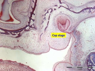


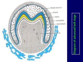


The document discusses the stages of tooth development from initiation to root formation. It describes the morphological stages as dental lamina, bud, cap, and bell stages. The histophysiological stages are initiation, proliferation, histodifferentiation, morphodifferentiation, and apposition. Key events include formation of the dental lamina and tooth buds, invagination of the enamel organ during the cap and bell stages, differentiation of ameloblasts and odontoblasts, and deposition of enamel and dentin. During the root stage, the cervical loop forms the root sheath to guide root formation.


